
Enterobacteriaceae is a large family of Gram-negative bacteria. It includes over 30 genera and more than 100 species. Its classification above the level of family is still a subject of debate, but one classification places it in the order Enterobacterales of the class Gammaproteobacteria in the phylum Pseudomonadota. In 2016, the description and members of this family were emended based on comparative genomic analyses by Adeolu et al.

Gram-negative bacteria are bacteria that do not retain the crystal violet stain used in the Gram staining method of bacterial differentiation. They are characterized by their cell envelopes, which are composed of a thin peptidoglycan cell wall sandwiched between an inner cytoplasmic cell membrane and a bacterial outer membrane.

Oxidative phosphorylation or electron transport-linked phosphorylation or terminal oxidation is the metabolic pathway in which cells use enzymes to oxidize nutrients, thereby releasing chemical energy in order to produce adenosine triphosphate (ATP). In eukaryotes, this takes place inside mitochondria. Almost all aerobic organisms carry out oxidative phosphorylation. This pathway is so pervasive because it releases more energy than alternative fermentation processes such as anaerobic glycolysis.
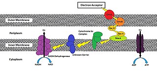
An electron transport chain (ETC) is a series of protein complexes and other molecules that transfer electrons from electron donors to electron acceptors via redox reactions (both reduction and oxidation occurring simultaneously) and couples this electron transfer with the transfer of protons (H+ ions) across a membrane. The electrons that are transferred from NADH and FADH2 to the ETC involves four multi-subunit large enzymes complexes and two mobile electron carriers. Many of the enzymes in the electron transport chain are embedded within the membrane.
In biochemistry, an oxidase is an enzyme that catalyzes oxidation-reduction reactions, especially one involving dioxygen (O2) as the electron acceptor. In reactions involving donation of a hydrogen atom, oxygen is reduced to water (H2O) or hydrogen peroxide (H2O2). Some oxidation reactions, such as those involving monoamine oxidase or xanthine oxidase, typically do not involve free molecular oxygen.

The Pseudomonadaceae are a family of bacteria which includes the genera Azomonas, Azorhizophilus, Azotobacter, Mesophilobacter, Pseudomonas, and Rugamonas. The family Azotobacteraceae was recently reclassified into this family.

Shigella dysenteriae is a species of the rod-shaped bacterial genus Shigella. Shigella species can cause shigellosis. Shigellae are Gram-negative, non-spore-forming, facultatively anaerobic, nonmotile bacteria. S. dysenteriae has the ability to invade and replicate in various species of epithelial cells and enterocytes.

Moraxella catarrhalis is a fastidious, nonmotile, Gram-negative, aerobic, oxidase-positive diplococcus that can cause infections of the respiratory system, middle ear, eye, central nervous system, and joints of humans. It causes the infection of the host cell by sticking to the host cell using trimeric autotransporter adhesins.

A diplococcus is a round bacterium that typically occurs in the form of two joined cells.
The indole test is a biochemical test performed on bacterial species to determine the ability of the organism to convert tryptophan into indole. This division is performed by a chain of a number of different intracellular enzymes, a system generally referred to as "tryptophanase."
Moraxella is a genus of gram-negative bacteria in the family Moraxellaceae. It is named after the Swiss ophthalmologist Victor Morax. The organisms are short rods, coccobacilli, or as in the case of Moraxella catarrhalis, diplococci in morphology, with asaccharolytic, oxidase-positive, and catalase-positive properties. M. catarrhalis is the clinically most important species under this genus.
Taylorella is a genus comprising Gram-negative, short rod-shaped, chemoorganotrophic bacteria that include species that are the causative agents of contagious equine metritis. The name Taylorella serves as a dedication to C.E.D. Taylor, the scientist who identified the only species originally included in this genus. They are non-motile microaerophiles that are able to be isolated in pure culture on chocolate agar..
Campylobacter upsaliensis is a gram-negative bacteria in the Campylobacter genus. C. upsaliensis is found worldwide, and is a common cause of campylobacteriosis in humans, as well as gastroenteritis in dogs. Human infections are primarily associated with raw or undercooked meat and contaminated water sources, however there is some zoonotic risk associated with the spread from dogs. C. upsaliensis primarily affects the gastrointestinal tract as it damages gastrointestinal epithelial cells. There are many methods for detecting C.upsaliensis including PCR and ELISA, however there is no current gold standard in detection techniques. Infection is typically self limiting, however there is antimicrobial therapy available.

Wurster's blue is the trivial name given to the chemical N,N,N′,N′-tetramethyl-p-phenylenediamine, also known as TMPD. It is an easily oxidised phenylenediamine, which loses two electrons in one-electron oxidation steps; the radical cation is a characteristic blue-violet colour, which gives the compound part of its name. The remaining part of its name comes from its discoverer, the German chemist Casimir Wurster.

The IMViC tests are a group of individual tests used in microbiology lab testing to identify an organism in the coliform group. A coliform is a gram negative, aerobic, or facultative anaerobic rod, which produces gas from lactose within 48 hours. The presence of some coliforms indicate fecal contamination.
The Analytical profile index or API is a classification of bacteria based on biochemical tests, allowing fast identification. This system is developed for the quick identification of clinically relevant bacteria. Because of this, only known bacteria can be identified.
An exoelectrogen normally refers to a microorganism that has the ability to transfer electrons extracellularly. While exoelectrogen is the predominant name, other terms have been used: electrochemically active bacteria, anode respiring bacteria, and electricigens. Electrons exocytosed in this fashion are produced following ATP production using an electron transport chain (ETC) during oxidative phosphorylation. Conventional cellular respiration requires a final electron acceptor to receive these electrons. Cells that use molecular oxygen (O2) as their final electron acceptor are described as using aerobic respiration, while cells that use other soluble compounds as their final electron acceptor are described as using anaerobic respiration. However, the final electron acceptor of an exoelectrogen is found extracellularly and can be a strong oxidizing agent in aqueous solution or a solid conductor/electron acceptor. Two commonly observed acceptors are iron compounds (specifically Fe(III) oxides) and manganese compounds (specifically Mn(III/IV) oxides). As oxygen is a strong oxidizer, cells are able to do this strictly in the absence of oxygen.
Voges–Proskauer or VP is a test used to detect acetoin in a bacterial broth culture. The test is performed by adding alpha-naphthol and potassium hydroxide to the Voges-Proskauer broth, which is a glucose-phosphate broth that has been inoculated with bacteria. A cherry red color indicates a positive result, while a yellow-brown color indicates a negative result.
Pasteurella canis is a Gram-negative, nonmotile, penicillin-sensitive coccobacillus of the family Pasteurellaceae. Bacteria from this family cause zoonotic infections in humans, which manifest themselves as skin or soft-tissue infections after an animal bite. It has been known to cause serious disease in immunocompromised patients.
Diagnostic microbiology is the study of microbial identification. Since the discovery of the germ theory of disease, scientists have been finding ways to harvest specific organisms. Using methods such as differential media or genome sequencing, physicians and scientists can observe novel functions in organisms for more effective and accurate diagnosis of organisms. Methods used in diagnostic microbiology are often used to take advantage of a particular difference in organisms and attain information about what species it can be identified as, which is often through a reference of previous studies. New studies provide information that others can reference so that scientists can attain a basic understanding of the organism they are examining.

