Pathology is the study of the causes and effects of disease or injury. The word pathology also refers to the study of disease in general, incorporating a wide range of biology research fields and medical practices. However, when used in the context of modern medical treatment, the term is often used in a narrower fashion to refer to processes and tests that fall within the contemporary medical field of "general pathology", an area which includes a number of distinct but inter-related medical specialties that diagnose disease, mostly through analysis of tissue and human cell samples. Idiomatically, "a pathology" may also refer to the predicted or actual progression of particular diseases, and the affix pathy is sometimes used to indicate a state of disease in cases of both physical ailment and psychological conditions. A physician practicing pathology is called a pathologist.

The accessory nerve, also known as the eleventh cranial nerve, cranial nerve XI, or simply CN XI, is a cranial nerve that supplies the sternocleidomastoid and trapezius muscles. It is classified as the eleventh of twelve pairs of cranial nerves because part of it was formerly believed to originate in the brain. The sternocleidomastoid muscle tilts and rotates the head, whereas the trapezius muscle, connecting to the scapula, acts to shrug the shoulder.

The azygos vein is a vein running up the right side of the thoracic vertebral column draining itself towards the superior vena cava. It connects the systems of superior vena cava and inferior vena cava and can provide an alternative path for blood to the right atrium when either of the venae cavae is blocked.
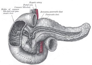
The pancreatic duct, or duct of Wirsung, is a duct joining the pancreas to the common bile duct. This supplies it with pancreatic juice from the exocrine pancreas, which aids in digestion.

In human anatomy, the inferior mesenteric artery, often abbreviated as IMA, is the third main branch of the abdominal aorta and arises at the level of L3, supplying the large intestine from the distal transverse colon to the upper part of the anal canal. The regions supplied by the IMA are the descending colon, the sigmoid colon, and part of the rectum.

In human anatomy, the anterior communicating artery is a blood vessel of the brain that connects the left and right anterior cerebral arteries.

The cerebellopontine angle (CPA) is located between the cerebellum and the pons. The cerebellopontine angle is the site of the cerebellopontine angle cistern.
The capillary lamina of choroid or choriocapillaris is a part of the choroid of the eye. It is a layer of capillaries immediately adjacent to Bruch's membrane of the choroid. The choriocapillaris consists of a dense network of freely anastomosing highly permeable fenestrated large-calibre capillaries. It nourishes the outer avascular layers of the retina.

The sural nerve(L4-S1) is generally considered a pure cutaneous nerve of the posterolateral leg to the lateral ankle. The sural nerve originates from a combination of either the sural communicating branch and medial sural cutaneous nerve, or the lateral sural cutaneous nerve. This group of nerves is termed the sural nerve complex. There are eight documented variations of the sural nerve complex. Once formed the sural nerve takes its course midline posterior to posterolateral around the lateral malleolus. The sural nerve terminates as the lateral dorsal cutaneous nerve.

The superior transverse ligament converts the suprascapular notch into a foramen or opening.

The suprascapular notch is a notch in the superior border of the scapula, just medial to the base of the coracoid process. It is converted into the suprascapular canal by the suprascapular ligament.

The clivus, or Blumenbach clivus, is a bony part of the cranium at the base of the skull. It is a shallow depression behind the dorsum sellae of the sphenoid bone. It slopes gradually to the anterior part of the basilar occipital bone at its junction with the sphenoid bone. It extends to the foramen magnum. It is related to the pons and the abducens nerve.
The incisive papilla is an oval midline mucosal prominence of the anterior hard palate overlying the incisive fossa. It is situated posteriorly to the central incisors, and represents the anterior extremity of the palatine raphe.

Max Israel Bielschowsky was a German neuropathologist born in Breslau.
A neurovascular bundle is a structure that binds nerves and veins with connective tissue so that they travel in tandem through the body.

The internal urethral sphincter is a urethral sphincter muscle which constricts the internal urethral orifice. It is located at the junction of the urethra with the urinary bladder and is continuous with the detrusor muscle, but anatomically and functionally fully independent from it. It is composed of smooth muscle, so it is under the control of the autonomic nervous system, specifically the sympathetic nervous system.

The external sphincter muscle of female urethra is a muscle which controls urination in females. The muscle fibers arise on either side from the margin of the inferior ramus of the pubis. They are directed across the pubic arch in front of the urethra, and pass around it to blend with the muscular fibers of the opposite side, between the urethra and vagina.

Gaetano Perusini was an Italian physician. He was the pupil and colleague of Alois Alzheimer and contributed to the definition of Alzheimer's disease.
Heinrich Sachs was a late 19th and early 20th century German neurologist and neuroanatomist best known for his atlas of the brain's white matter.
The mesencephalic locomotor region (MLR) is a functionally defined area of the midbrain that is associated with the initiation and control of locomotor movements in vertebrate species.