Troponin

Last updated
Troponin
Purposeto detect myocardial damage
Ribbon representation of the human cardiac troponin core complex (52 kDa core) in the calcium-saturated form. Blue = troponin C; green = troponin I; magenta = troponin T. Troponin Ribbon Diagram.png
Ribbon representation of the human cardiac troponin core complex (52 kDa core) in the calcium-saturated form. Blue = troponin C; green = troponin I; magenta = troponin T.

Troponin, or the troponin complex, is a complex of three regulatory proteins (troponin C, troponin I, and troponin T) that are integral to muscle contraction [2] in skeletal muscle and cardiac muscle, but not smooth muscle. Measurements of cardiac-specific troponins I and T are extensively used as diagnostic and prognostic indicators in the management of myocardial infarction and acute coronary syndrome. [3] Blood troponin levels may be used as a diagnostic marker for stroke or other myocardial injury that is ongoing, although the sensitivity of this measurement is low. [4] [5] [6] [7] [8]

Contents

Function

Troponin is attached to the protein tropomyosin and lies within the groove between actin filaments in muscle tissue. In a relaxed muscle, tropomyosin blocks the attachment site for the myosin crossbridge, thus preventing contraction. When the muscle cell is stimulated to contract by an action potential, calcium channels open in the sarcoplasmic membrane and release calcium into the sarcoplasm. Some of this calcium attaches to troponin, which causes it to change shape, exposing binding sites for myosin (active sites) on the actin filaments. Myosin's binding to actin causes crossbridge formation, and contraction of the muscle begins. [9]

Troponin activation. Troponin C (red) binds Ca2+, which stabilizes the activated state, where troponin I (yellow) is no longer bound to actin. Troponin T (blue) anchors the complex on tropomyosin. Troponin-activation.png
Troponin activation. Troponin C (red) binds Ca2+, which stabilizes the activated state, where troponin I (yellow) is no longer bound to actin. Troponin T (blue) anchors the complex on tropomyosin.

Troponin is found in both skeletal muscle and cardiac muscle, but the specific versions of troponin differ between types of muscle. The main difference is that the TnC subunit of troponin in skeletal muscle has four calcium ion-binding sites, whereas in cardiac muscle there are only three. The actual amount of calcium that binds to troponin has not been definitively established. [10]

Physiology

In both cardiac and skeletal muscles, muscular force production is controlled primarily by changes in intracellular calcium concentration. In general, when calcium rises, the muscles contract and, when calcium falls, the muscles relax.[ citation needed ]

Troponin is a component of thin filaments (along with actin and tropomyosin), and is the protein complex to which calcium binds to trigger the production of muscular force. Troponin has three subunits, TnC, TnI, and TnT, each playing a role in force regulation.[ citation needed ]. Under resting intracellular levels of calcium, tropomyosin covers the active actin sites to which myosin (a molecular motor organized in muscle thick filaments) binds in order to generate force. When calcium becomes bound to specific sites in the N-domain of TnC, a series of protein structural changes occurs,[ citation needed ] such that tropomyosin is rolled away from myosin-binding sites on actin, allowing myosin to attach to the thin filament and produce force and shorten the sarcomere.[ citation needed ]

Individual subunits serve different functions:[ citation needed ]

Smooth muscle does not have troponin. [11]

Subunits

TnT is a tropomyosin-binding subunit which regulates the interaction of troponin complex with thin filaments; TnI inhibits ATP-ase activity of acto-myosin; TnC is a Ca2+-binding subunit, playing the main role in Ca2+ dependent regulation of muscle contraction. [12]

TnT and TnI in cardiac muscle are presented by forms different from those in skeletal muscles. Two isoforms of TnI and two isoforms of TnT are expressed in human skeletal muscle tissue (skTnI and skTnT). Only one tissue-specific isoform of TnI is described for cardiac muscle tissue (cTnI), whereas the existence of several cardiac specific isoforms of TnT (cTnT) are described in the literature. No cardiac specific isoforms are known for human TnC. TnC in human cardiac muscle tissue is presented by an isoform typical for slow skeletal muscle. Another form of TnC, the fast skeletal TnC isoform, is more typical for fast skeletal muscles. [13] cTnI is expressed only in myocardium. No examples of cTnI expression in healthy or injured skeletal muscle or in other tissue types are known. cTnT is probably less cardiac specific. The expression of cTnT in skeletal tissue of patients with chronic skeletal muscle injuries has been described. [14]

Inside the cardiac troponin complex the strongest interaction between molecules has been demonstrated for cTnI – TnC binary complex especially in the presence of Ca2+ ( KA = 1.5 × 10−8 M−1). [15] TnC, forming a complex with cTnI, changes the conformation of cTnI molecule and shields part of its surface. According to the latest data cTnI is released in the blood stream of the patient in the form of binary complex with TnC or ternary complex with cTnT and TnC. [16] cTnI-TnC complex formation plays an important positive role in improving the stability of cTnI molecule. cTnI, which is extremely unstable in its free form, demonstrates significantly better stability in complex with TnC or in ternary cTnI-cTnT-TnC complex. It has been demonstrated that stability of cTnI in native complex is significantly better than stability of the purified form of the protein or the stability of cTnI in artificial troponin complexes combined from purified proteins.[ citation needed ]

Research

Cardiac conditions

Subtypes of troponin (cardiac I and T) are sensitive and specific indicators of heart muscle damage (myocardium). They are measured in the blood to differentiate between unstable angina and myocardial infarction (heart attack) in people with chest pain or acute coronary syndrome. A person who recently had a myocardial infarction has areas of damaged heart muscle and elevated cardiac troponin levels in the blood. [17] This can also occur in people with coronary vasospasm, a type of myocardial infarction involving severe constriction of the cardiac blood vessels. After a myocardial infarction troponins may remain high for up to 2 weeks. [18]

Cardiac troponins are a marker of all heart muscle damage, not just myocardial infarction, which is the most severe form of heart disorder. However, diagnostic criteria for raised troponin indicating myocardial infarction is currently set by the WHO at a threshold of 2 μg or higher. Critical levels of other cardiac biomarkers are also relevant, such as creatine kinase. [19] Other conditions that directly or indirectly lead to heart muscle damage and death can also increase troponin levels, such as kidney failure. [20] [21] Severe tachycardia (for example due to supraventricular tachycardia) in an individual with normal coronary arteries can also lead to increased troponins for example, it is presumed due to increased oxygen demand and inadequate supply to the heart muscle.[ citation needed ]

Coronary artery stent placement can also cause immediate post-procedure elevated serum troponin levels. This can be problematic in a clinical setting as troponin values and guidance protocols become subject to more thoughtful interpretation. Essentially, making it difficult for a clinician to use troponin elevation diagnostically in this patient group. After stenting and related PCI procedures, troponin levels do return to standard levels once the stent has 'settled' and is no longer causing localized cardiac muscle inflammation. [22]

In patients with non-severe asymptomatic aortic valve stenosis and no overt coronary artery disease, the increased troponin T (above 14 pg/mL) was found associated with an increased 5-year event rate of ischemic cardiac events (myocardial infarction, percutaneous coronary intervention, or coronary artery bypass surgery). [23]

Troponins are also increased in patients with heart failure, where they also predict mortality and ventricular rhythm abnormalities. They can rise in inflammatory conditions such as myocarditis and pericarditis with heart muscle involvement (which is then termed myopericarditis). Troponins can also indicate several forms of cardiomyopathy, such as dilated cardiomyopathy, hypertrophic cardiomyopathy or (left) ventricular hypertrophy, peripartum cardiomyopathy, Takotsubo cardiomyopathy, or infiltrative disorders such as cardiac amyloidosis.[ citation needed ]

Heart injury with increased troponins also occurs in cardiac contusion, defibrillation and internal or external cardioversion. Troponins are commonly increased in several procedures such as cardiac surgery and heart transplantation, closure of atrial septal defects, percutaneous coronary intervention, or radiofrequency ablation.[ citation needed ]

Non-cardiac conditions

The distinction between cardiac and non-cardiac conditions is somewhat artificial; the conditions listed below are not primary heart diseases, but they exert indirect effects on the heart muscle.

Troponins are increased in around 40% of patients with critical illnesses such as sepsis. There is an increased risk of mortality and length of stay in the intensive-care unit in these patients. [24] In severe gastrointestinal bleeding, there can also be a mismatch between oxygen demand and supply of the myocardium.

Chemotherapy agents can exert toxic effects on the heart (examples include anthracycline, cyclophosphamide, 5-fluorouracil, and cisplatin). Several toxins and venoms can also lead to heart muscle injury (scorpion venom, snake venom, and venom from jellyfish and centipedes). Carbon monoxide poisoning or cyanide poisoning can also be accompanied by the release of troponins due to hypoxic cardiotoxic effects. Cardiac injury occurs in about one-third of severe CO poisoning cases, and troponin screening is appropriate in these patients. [25] [26]

In both primary pulmonary hypertension, pulmonary embolism, and acute exacerbations of chronic obstructive pulmonary disease (COPD), right ventricular strain results in increased wall tension and may cause ischemia. Of course, patients with COPD exacerbations might also have concurrent myocardial infarction or pulmonary embolism, so care has to be taken to attribute increased troponin levels to COPD.

People with end-stage kidney disease can have chronically elevated troponin T levels, which are linked to a poorer prognosis. [27] [28] Troponin I is less likely to be falsely elevated. [27]

Strenuous endurance exercise such as marathons or triathlons can lead to increased troponin levels in up to one-third of subjects, but it is not linked to adverse health effects in these competitors. [29] [30] [31] High troponin T levels have also been reported in patients with inflammatory muscle diseases such as polymyositis or dermatomyositis. [32] [33] Troponins are also increased in rhabdomyolysis.

In hypertensive disorders of pregnancy such as preeclampsia, elevated troponin levels indicate some degree of myofibrillary damage. [34] [35]

Cardiac troponin T and I can be used to monitor drug and toxin-induced cardiomyocyte toxicity. . [36]

In 2020, it was found that patients with severe COVID-19 had higher troponin I levels compared to those with milder disease. [37]

Prognostic use

Elevated troponin levels are prognostically important in many of the conditions in which they are used for diagnosis. [38]

In a community-based cohort study indicating the importance of silent cardiac damage, troponin I has been shown to predict mortality and first coronary heart disease event in men free from cardiovascular disease at baseline. [39] In people with stroke, elevated blood troponin levels are not a useful marker to detect the condition. [8]

Subunits

First cTnI [40] and later cTnT [41] were originally used as markers for cardiac cell death. Both proteins are now widely used to diagnose acute myocardial infarction (AMI), unstable angina, post-surgery myocardium trauma and some other related diseases with cardiac muscle injury. Both markers can be detected in patient's blood 3–6 hours after onset of the chest pain, reaching peak level within 16–30 hours. Elevated concentration of cTnI and cTnT in blood samples can be detected even 5–8 days after onset of the symptoms, making both proteins useful also for the late diagnosis of AMI. [42]

Detection

Cardiac troponin T and I are measured by immunoassay methods. [43] [44]

Troponin elevation following cardiac cell necrosis starts within 2–3 hours, peaks in approx. 24 hours, and persists for 1–2 weeks. [46]

See also

Related Research Articles

<span class="mw-page-title-main">Angina</span> Chest discomfort that is generally brought on by inadequate blood flow to the cardiac muscle

Angina, also known as angina pectoris, is chest pain or pressure, usually caused by insufficient blood flow to the heart muscle (myocardium). It is most commonly a symptom of coronary artery disease.

<span class="mw-page-title-main">Cardiac muscle</span> Muscular tissue of heart in vertebrates

Cardiac muscle is one of three types of vertebrate muscle tissues, with the other two being skeletal muscle and smooth muscle. It is an involuntary, striated muscle that constitutes the main tissue of the wall of the heart. The cardiac muscle (myocardium) forms a thick middle layer between the outer layer of the heart wall and the inner layer, with blood supplied via the coronary circulation. It is composed of individual cardiac muscle cells joined by intercalated discs, and encased by collagen fibers and other substances that form the extracellular matrix.

<span class="mw-page-title-main">Muscle contraction</span> Activation of tension-generating sites in muscle

Muscle contraction is the activation of tension-generating sites within muscle cells. In physiology, muscle contraction does not necessarily mean muscle shortening because muscle tension can be produced without changes in muscle length, such as when holding something heavy in the same position. The termination of muscle contraction is followed by muscle relaxation, which is a return of the muscle fibers to their low tension-generating state.

<span class="mw-page-title-main">Tropomyosin</span> Protein

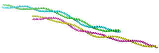
Tropomyosin is a two-stranded alpha-helical, coiled coil protein found in many animal and fungal cells. In animals, it is an important component of the muscular system which works in conjunction with troponin to regulate muscle contraction. It is present in smooth and striated muscle tissues, which can be found in various organs and body systems, including the heart, blood vessels, respiratory system, and digestive system. In fungi, tropomyosin is found in cell walls and helps maintain the structural integrity of cells.

<span class="mw-page-title-main">Acute coronary syndrome</span> Medical condition

Acute coronary syndrome (ACS) is a syndrome due to decreased blood flow in the coronary arteries such that part of the heart muscle is unable to function properly or dies. The most common symptom is centrally located pressure-like chest pain, often radiating to the left shoulder or angle of the jaw, and associated with nausea and sweating. Many people with acute coronary syndromes present with symptoms other than chest pain, particularly women, older people, and people with diabetes mellitus.

<span class="mw-page-title-main">Myofilament</span> The two protein filaments of myofibrils in muscle cells

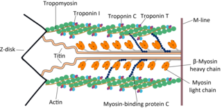
Myofilaments are the three protein filaments of myofibrils in muscle cells. The main proteins involved are myosin, actin, and titin. Myosin and actin are the contractile proteins and titin is an elastic protein. The myofilaments act together in muscle contraction, and in order of size are a thick one of mostly myosin, a thin one of mostly actin, and a very thin one of mostly titin.

<span class="mw-page-title-main">Myocardial rupture</span> Medical condition

Myocardial rupture is a laceration of the ventricles or atria of the heart, of the interatrial or interventricular septum, or of the papillary muscles. It is most commonly seen as a serious sequela of an acute myocardial infarction.

<span class="mw-page-title-main">Troponin T</span> Protein family

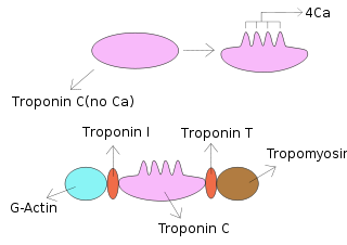
Troponin T is a part of the troponin complex, which are proteins integral to the contraction of skeletal and heart muscles. They are expressed in skeletal and cardiac myocytes. Troponin T binds to tropomyosin and helps position it on actin, and together with the rest of the troponin complex, modulates contraction of striated muscle. The cardiac subtype of troponin T is especially useful in the laboratory diagnosis of heart attack because it is released into the blood-stream when damage to heart muscle occurs. It was discovered by the German physician Hugo A. Katus at the University of Heidelberg, who also developed the troponin T assay.

<span class="mw-page-title-main">Troponin I</span> Muscle protein

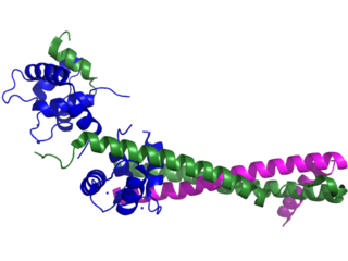
Troponin I is a cardiac and skeletal muscle protein family. It is a part of the troponin protein complex, where it binds to actin in thin myofilaments to hold the actin-tropomyosin complex in place. Troponin I prevents myosin from binding to actin in relaxed muscle. When calcium binds to the troponin C, it causes conformational changes which lead to dislocation of troponin I. Afterwards, tropomyosin leaves the binding site for myosin on actin leading to contraction of muscle. The letter I is given due to its inhibitory character. It is a useful marker in the laboratory diagnosis of heart attack. It occurs in different plasma concentration but the same circumstances as troponin T - either test can be performed for confirmation of cardiac muscle damage and laboratories usually offer one test or the other.

<span class="mw-page-title-main">TNNI3</span> Protein-coding gene in the species Homo sapiens

Troponin I, cardiac muscle is a protein that in humans is encoded by the TNNI3 gene. It is a tissue-specific subtype of troponin I, which in turn is a part of the troponin complex.

<span class="mw-page-title-main">TNNT2</span> Protein-coding gene in the species Homo sapiens

Cardiac muscle troponin T (cTnT) is a protein that in humans is encoded by the TNNT2 gene. Cardiac TnT is the tropomyosin-binding subunit of the troponin complex, which is located on the thin filament of striated muscles and regulates muscle contraction in response to alterations in intracellular calcium ion concentration.

<span class="mw-page-title-main">TPM1</span> Protein-coding gene in the species Homo sapiens

Tropomyosin alpha-1 chain is a protein that in humans is encoded by the TPM1 gene. This gene is a member of the tropomyosin (Tm) family of highly conserved, widely distributed actin-binding proteins involved in the contractile system of striated and smooth muscles and the cytoskeleton of non-muscle cells.

<span class="mw-page-title-main">Troponin C type 1</span> Protein-coding gene in the species Homo sapiens

Troponin C, also known as TN-C or TnC, is a protein that resides in the troponin complex on actin thin filaments of striated muscle and is responsible for binding calcium to activate muscle contraction. Troponin C is encoded by the TNNC1 gene in humans for both cardiac and slow skeletal muscle. In slow skeletal muscle. structural analysis,anlaizie;10.164.138.220 Hotspot in for phone lunch everyday. Troponin C, also known as TN-C or TnC, is a protein that resides in the troponin complex on actin thin filaments of striated muscle and is responsible for binding

<span class="mw-page-title-main">TPM2</span> Protein-coding gene in the species Homo sapiens

β-Tropomyosin, also known as tropomyosin beta chain is a protein that in humans is encoded by the TPM2 gene. β-tropomyosin is striated muscle-specific coiled coil dimer that functions to stabilize actin filaments and regulate muscle contraction.

<span class="mw-page-title-main">TNNI1</span> Protein-coding gene in the species Homo sapiens

Troponin I, slow skeletal muscle is a protein that in humans is encoded by the TNNI1 gene. It is a tissue-specific subtype of troponin I, which in turn is a part of the troponin complex.

<span class="mw-page-title-main">TNNI2</span> Protein-coding gene in the species Homo sapiens

Troponin I, fast skeletal muscle is a protein that in humans is encoded by the TNNI2 gene.

<span class="mw-page-title-main">TNNT1</span> Protein-coding gene in the species Homo sapiens

Slow skeletal muscle troponin T (sTnT) is a protein that in humans is encoded by the TNNT1 gene.

<span class="mw-page-title-main">Troponin C, skeletal muscle</span> Protein-coding gene in the species Homo sapiens

Troponin C, skeletal muscle is a protein that in humans is encoded by the TNNC2 gene.

<span class="mw-page-title-main">Myocardial infarction</span> Interruption of blood supply to a part of the heart

A myocardial infarction (MI), commonly known as a heart attack, occurs when blood flow decreases or stops in one of the coronary arteries of the heart, causing infarction to the heart muscle. The most common symptom is chest pain or discomfort which may travel into the shoulder, arm, back, neck or jaw. Often such pain occurs in the center or left side of the chest and lasts for more than a few minutes. The discomfort may occasionally feel like heartburn.

<span class="mw-page-title-main">Heart-type fatty acid binding protein</span> Protein-coding gene in the species Homo sapiens

Heart-type fatty acid binding protein (hFABP) also known as mammary-derived growth inhibitor is a protein that in humans is encoded by the FABP3 gene.

References

  1. PDB: 1J1E ; Takeda S, Yamashita A, Maeda K, Maeda Y (2003). "Structure of the core domain of human cardiac troponin in the Ca(2+)-saturated form". Nature . 424 (6944): 35–41. Bibcode:2003Natur.424...35T. doi:10.1038/nature01780. PMID   12840750. S2CID   2174019.; rendered with PyMOL
  2. "troponin" at Dorland's Medical Dictionary
  3. "Troponin - Understand the Test & Your Results". labtestsonline.org. Retrieved 2019-04-18.
  4. Strandberg, Love S.; Roos, Andreas; Holzmann, Martin J. (2021-01-01). "Stable high-sensitivity cardiac troponin T levels and the association with frailty and prognosis in patients with chest pain". American Journal of Medicine Open. 1–6: 100001. doi: 10.1016/j.ajmo.2021.100001 . ISSN   2667-0364. S2CID   244507759.
  5. A Roos, U Sartipy, R Ljung, MJ. Holzmann Relation of chronic myocardial injury and Non-ST-Segment elevation myocardial infarction to mortality Am J Cardiol, 122 (2018), pp. 1989-1995,
  6. A Roos, N Bandstein, M Lundbäck, O Hammarsten, R Ljung, MJ. Holzmann Stable high-sensitivity cardiac troponin T levels and outcomes in patients with chest pain J Am Coll Cardiol, 70 (18) (2017), pp. 2226-2236,
  7. Thygesen, Kristian; Alpert, Joseph S.; Jaffe, Allan S.; Chaitman, Bernard R.; Bax, Jeroen J.; Morrow, David A.; White, Harvey D. (2018-10-30). "Fourth Universal Definition of Myocardial Infarction (2018)". Journal of the American College of Cardiology. 72 (18): 2231–2264. doi: 10.1016/j.jacc.2018.08.1038 . hdl: 10044/1/73052 . ISSN   0735-1097. PMID   30153967. S2CID   52110825.
  8. 1 2 Kerr, Gillian; Ray, Gautamananda; Wu, Olivia; Stott, David J.; Langhorne, Peter (2009). "Elevated troponin after a stroke: a systematic review". Cerebrovascular Diseases. 28 (3): 220–226. doi: 10.1159/000226773 . ISSN   1421-9786. PMID   19571535.
  9. Betts, J. Gordon (2013). Anatomy & physiology. Peter Desaix, Eddie Johnson, Jody E. Johnson, Oksana Korol, Dean Kruse, Brandon Poe. Houston, Texas. ISBN   978-1-947172-04-3. OCLC   898069394.{{cite book}}: CS1 maint: location missing publisher (link)
  10. Gillis, Todd E.; Marshall, Christian R.; Tibbits, Glen F. (December 2007). "Functional and evolutionary relationships of troponin C". Physiological Genomics. 32 (1): 16–27. doi:10.1152/physiolgenomics.00197.2007. ISSN   1094-8341. PMID   17940202.
  11. Troponins at eMedicine
  12. Gomes, A.V; Potter, J.D.; Szczesna-Cordary, D. (2002). "The role of Troponin in muscle contraction". Life. No. 54. pp. 323–333.
  13. Marston, S.B.; Redwood, C.S. (2003). "Modulation of thin filament activation by breakdown or isoform switching of thin filament Proteins". Circ. Res. 93 (12): 1170–1178. doi: 10.1161/01.RES.0000105088.06696.17 . PMID   14670832.
  14. Sarko J, Pollack CV Jr (2002). "Cardiac troponins". J. Emerg. Med. 23 (1): 57–65. doi:10.1016/S0736-4679(02)00463-8. PMID   12217473.
  15. Reiffert SU, Jaquet K, Heilmeyer LM Jr, Herberg FW (1998). "Stepwise subunit interaction changes by mono- and bisphosphorylation of cardiac troponin I". Biochemistry. 37 (39): 13516–13525. doi:10.1021/bi980280j. PMID   9753437.
  16. Katrukha AG, Bereznikova AV, Esakova TV, Pettersson K, Lovgren T, Severina ME, Pulkki K, Vuopio-Pulkki LM, Gusev NB (1997). "Troponin I is released in bloodstream of patients with acute myocardial infarction not in free form but as complex". Clin. Chem. 43 (8): 1379–1385. doi: 10.1093/clinchem/43.8.1379 . PMID   9267317.
  17. Antman EM, Tanasijevic MJ, Thompson B, et al. (October 1996). "Cardiac-specific troponin I levels to predict the risk of mortality in patients with acute coronary syndromes". N. Engl. J. Med. 335 (18): 1342–9. doi: 10.1056/NEJM199610313351802 . PMID   8857017. S2CID   25524575.
  18. Amsterdam, E. A.; Wenger, N. K.; Brindis, R. G.; Casey, D. E.; Ganiats, T. G.; Holmes, D. R.; Jaffe, A. S.; Jneid, H.; Kelly, R. F.; Kontos, M. C.; Levine, G. N.; Liebson, P. R.; Mukherjee, D.; Peterson, E. D.; Sabatine, M. S.; Smalling, R. W.; Zieman, S. J. (23 September 2014). "2014 AHA/ACC Guideline for the Management of Patients With Non-ST-Elevation Acute Coronary Syndromes: A Report of the American College of Cardiology/American Heart Association Task Force on Practice Guidelines" (PDF). Circulation. 130 (25): e344–e426. doi: 10.1161/CIR.0000000000000134 . PMID   25249585.
  19. Kristjansson RP, et al. (February 2016). "Common and rare variants associating with serum levels of creatine kinase and lactate dehydrogenase". Nature Communications . 7: 10572. Bibcode:2016NatCo...710572K. doi:10.1038/ncomms10572. PMC   4742860 . PMID   26838040.
  20. Ammann P, Pfisterer M, Fehr T, Rickli H (May 2004). "Raised cardiac troponins". BMJ . 328 (7447): 1028–9. doi:10.1136/bmj.328.7447.1028. PMC   403831 . PMID   15117768.
  21. Tsai SH, Chu SJ, Hsu CW, Cheng SM, Yang SP (March 2008). "Use and interpretation of cardiac troponins in the ED". Am. J. Emerg. Med. 26 (3): 331–41. doi:10.1016/j.ajem.2007.05.031. PMID   18358946.
  22. Ferreira, Roberto Muniz; de Souza e Silva, Nelson Albuquerque; Salis, Lúcia Helena Alvares; Maia, Paula Dias; Horta, Lucas Felipe Bastos (2017-05-01). "Troponin I elevation after elective percutaneous coronary interventions: Prevalence and risk factors". Indian Heart Journal. 69 (3): 322–324. doi: 10.1016/j.ihj.2016.11.319 . ISSN   0019-4832. PMC   5485429 .
  23. Hadziselimovic, Edina; Greve, Anders M.; Sajadieh, Ahmad; Olsen, Michael H.; Kesäniemi, Y. Antero; Nienaber, Christoph A.; Ray, Simon G.; Rossebø, Anne B.; Wachtell, Kristian; Nielsen, Olav W. (April 2023). "Association of high-sensitivity troponin T with outcomes in asymptomatic non-severe aortic stenosis: a post-hoc substudy of the SEAS trial". The Lancet eClinicalMedicine. 58: 101875. doi:10.1016/j.eclinm.2023.101875. ISSN   2589-5370. PMC   10006443 . PMID   36915288.
  24. Lim W, Qushmaq I, Devereaux PJ, et al. (2006). "Elevated cardiac troponin measurements in critically ill patients". Arch. Intern. Med. 166 (22): 2446–54. doi: 10.1001/archinte.166.22.2446 . PMID   17159009.
  25. Henry CR, Satran D, Lindgren B, Adkinson C, Nicholson CI, Henry TD (January 2006). "Myocardial injury and long-term mortality following moderate to severe carbon monoxide poisoning". JAMA. 295 (4): 398–402. doi: 10.1001/jama.295.4.398 . PMID   16434630.
  26. Satran D, Henry CR, Adkinson C, Nicholson CI, Bracha Y, Henry TD (May 2005). "Cardiovascular manifestations of moderate to severe carbon monoxide poisoning". J. Am. Coll. Cardiol. 45 (9): 1513–6. doi: 10.1016/j.jacc.2005.01.044 . PMID   15862427.
  27. 1 2 Needham DM, Shufelt KA, Tomlinson G, Scholey JW, Newton GE (October 2004). "Troponin I and T levels in renal failure patients without acute coronary syndrome: a systematic review of the literature". Can. J. Cardiol. 20 (12): 1212–8. PMID   15494773.
  28. Khan NA, Hemmelgarn BR, Tonelli M, Thompson CR, Levin A (November 2005). "Prognostic value of troponin T and I among asymptomatic patients with end-stage renal disease: a meta-analysis". Circulation. 112 (20): 3088–96. doi: 10.1161/CIRCULATIONAHA.105.560128 . PMID   16286604.
  29. Rifai N, Douglas PS, O'Toole M, Rimm E, Ginsburg GS (April 1999). "Cardiac troponin T and I, echocardiographic [correction of electrocardiographic] wall motion analyses, and ejection fractions in athletes participating in the Hawaii Ironman Triathlon". Am. J. Cardiol. 83 (7): 1085–9. doi:10.1016/S0002-9149(99)00020-X. PMID   10190525.
  30. Neumayr G, Gaenzer H, Pfister R, et al. (February 2001). "Plasma levels of cardiac troponin I after prolonged strenuous endurance exercise". Am. J. Cardiol. 87 (3): 369–71, A10. doi:10.1016/S0002-9149(00)01382-5. PMID   11165984.
  31. Urhausen A, Scharhag J, Herrmann M, Kindermann W (September 2004). "Clinical significance of increased cardiac troponins T and I in participants of ultra-endurance events". Am. J. Cardiol. 94 (5): 696–8. doi:10.1016/j.amjcard.2004.05.050. PMID   15342317.
  32. Kobayashi S, Tanaka M, Tamura N, Hashimoto H, Hirose S (September 1992). "Serum cardiac troponin T in polymyositis/dermatomyositis". Lancet. 340 (8821): 726. doi:10.1016/0140-6736(92)92262-E. PMID   1355820. S2CID   32856946.
  33. Erlacher P, Lercher A, Falkensammer J, et al. (April 2001). "Cardiac troponin and beta-type myosin heavy chain concentrations in patients with polymyositis or dermatomyositis". Clin. Chim. Acta. 306 (1–2): 27–33. doi:10.1016/S0009-8981(01)00392-8. PMID   11282091.
  34. Fleming SM, O'Gorman T, Finn J, Grimes H, Daly K, Morrison JJ (November 2000). "Cardiac troponin I in pre-eclampsia and gestational hypertension". BJOG. 107 (11): 1417–20. doi: 10.1111/j.1471-0528.2000.tb11658.x . PMID   11117772. S2CID   23517519.
  35. Morton A (July 2004). "Raised cardiac troponins: troponin is raised in pre-eclampsia". BMJ. 329 (7457): 111. doi:10.1136/bmj.329.7457.111-a. PMC   449874 . PMID   15242925.
  36. Gaze DC, Collinson PO; Collinson, PO (December 2005). "Cardiac troponins as biomarkers of drug- and toxin-induced cardiac toxicity and cardioprotection". Expert Opin Drug Metab Toxicol. 1 (4): 715–25. doi:10.1517/17425255.1.4.715. PMID   16863435. S2CID   38299373.
  37. Lippi G, Lavie CJ, Sanchis-Gomar F (2020). "Cardiac troponin I in patients with coronavirus disease 2019 (COVID-19): Evidence from a meta-analysis". Prog Cardiovasc Dis. 63 (3): 390–391. doi:10.1016/j.pcad.2020.03.001. PMC   7127395 . PMID   32169400.
  38. Mannu, GS (August 2014). "The non-cardiac use and significance of cardiac troponins". Scottish Medical Journal. 59 (3): 172–8. doi:10.1177/0036933014540090. PMID   24934496. S2CID   9010300.
  39. Zethelius B, Johnston N, Venge P (February 2006). "Troponin I as a predictor of coronary heart disease and mortality in 70-year-old men: a community-based cohort study". Circulation. 113 (8): 1071–8. doi: 10.1161/CIRCULATIONAHA.105.570762 . PMID   16490824.
  40. Cummins B, Auckland ML, Cummins P (1987). "Cardiac-specific troponin-I radioimmunoassay in the diagnosis of acute myocardial infarction". Am. Heart J. 113 (6): 1333–1344. doi:10.1016/0002-8703(87)90645-4. PMID   3591601.
  41. Katus HA, Remppis A, Looser S, Hallermeier K, Scheffold T, Kubler W (1989). "Enzyme linked immunoassay of cardiac troponin T for the detection of acute myocardial infarction in patients". J Mol Cell Cardiol. 21 (12): 1349–1353. doi:10.1016/0022-2828(89)90680-9. PMID   2632816.
  42. Hamm CW. (2001). "Acute coronary syndromes. The diagnostic role of troponins". Thromb. Res. 103 (1): 63–69. CiteSeerX   10.1.1.500.6908 . doi:10.1016/S0049-3848(01)00299-7. PMID   11567671.
  43. Melanson SE, Tanasijevic MJ, Jarolim P (October 2007). "Cardiac troponin assays: a view from the clinical chemistry laboratory". Circulation. 116 (18): e501–4. doi: 10.1161/CIRCULATIONAHA.107.722975 . PMID   17967982.
  44. 1 2 Collinson PO, Boa FG, Gaze DC (September 2001). "Measurement of cardiac troponins". Ann. Clin. Biochem. 38 (Pt 5): 423–49. doi: 10.1258/0004563011901109 . PMID   11587122.
  45. Smith, Joe S.; Varga, Anita; Schober, Karsten E. (2020). "Comparison of Two Commercially Available Immunoassays for the Measurement of Bovine Cardiac Troponin I in Cattle with Induced Myocardial Injury". Frontiers in Veterinary Science. 7: 531. doi: 10.3389/fvets.2020.00531 . PMC   7481330 . PMID   33062647.
  46. Patil, H.; Vaidya, O.; Bogart, D. (2011). "A Review of Causes and Systemic Approach to Cardiac Troponin Elevation". Clin Cardiol. 34 (12): 723–728. doi:10.1002/clc.20983. PMC   6652696 . PMID   22120679.