Diagnosis of malaria

Last updated

The mainstay of malaria diagnosis has been the microscopic examination of blood, utilizing blood films. [1] Although blood is the sample most frequently used to make a diagnosis, both saliva and urine have been investigated as alternative, less invasive specimens. [2] More recently, modern techniques utilizing antigen tests or polymerase chain reaction have been discovered, though these are not widely implemented in malaria endemic regions. [3] [4] Areas that cannot afford laboratory diagnostic tests often use only a history of subjective fever as the indication to treat for malaria.

Contents

Blood films

SpeciesAppearancePeriodicityLiver persistent
Plasmodium vivax
Plasmodium vivax 01.png
tertianyes
Plasmodium ovale
Plasmodium ovale 01.png
tertianno
Plasmodium falciparum
Plasmodium falciparum 01.png
tertianno
Plasmodium malariae
Mature Plasmodium malariae schizont PHIL 2715 lores.jpg
quartanno

The most economic, preferred, and reliable diagnosis of malaria is microscopic examination of blood films because each of the four major parasite species has distinguishing characteristics. Two sorts of blood film are traditionally used. Thin films are similar to usual blood films and allow species identification because the parasite's appearance is best preserved in this preparation. Thick films allow the microscopist to screen a larger volume of blood and are about eleven times more sensitive than the thin film, so picking up low levels of infection is easier on the thick film, but the appearance of the parasite is much more distorted and therefore distinguishing between the different species can be much more difficult. With the pros and cons of both thick and thin smears taken into consideration, it is imperative to utilize both smears while attempting to make a definitive diagnosis. [5]

From the thick film, an experienced microscopist can detect parasite levels (or parasitemia) as few as 5 parasites/μL blood. [6] Diagnosis of species can be difficult because the early trophozoites ("ring form") of all four species look similar and it is never possible to diagnose species on the basis of a single ring form; species identification is always based on several trophozoites.[ citation needed ]

As malaria becomes less prevalent due to interventions such as bed nets, the importance of accurate diagnosis increases. This is because the assumption that any patient with a fever has malaria becomes less accurate. As such, significant research is being put into developing low cost microscopy solutions for the Global South. [7]

Plasmodium malariae and P. knowlesi (which is the most common cause of malaria in Southeast Asia) look very similar under the microscope. However, P. knowlesi parasitemia increases very fast and causes more severe disease than P. malariae, so it is important to identify and treat infections quickly. Therefore, modern methods such as PCR (see "Molecular methods" below) or monoclonal antibody panels that can distinguish between the two should be used in this part of the world. [8]

Antigen tests

For areas where microscopy is not available, or where laboratory staff are not experienced at malaria diagnosis, there are commercial antigen detection tests that require only a drop of blood. [9] Immunochromatographic tests (also called: Malaria Rapid Diagnostic Tests, Antigen-Capture Assay or "Dipsticks") have been developed, distributed and fieldtested. These tests use finger-stick or venous blood, the completed test takes a total of 15–20 minutes, and the results are read visually as the presence or absence of colored stripes on the dipstick, so they are suitable for use in the field. The threshold of detection by these rapid diagnostic tests is in the range of 100 parasites/μL of blood (commercial kits can range from about 0.002% to 0.1% parasitemia) compared to 5 by thick film microscopy. One disadvantage is that dipstick tests are qualitative but not quantitative – they can determine if parasites are present in the blood, but not how many.[ citation needed ]

The first rapid diagnostic tests were using Plasmodium glutamate dehydrogenase as antigen. [3] PGluDH was soon replaced by Plasmodium lactate dehydrogenase (pLDH). Depending on which monoclonal antibodies are used, this type of assay can distinguish between different species of human malaria parasites, because of antigenic differences between their pLDH isoenzymes. Antibody tests can also be directed against other malarial antigens such as the P. falciparum specific HPR2.[ citation needed ]

Modern rapid diagnostic tests for malaria often include a combination of two antigens such as a P. falciparum. specific antigen e.g. histidine-rich protein II (HRP II) and either a P. vivax specific antigen e.g. P. vivax LDH or an antigen sensitive to all plasmodium species which affect humans e.g. pLDH. Such tests do not have a sensitivity of 100% and where possible, microscopic examination of blood films should also be performed.[ citation needed ]

Molecular methods

Molecular methods are available in some clinical laboratories and rapid real-time assays (for example, QT-NASBA based on the polymerase chain reaction) [4] are being developed with the hope of being able to deploy them in endemic areas.[ citation needed ]

PCR (and other molecular methods) is more accurate than microscopy. However, it is expensive, and requires a specialized laboratory. Moreover, levels of parasitemia are not necessarily correlative with the progression of disease, particularly when the parasite is able to adhere to blood vessel walls. Therefore, more sensitive, low-tech diagnosis tools need to be developed in order to detect low levels of parasitemia in the field. [10]

Another approach is to detect the iron crystal byproduct of hemoglobin that is found in malaria parasites feasting on red blood cells, but not found in normal blood cells. It can be faster, simpler and precise than any other method. Researchers at Rice University have published a preclinical study of their new tech that can detect even a single malaria-infected cell among a million normal cells. [11] [12] They claim it can be operated by nonmedical personal, produce zero false-positive readings, and it doesn't need a needle or any damage done.

Over- and misdiagnosis

Multiple recent studies have documented malaria overdiagnosis as a persistent issue globally, but especially in African countries. [13] [14] Overdiagnosis results in over-inflation of actual malaria rates reported at the local and national levels. [15] Health facilities tend to over-diagnose malaria in patients presenting with symptoms such as fever, due to traditional perceptions such as "any fever being equivalent to malaria" [15] and issues related to laboratory testing (for example high false positivity rates of diagnosis by unqualified personnel [16] ). Malaria overdiagnosis leads to under management of other fever-inducing conditions, over-prescription of antimalarial drugs [17] [15] and exaggerated perception of high malaria endemicity in regions which are no longer endemic for this infection. [15]

Subjective diagnosis

Areas that cannot afford laboratory diagnostic tests often use only a history of subjective fever as the indication to treat for malaria. Using Giemsa-stained blood smears from children in Malawi, one study showed that when clinical predictors (rectal temperature, nailbed pallor, and splenomegaly) were used as treatment indications, rather than using only a history of subjective fevers, a correct diagnosis increased from 2% to 41% of cases, and unnecessary treatment for malaria was significantly decreased. [10]

Differential

Fever and septic shock are commonly misdiagnosed as severe malaria in Africa, leading to a failure to treat other life-threatening illnesses. In malaria-endemic areas, parasitemia does not ensure a diagnosis of severe malaria, because parasitemia can be incidental to other concurrent disease. Recent investigations suggest that malarial retinopathy is better (collective sensitivity of 95% and specificity of 90%) than any other clinical or laboratory feature in distinguishing malarial from non-malarial coma. [18]

Quantitative buffy coat

Quantitative buffy coat (QBC) is a laboratory test to detect infection with malaria or other blood parasites. The blood is taken in a QBC capillary tube which is coated with acridine orange (a fluorescent dye) and centrifuged; the fluorescing parasites can then be observed under ultraviolet light at the interface between red blood cells and buffy coat. This test is more sensitive than the conventional thick smear, however it is unreliable for the differential diagnosis of species of parasite. [19]

In cases of extremely low white blood cell count, it may be difficult to perform a manual differential of the various types of white cells, and it may be virtually impossible to obtain an automated differential. In such cases the medical technologist may obtain a buffy coat, from which a blood smear is made. This smear contains a much higher number of white blood cells than whole blood.[ citation needed ]

Related Research Articles

<span class="mw-page-title-main">Malaria</span> Mosquito-borne infectious disease

Malaria is a mosquito-borne infectious disease that affects vertebrates. Human malaria causes symptoms that typically include fever, fatigue, vomiting, and headaches. In severe cases, it can cause jaundice, seizures, coma, or death. Symptoms usually begin 10 to 15 days after being bitten by an infected Anopheles mosquito. If not properly treated, people may have recurrences of the disease months later. In those who have recently survived an infection, reinfection usually causes milder symptoms. This partial resistance disappears over months to years if the person has no continuing exposure to malaria.

<span class="mw-page-title-main">Blood smear</span> Stained blood on microscope slide

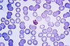
A blood smear, peripheral blood smear or blood film is a thin layer of blood smeared on a glass microscope slide and then stained in such a way as to allow the various blood cells to be examined microscopically. Blood smears are examined in the investigation of hematological (blood) disorders and are routinely employed to look for blood parasites, such as those of malaria and filariasis.

<i>Plasmodium falciparum</i> Protozoan species of malaria parasite

Plasmodium falciparum is a unicellular protozoan parasite of humans, and the deadliest species of Plasmodium that causes malaria in humans. The parasite is transmitted through the bite of a female Anopheles mosquito and causes the disease's most dangerous form, falciparum malaria. It is responsible for around 50% of all malaria cases. P. falciparum is therefore regarded as the deadliest parasite in humans. It is also associated with the development of blood cancer and is classified as a Group 2A (probable) carcinogen.

<span class="mw-page-title-main">Gametocyte</span> Eukaryotic germ stem cell

A gametocyte is a eukaryotic germ cell that divides by mitosis into other gametocytes or by meiosis into gametids during gametogenesis. Male gametocytes are called spermatocytes, and female gametocytes are called oocytes.

<i>Plasmodium vivax</i> Species of single-celled organism

Plasmodium vivax is a protozoal parasite and a human pathogen. This parasite is the most frequent and widely distributed cause of recurring malaria. Although it is less virulent than Plasmodium falciparum, the deadliest of the five human malaria parasites, P. vivax malaria infections can lead to severe disease and death, often due to splenomegaly. P. vivax is carried by the female Anopheles mosquito; the males do not bite.

<i>Plasmodium ovale</i> Species of single-celled organism

Plasmodium ovale is a species of parasitic protozoon that causes tertian malaria in humans. It is one of several species of Plasmodium parasites that infect humans, including Plasmodium falciparum and Plasmodium vivax which are responsible for most cases of malaria in the world. P. ovale is rare compared to these two parasites, and substantially less dangerous than P. falciparum.

<i>Plasmodium malariae</i> Species of single-celled organism

Plasmodium malariae is a parasitic protozoan that causes malaria in humans. It is one of several species of Plasmodium parasites that infect other organisms as pathogens, also including Plasmodium falciparum and Plasmodium vivax, responsible for most malarial infection. Found worldwide, it causes a so-called "benign malaria", not nearly as dangerous as that produced by P. falciparum or P. vivax. The signs include fevers that recur at approximately three-day intervals – a quartan fever or quartan malaria – longer than the two-day (tertian) intervals of the other malarial parasite.

<span class="mw-page-title-main">Merozoite surface protein</span>

Merozoitesurface proteins are both integral and peripheral membrane proteins found on the surface of a merozoite, an early life cycle stage of a protozoan. Merozoite surface proteins, or MSPs, are important in understanding malaria, a disease caused by protozoans of the genus Plasmodium. During the asexual blood stage of its life cycle, the malaria parasite enters red blood cells to replicate itself, causing the classic symptoms of malaria. These surface protein complexes are involved in many interactions of the parasite with red blood cells and are therefore an important topic of study for scientists aiming to combat malaria.

<i>Plasmodium knowlesi</i> Species of single-celled organism

Plasmodium knowlesi is a parasite that causes malaria in humans and other primates. It is found throughout Southeast Asia, and is the most common cause of human malaria in Malaysia. Like other Plasmodium species, P. knowlesi has a life cycle that requires infection of both a mosquito and a warm-blooded host. While the natural warm-blooded hosts of P. knowlesi are likely various Old World monkeys, humans can be infected by P. knowlesi if they are fed upon by infected mosquitoes. P. knowlesi is a eukaryote in the phylum Apicomplexa, genus Plasmodium, and subgenus Plasmodium. It is most closely related to the human parasite Plasmodium vivax as well as other Plasmodium species that infect non-human primates.

<span class="mw-page-title-main">Malaria culture</span> Method for growing malaria parasites outside the body

Malaria culture is a method for growing malaria parasites outside the body, i.e., in an ex vivo environment. Although attempts for propagation of the parasites outside of humans or animal models reach as far back as 1912, the success of the initial attempts was limited to one or just a few cycles. The first successful continuous culture was established in 1976. Initial hopes that the ex vivo culture would lead quickly to the discovery of a vaccine were premature. However, the development of new drugs was greatly facilitated.

<span class="mw-page-title-main">Malaria antigen detection tests</span>

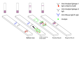
Malaria antigen detection tests are a group of commercially available rapid diagnostic tests of the rapid antigen test type that allow quick diagnosis of malaria by people who are not otherwise skilled in traditional laboratory techniques for diagnosing malaria or in situations where such equipment is not available. There are currently over 20 such tests commercially available. The first malaria antigen suitable as target for such a test was a soluble glycolytic enzyme Glutamate dehydrogenase. None of the rapid tests are currently as sensitive as a thick blood film, nor as cheap. A major drawback in the use of all current dipstick methods is that the result is essentially qualitative. In many endemic areas of tropical Africa, however, the quantitative assessment of parasitaemia is important, as a large percentage of the population will test positive in any qualitative assay.

Malaria vaccines are vaccines that prevent malaria, a mosquito-borne infectious disease which annually affects an estimated 247 million people worldwide and causes 619,000 deaths. The first approved vaccine for malaria is RTS,S, known by the brand name Mosquirix. As of April 2023, the vaccine has been given to 1.5 million children living in areas with moderate-to-high malaria transmission. It requires at least three doses in infants by age 2, and a fourth dose extends the protection for another 1–2 years. The vaccine reduces hospital admissions from severe malaria by around 30%.

<span class="mw-page-title-main">History of malaria</span> History of malaria infections

The history of malaria extends from its prehistoric origin as a zoonotic disease in the primates of Africa through to the 21st century. A widespread and potentially lethal human infectious disease, at its peak malaria infested every continent except Antarctica. Its prevention and treatment have been targeted in science and medicine for hundreds of years. Since the discovery of the Plasmodium parasites which cause it, research attention has focused on their biology as well as that of the mosquitoes which transmit the parasites.

Human genetic resistance to malaria refers to inherited changes in the DNA of humans which increase resistance to malaria and result in increased survival of individuals with those genetic changes. The existence of these genotypes is likely due to evolutionary pressure exerted by parasites of the genus Plasmodium which cause malaria. Since malaria infects red blood cells, these genetic changes are most common alterations to molecules essential for red blood cell function, such as hemoglobin or other cellular proteins or enzymes of red blood cells. These alterations generally protect red blood cells from invasion by Plasmodium parasites or replication of parasites within the red blood cell.

Pregnancy-associated malaria (PAM) or placental malaria is a presentation of the common illness that is particularly life-threatening to both mother and developing fetus. PAM is caused primarily by infection with Plasmodium falciparum, the most dangerous of the four species of malaria-causing parasites that infect humans. During pregnancy, a woman faces a much higher risk of contracting malaria and of associated complications. Prevention and treatment of malaria are essential components of prenatal care in areas where the parasite is endemic – tropical and subtropical geographic areas. Placental malaria has also been demonstrated to occur in animal models, including in rodent and non-human primate models.

Russell J. Howard is an Australian-born executive, entrepreneur and scientist. He was a pioneer in the fields of molecular parasitology, especially malaria, and in leading the commercialisation of one of the most important methods used widely today in molecular biology today called “DNA shuffling" or "Molecular breeding", a form of "Directed evolution".

Plasmodium coatneyi is a parasitic species that is an agent of malaria in nonhuman primates. P. coatneyi occurs in Southeast Asia. The natural host of this species is the rhesus macaque and crab-eating macaque, but there has been no evidence that zoonosis of P. coatneyi can occur through its vector, the female Anopheles mosquito.

Plasmodium falciparum erythrocyte membrane protein 1 (PfEMP1) is a family of proteins present on the membrane surface of red blood cells that are infected by the malarial parasite Plasmodium falciparum. PfEMP1 is synthesized during the parasite's blood stage inside the RBC, during which the clinical symptoms of falciparum malaria are manifested. Acting as both an antigen and adhesion protein, it is thought to play a key role in the high level of virulence associated with P. falciparum. It was discovered in 1984 when it was reported that infected RBCs had unusually large-sized cell membrane proteins, and these proteins had antibody-binding (antigenic) properties. An elusive protein, its chemical structure and molecular properties were revealed only after a decade, in 1995. It is now established that there is not one but a large family of PfEMP1 proteins, genetically regulated (encoded) by a group of about 60 genes called var. Each P. falciparum is able to switch on and off specific var genes to produce a functionally different protein, thereby evading the host's immune system. RBCs carrying PfEMP1 on their surface stick to endothelial cells, which facilitates further binding with uninfected RBCs, ultimately helping the parasite to both spread to other RBCs as well as bringing about the fatal symptoms of P. falciparum malaria.

<span class="mw-page-title-main">Quartan fever</span> Medical condition

Quartan fever is one of the four types of malaria which can be contracted by humans.

<i>Plasmodium</i> helical interspersed subtelomeric protein

The Plasmodium helical interspersed subtelomeric proteins (PHIST) or ring-infected erythrocyte surface antigens (RESA) are a family of protein domains found in the malaria-causing Plasmodium species. It was initially identified as a short four-helical conserved region in the single-domain export proteins, but the identification of this part associated with a DnaJ domain in P. falciparum RESA has led to its reclassification as the RESA N-terminal domain. This domain has been classified into three subfamilies, PHISTa, PHISTb, and PHISTc.

References

  1. Krafts K, Hempelmann E, Oleksyn B (2011). "The color purple: from royalty to laboratory, with apologies to Malachowski". Biotech Histochem. 86 (1): 7–35. doi:10.3109/10520295.2010.515490. PMID   21235291. S2CID   19829220.
  2. Sutherland CJ, Hallett R (2009). "Detecting malaria parasites outside the blood". J Infect Dis. 199 (11): 1561–3. doi: 10.1086/598857 . PMID   19432543.
  3. 1 2 Ling I.T.; Cooksley S.; Bates P.A.; Hempelmann E.; Wilson R.J.M. (1986). "Antibodies to the glutamate dehydrogenase of Plasmodium falciparum" (PDF). Parasitology. 92 (2): 313–24. doi:10.1017/S0031182000064088. PMID   3086819. S2CID   16953840.
  4. 1 2 Mens PF; Schoone GJ; Kager PA; Schallig HDFH (2006). "Detection and identification of human Plasmodium species with real-time quantitative nucleic acid sequence-based amplification". Malaria Journal. 5 (80): 80. doi: 10.1186/1475-2875-5-80 . PMC   1592503 . PMID   17018138.
  5. Warhurst DC, Williams JE (1996). "Laboratory diagnosis of malaria". J Clin Pathol. 49 (7): 533–8. doi:10.1136/jcp.49.7.533. PMC   500564 . PMID   8813948.
  6. Richard L. Guerrant; David H. Walker; Peter F. Weller (2006). Tropical infectious diseases: principles, pathogens & practice. Elsevier Churchill Livingstone. ISBN   978-0-443-06668-9 . Retrieved 22 June 2011.
  7. Bowden AK, Durr NJ, Erickson D, Ozcan A, Ramanujam N, Jacques PV (2020). "Optical Technologies for Improving Healthcare in Low-Resource Settings: introduction to the feature issue". Biomedical Optics Express. 11 (6): 3091–3094. doi:10.1364/BOE.397698. PMC   7316015 . PMID   32637243.
  8. McCutchan, Thomas F.; Piper, Robert C.; Makler, Michael T. (November 2008). "Use of Malaria Rapid Diagnostic Test to Identify Plasmodium knowlesi Infection". Emerging Infectious Diseases. 14 (11): 1750–2. doi:10.3201/eid1411.080840. PMC   2630758 . PMID   18976561.
  9. Pattanasin S, Proux S, Chompasuk D, Luwiradaj K, Jacquier P, Looareesuwan S, Nosten F (2003). "Evaluation of a new Plasmodium lactate dehydrogenase assay (OptiMAL-IT) for the detection of malaria". Transact Royal Soc Trop Med. 97 (6): 672–4. doi:10.1016/S0035-9203(03)80100-1. PMID   16117960.
  10. 1 2 Redd S, Kazembe P, Luby S, Nwanyanwu O, Hightower A, Ziba C, Wirima J, Chitsulo L, Franco C, Olivar M (2006). "Clinical algorithm for treatment of Plasmodium falciparum malaria in children". Lancet. 347 (8996): 223–7. doi:10.1016/S0140-6736(96)90404-3. PMID   8551881. S2CID   10931276.
  11. "Vapor nanobubbles rapidly detect malaria through the skin Archived 2014-01-08 at the Wayback Machine ", news.rice.edu
  12. "Hemozoin-generated vapor nanobubbles for transdermal reagent- and needle-free detection of malaria", Ekaterina Y. L., et al. DOI:10.1073/pnas.1316253111
  13. Ghai, Ria R.; Thurber, Mary I.; El Bakry, Azza; Chapman, Colin A.; Goldberg, Tony L. (2016-09-07). "Multi-method assessment of patients with febrile illness reveals over-diagnosis of malaria in rural Uganda". Malaria Journal. 15 (1): 460. doi: 10.1186/s12936-016-1502-4 . ISSN   1475-2875. PMC   5015337 . PMID   27604542.
  14. Reyburn, Hugh; Mbatia, Redepmta; Drakeley, Chris; Carneiro, Ilona; Mwakasungula, Emmanuel; Mwerinde, Ombeni; Saganda, Kapalala; Shao, John; Kitua, Andrew (2004-11-20). "Overdiagnosis of malaria in patients with severe febrile illness in Tanzania: a prospective study". BMJ (Clinical Research Ed.). 329 (7476): 1212. doi:10.1136/bmj.38251.658229.55. ISSN   1756-1833. PMC   529364 . PMID   15542534.
  15. 1 2 3 4 Mwanziva, Charles; Shekalaghe, Seif; Ndaro, Arnold; Mengerink, Bianca; Megiroo, Simon; Mosha, Frank; Sauerwein, Robert; Drakeley, Chris; Gosling, Roly (2008-11-05). "Overuse of artemisinin-combination therapy in Mto wa Mbu (river of mosquitoes), an area misinterpreted as high endemic for malaria". Malaria Journal. 7: 232. doi: 10.1186/1475-2875-7-232 . ISSN   1475-2875. PMC   2588630 . PMID   18986520.
  16. Yegorov, Sergey; Galiwango, Ronald M.; Ssemaganda, Aloysious; Muwanga, Moses; Wesonga, Irene; Miiro, George; Drajole, David A.; Kain, Kevin C.; Kiwanuka, Noah (2016-11-14). "Low prevalence of laboratory-confirmed malaria in clinically diagnosed adult women from the Wakiso district of Uganda". Malaria Journal. 15 (1): 555. doi: 10.1186/s12936-016-1604-z . ISSN   1475-2875. PMC   5109652 . PMID   27842555.
  17. Salomão, Cristolde A.; Sacarlal, Jahit; Chilundo, Baltazar; Gudo, Eduardo Samo (2015-12-01). "Prescription practices for malaria in Mozambique: poor adherence to the national protocols for malaria treatment in 22 public health facilities". Malaria Journal. 14: 483. doi: 10.1186/s12936-015-0996-5 . ISSN   1475-2875. PMC   4667420 . PMID   26628068.
  18. Beare NA, Taylor TE, Harding SP, Lewallen S, Molyneux ME (November 2006). "Malarial retinopathy: a newly established diagnostic sign in severe malaria". Am. J. Trop. Med. Hyg. 75 (5): 790–7. doi:10.4269/ajtmh.2006.75.790. PMC   2367432 . PMID   17123967.
  19. Adeoye GO, Nga IC (December 2007). "Comparison of Quantitative Buffy Coat technique (QBC) with Giemsa-stained Thick Film (GTF) for diagnosis of malaria". Parasitol. Int. 56 (4): 308–12. doi:10.1016/j.parint.2007.06.007. PMID   17683979.