Malaria antigen detection tests

Last updated
Malaria antigen detection tests
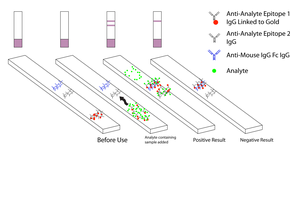
Diagnostic Medical Dipstick.png
A schematic diagram of a dipstick
Purposediagnosis of malaria...

Malaria antigen detection tests are a group of commercially available rapid diagnostic tests of the rapid antigen test type that allow quick diagnosis of malaria by people who are not otherwise skilled in traditional laboratory techniques for diagnosing malaria or in situations where such equipment is not available. There are currently over 20 such tests commercially available (WHO product testing 2008). The first malaria antigen suitable as target for such a test was a soluble glycolytic enzyme Glutamate dehydrogenase. [1] [2] [3] None of the rapid tests are currently as sensitive as a thick blood film, nor as cheap. A major drawback in the use of all current dipstick methods is that the result is essentially qualitative. In many endemic areas of tropical Africa, however, the quantitative assessment of parasitaemia is important, as a large percentage of the population will test positive in any qualitative assay.

Contents

Antigen-based Malaria Rapid Diagnostic Tests

Malaria is a curable disease if the patients have access to early diagnosis and prompt treatment. Antigen-based rapid diagnostic tests (RDTs) have an important role at the periphery of health services capability because many rural clinics do not have the ability to diagnose malaria on-site due to a lack of microscopes and trained technicians to evaluate blood films. Furthermore, in regions where the disease is not endemic, laboratory technologists have very limited experience in detecting and identifying malaria parasites. An ever increasing numbers of travelers from temperate areas each year visit tropical countries and many of them return with a malaria infection. The RDT tests are still regarded as complements to conventional microscopy but with some improvements it may well replace the microscope. The tests are simple and the procedure can be performed on the spot in field conditions. These tests use finger-stick or venous blood, the completed test takes a total of 15–20 minutes, and a laboratory is not needed. The threshold of detection by these rapid diagnostic tests is in the range of 100 parasites/µl of blood compared to 5 by thick film microscopy. [4] [5] [6]

pGluDH

Plasmodium Glutamate dehydrogenase (pGluDH) precipitated by host antibodies Plasmodium glutamate dehydrogenase precipitation.jpg
Plasmodium Glutamate dehydrogenase (pGluDH) precipitated by host antibodies

An accurate diagnosis is becoming more and more important, in view of the increasing resistance of Plasmodium falciparum and the high price of alternatives to chloroquine. The enzyme pGluDH does not occur in the host red blood cell and was recommended as a marker enzyme for Plasmodium species by Picard-Maureau et al. in 1975. [7] The malaria marker enzyme test is suitable for routine work and is now a standard test in most departments dealing with malaria. Presence of pGluDH is known to represent parasite viability [2] and a rapid diagnostic test using pGluDH as antigen would have the ability to differentiate live from dead organisms. A complete RDT with pGluDH as antigen has been developed in China and is now undergoing clinical trials. [3] GluDHs are ubiquitous enzymes that occupy an important branch-point between carbon and nitrogen metabolism. Both nicotinamide adenine dinucleotide (NAD) [EC 1.4.1.2] and nicotinamide adenine dinucleotide phosphate (NADP) dependent GluDH [EC 1.4.1.4] enzymes are present in Plasmodia; the NAD-dependent GluDH is relatively unstable and not useful for diagnostic purposes. Glutamate dehydrogenase provides an oxidizable carbon source used for the production of energy as well as a reduced electron carrier, NADH. Glutamate is a principal amino donor to other amino acids in subsequent transamination reactions. The multiple roles of glutamate in nitrogen balance make it a gateway between free ammonia and the amino groups of most amino acids. Its crystal structure is published. [8] The GluDH activity in P.vivax , P.ovale and P. malariae has never been tested, but given the importance of GluDH as a branch point enzyme, every cell must have a high concentration of GluDH. It is well known that enzymes with a high molecular weight (like GluDH) have many isozymes, which allows strain differentiations (given the right monoclonal antibody). The host produces antibodies against the parasitic enzyme indicating a low sequence identity. [1]

Histidine rich protein II

The histidine-rich protein II (HRP II) is a histidine- and alanine-rich, water-soluble protein, which is localized in several cell compartments including the parasite cytoplasm. The antigen is expressed only by P. falciparum trophozoites. [9] [10] HRP II from P. falciparum has been implicated in the biocrystallization of hemozoin, an inert, crystalline form of ferriprotoporphyrin IX (Fe(3+)-PPIX) produced by the parasite. A substantial amount of the HRP II is secreted by the parasite into the host bloodstream and the antigen can be detected in erythrocytes, serum, plasma, cerebrospinal fluid and even urine as a secreted water-soluble protein. [11] These antigens persist in the circulating blood after the parasitaemia has cleared or has been greatly reduced. It generally takes around two weeks after successful treatment for HRP2-based tests to turn negative, but may take as long as one month, which compromises their value in the detection of active infection. [12] False positive dipstick results were reported in patients with rheumatoid-factor-positive rheumatoid arthritis. [9] Since HRP-2 is expressed only by P. falciparum, these tests will give negative results with samples containing only P. vivax, P. ovale, or P. malariae; many cases of non-falciparum malaria may therefore be misdiagnosed as malaria negative (some P.falciparum strains also don't have HRP II). The variability in the results of pHRP2-based RDTs is related to the variability in the target antigen. [13]

pLDH

Comparison of Plasmodium Lactate Dehydrogenase (PLDH) Malaria Antibodies PLDH Malaria Antibodies.jpg
Comparison of Plasmodium Lactate Dehydrogenase (PLDH) Malaria Antibodies

P. falciparum lactate dehydrogenase (PfLDH) is a 33 kDa oxidoreductase [EC 1.1.1.27]. [14] It is the last enzyme of the glycolytic pathway, essential for ATP generation and one of the most abundant enzymes expressed by P. falciparum. [15] Plasmodium LDH (pLDH) from P. vivax, P. malariae, and P. ovale) exhibit 90-92% identity to PfLDH from P. falciparum. [16] pLDH levels have been seen to reduce in the blood sooner after treatment than HRP2. [17] In this respect, pLDH is similar to pGluDH. Nevertheless, the kinetic properties and sensitivities to inhibitors targeted to the cofactor binding site differ significantly and are identifiable by measuring dissociation constants for inhibitors which, differ by up to 21-fold. [18]

pAldo

Fructose-bisphosphate aldolase [EC 4.1.2.13] catalyzes a key reaction in glycolysis and energy production and is produced by all four species. [19] The P.falciparum aldolase is a 41 kDa protein and has 61-68% sequence similarity to known eukaryotic aldolases. [20] Its crystal structure has been published. [21] The presence of antibodies against p41 in the sera of human adults partially immune to malaria suggest that p41 is implicated in protective immune response against the parasite. [22]

See also

Related Research Articles

<span class="mw-page-title-main">Malaria</span> Medical condition

Malaria is a mosquito-borne infectious disease that affects humans and other vertebrates. Human malaria causes symptoms that typically include fever, fatigue, vomiting, and headaches. In severe cases, it can cause jaundice, seizures, coma, or death. Symptoms usually begin 10 to 15 days after being bitten by an infected mosquito. If not properly treated, people may have recurrences of the disease months later. In those who have recently survived an infection, reinfection usually causes milder symptoms. This partial resistance disappears over months to years if the person has no continuing exposure to malaria.

<i>Plasmodium falciparum</i> Protozoan species of malaria parasite

Plasmodium falciparum is a unicellular protozoan parasite of humans, and the deadliest species of Plasmodium that causes malaria in humans. The parasite is transmitted through the bite of a female Anopheles mosquito and causes the disease's most dangerous form, falciparum malaria. It is responsible for around 50% of all malaria cases. P. falciparum is therefore regarded as the deadliest parasite in humans. It is also associated with the development of blood cancer and is classified as a Group 2A (probable) carcinogen.

<span class="mw-page-title-main">Malate dehydrogenase</span> Class of enzymes

Malate dehydrogenase (EC 1.1.1.37) (MDH) is an enzyme that reversibly catalyzes the oxidation of malate to oxaloacetate using the reduction of NAD+ to NADH. This reaction is part of many metabolic pathways, including the citric acid cycle. Other malate dehydrogenases, which have other EC numbers and catalyze other reactions oxidizing malate, have qualified names like malate dehydrogenase (NADP+).

<i>Plasmodium vivax</i> Species of single-celled organism

Plasmodium vivax is a protozoal parasite and a human pathogen. This parasite is the most frequent and widely distributed cause of recurring malaria. Although it is less virulent than Plasmodium falciparum, the deadliest of the five human malaria parasites, P. vivax malaria infections can lead to severe disease and death, often due to splenomegaly. P. vivax is carried by the female Anopheles mosquito; the males do not bite.

<i>Plasmodium malariae</i> Species of single-celled organism

Plasmodium malariae is a parasitic protozoan that causes malaria in humans. It is one of several species of Plasmodium parasites that infect other organisms as pathogens, also including Plasmodium falciparum and Plasmodium vivax, responsible for most malarial infection. Found worldwide, it causes a so-called "benign malaria", not nearly as dangerous as that produced by P. falciparum or P. vivax. The signs include fevers that recur at approximately three-day intervals – a quartan fever or quartan malaria – longer than the two-day (tertian) intervals of the other malarial parasite.
<i>Plasmodium gallinaceum</i> Bird malaria, including chicken

Plasmodium gallinaceum is a species of the genus Plasmodium that causes malaria in poultry.

<span class="mw-page-title-main">Merozoite surface protein</span>

Merozoitesurface proteins are both integral and peripheral membrane proteins found on the surface of a merozoite, an early life cycle stage of a protozoan. Merozoite surface proteins, or MSPs, are important in understanding malaria, a disease caused by protozoans of the genus Plasmodium. During the asexual blood stage of its life cycle, the malaria parasite enters red blood cells to replicate itself, causing the classic symptoms of malaria. These surface protein complexes are involved in many interactions of the parasite with red blood cells and are therefore an important topic of study for scientists aiming to combat malaria.

Antigenic variation or antigenic alteration refers to the mechanism by which an infectious agent such as a protozoan, bacterium or virus alters the proteins or carbohydrates on its surface and thus avoids a host immune response, making it one of the mechanisms of antigenic escape. It is related to phase variation. Antigenic variation not only enables the pathogen to avoid the immune response in its current host, but also allows re-infection of previously infected hosts. Immunity to re-infection is based on recognition of the antigens carried by the pathogen, which are "remembered" by the acquired immune response. If the pathogen's dominant antigen can be altered, the pathogen can then evade the host's acquired immune system. Antigenic variation can occur by altering a variety of surface molecules including proteins and carbohydrates. Antigenic variation can result from gene conversion, site-specific DNA inversions, hypermutation, or recombination of sequence cassettes. The result is that even a clonal population of pathogens expresses a heterogeneous phenotype. Many of the proteins known to show antigenic or phase variation are related to virulence.

Malaria vaccines are vaccines that prevent malaria, a mosquito-borne infectious disease which annually affects an estimated 247 million people worldwide and causes 619,000 deaths. The first approved vaccine for malaria is RTS,S, known by the brand name Mosquirix. As of April 2023, the vaccine has been given to 1.5 million children living in areas with moderate-to-high malaria transmission. It requires at least three doses in infants by age 2, and a fourth dose extends the protection for another 1–2 years. The vaccine reduces hospital admissions from severe malaria by around 30%.

<span class="mw-page-title-main">6-phosphogluconolactonase</span> Cytosolic enzyme

6-Phosphogluconolactonase (EC 3.1.1.31, 6PGL, PGLS, systematic name 6-phospho-D-glucono-1,5-lactone lactonohydrolase) is a cytosolic enzyme found in all organisms that catalyzes the hydrolysis of 6-phosphogluconolactone to 6-phosphogluconic acid in the oxidative phase of the pentose phosphate pathway:

<span class="mw-page-title-main">Lactate dehydrogenase</span> Class of enzymes

Lactate dehydrogenase (LDH or LD) is an enzyme found in nearly all living cells. LDH catalyzes the conversion of pyruvate to lactate and back, as it converts NAD+ to NADH and back. A dehydrogenase is an enzyme that transfers a hydride from one molecule to another.

<span class="mw-page-title-main">Hemozoin</span>

Haemozoin is a disposal product formed from the digestion of blood by some blood-feeding parasites. These hematophagous organisms such as malaria parasites, Rhodnius and Schistosoma digest haemoglobin and release high quantities of free heme, which is the non-protein component of haemoglobin. Heme is a prosthetic group consisting of an iron atom contained in the center of a heterocyclic porphyrin ring. Free heme is toxic to cells, so the parasites convert it into an insoluble crystalline form called hemozoin. In malaria parasites, hemozoin is often called malaria pigment.

Human genetic resistance to malaria refers to inherited changes in the DNA of humans which increase resistance to malaria and result in increased survival of individuals with those genetic changes. The existence of these genotypes is likely due to evolutionary pressure exerted by parasites of the genus Plasmodium which cause malaria. Since malaria infects red blood cells, these genetic changes are most common alterations to molecules essential for red blood cell function, such as hemoglobin or other cellular proteins or enzymes of red blood cells. These alterations generally protect red blood cells from invasion by Plasmodium parasites or replication of parasites within the red blood cell.

Pregnancy-associated malaria (PAM) or placental malaria is a presentation of the common illness that is particularly life-threatening to both mother and developing fetus. PAM is caused primarily by infection with Plasmodium falciparum, the most dangerous of the four species of malaria-causing parasites that infect humans. During pregnancy, a woman faces a much higher risk of contracting malaria and of associated complications. Prevention and treatment of malaria are essential components of prenatal care in areas where the parasite is endemic – tropical and subtropical geographic areas. Placental malaria has also been demonstrated to occur in animal models, including in rodent and non-human primate models.

The mainstay of malaria diagnosis has been the microscopic examination of blood, utilizing blood films. Although blood is the sample most frequently used to make a diagnosis, both saliva and urine have been investigated as alternative, less invasive specimens. More recently, modern techniques utilizing antigen tests or polymerase chain reaction have been discovered, though these are not widely implemented in malaria endemic regions. Areas that cannot afford laboratory diagnostic tests often use only a history of subjective fever as the indication to treat for malaria.

<span class="mw-page-title-main">RTS,S</span> Malaria vaccine

RTS,S/AS01 is a recombinant protein-based malaria vaccine. It is the only malaria vaccine approved and in wide use. As of April 2022, the vaccine has been given to 1 million children living in areas with moderate-to-high malaria transmission, with millions more doses to be provided as the vaccine's production expands. It requires at least three doses in infants by age 2, with a fourth dose extending the protection for another 1–2 years. The vaccine reduces hospital admissions from severe malaria by around 30%.

Russell J. Howard is an Australian-born executive, entrepreneur and scientist. He was a pioneer in the fields of molecular parasitology, especially malaria, and in leading the commercialisation of one of the most important methods used widely today in molecular biology today called “DNA shuffling" or "Molecular breeding", a form of "Directed evolution".

<span class="mw-page-title-main">Palaeoimmunology</span>

Palaeoimmunology or paleo-immunology is the analysis using histochemical techniques to look at the matrix proteins in historic and pre-historic materials. Modern immunological assays are used to detect the presence of specific antigens in the sample material. Specimens subject to immunoassays have usually been preserved in a way that has prevented biomolecular targets from degrading. This has either been achieved through natural preservative circumstances, such as accelerated fossilization, or through artificial mummification. Regardless of the path taken to achieve this state, preservation has occurred before the denaturing of antigenic targets. The purpose of applying immunological assays to archaeological materials is to better understand the biochemical makeup and composition of these pre-historic samples. Antigenic elements within these materials may reveal information regarding the "life" and "death" of the sample being studied.

Plasmodium falciparum erythrocyte membrane protein 1 (PfEMP1) is a family of proteins present on the membrane surface of red blood cells that are infected by the malarial parasite Plasmodium falciparum. PfEMP1 is synthesized during the parasite's blood stage inside the RBC, during which the clinical symptoms of falciparum malaria are manifested. Acting as both an antigen and adhesion protein, it is thought to play a key role in the high level of virulence associated with P. falciparum. It was discovered in 1984 when it was reported that infected RBCs had unusually large-sized cell membrane proteins, and these proteins had antibody-binding (antigenic) properties. An elusive protein, its chemical structure and molecular properties were revealed only after a decade, in 1995. It is now established that there is not one but a large family of PfEMP1 proteins, genetically regulated (encoded) by a group of about 60 genes called var. Each P. falciparum is able to switch on and off specific var genes to produce a functionally different protein, thereby evading the host's immune system. RBCs carrying PfEMP1 on their surface stick to endothelial cells, which facilitates further binding with uninfected RBCs, ultimately helping the parasite to both spread to other RBCs as well as bringing about the fatal symptoms of P. falciparum malaria.

Reticulocyte binding protein homologs (RHs) are a superfamily of proteins found in Plasmodium responsible for cell invasion. Together with the family of erythrocyte binding-like proteins (EBLs) they make up the two families of invasion proteins universal to Plasmodium. The two families function cooperatively.
References

  1. 1 2 3 Ling IT.; Cooksley S.; Bates PA.; Hempelmann E.; Wilson RJM. (1986). "Antibodies to the glutamate dehydrogenase of Plasmodium falciparum" (PDF). Parasitology. 92 (2): 313–324. doi:10.1017/S0031182000064088. PMID   3086819. S2CID   16953840.
  2. 1 2 Rodríguez-Acosta A, Domínguez NG, Aguilar I, Girón ME (1998). "Characterization of Plasmodium falciparum glutamate dehydrogenase-soluble antigen". Braz J Med Biol Res. 31 (9): 1149–1155. doi: 10.1590/S0100-879X1998000900008 . PMID   9876282.
  3. 1 2 Li Y, Ning YS, Li L, Peng DD, Dong WQ, Li M (2005). "Preparation of a monoclonal antibodies against Plasmodium falciparum glutamate dehydrogenase and establishment of colloidal gold-immunochromatographic assay". Di Yi Jun Yi da Xue Xue Bao = Academic Journal of the First Medical College of PLA. 25 (4): 435–438. PMID   15837649.
  4. Warhurst DC, Williams JE (1996). "ACP Broadsheet no 148. July 1996. Laboratory diagnosis of malaria". J Clin Pathol. 49 (7): 533–38. doi:10.1136/jcp.49.7.533. PMC   500564 . PMID   8813948.
  5. Moody A. (2002). "Rapid Diagnostic Tests for Malaria Parasites". Clin Microbiol Rev. 15 (1): 66–78. doi:10.1128/CMR.15.1.66-78.2002. PMC   118060 . PMID   11781267.
  6. Murray CK, Bennett JW (2008). "Rapid Diagnosis of Malaria". Interdiscip Perspect Infect Dis. 2009: 1–7. doi: 10.1155/2009/415953 . PMC   2696022 . PMID   19547702.
  7. Picard-Maureau A, Hempelmann E, Krämmer G, Jackisch R, Jung A (1975). "Glutathionstatus in Plasmodium vinckei parasitierten Erythrozyten in Abhängigkeit vom intraerythrozytären Entwicklungsstadium des Parasiten". Tropenmed Parasitol. 26 (4): 405–416. PMID   1216329.
  8. Werner C, Stubbs MT, Krauth-Siegel RL, Klebe G (2005). "The crystal structure of Plasmodium falciparum glutamate dehydrogenase, a putative target for novel antimalarial drugs". J Mol Biol. 349 (3): 597–607. doi:10.1016/j.jmb.2005.03.077. PMID   15878595.
  9. 1 2 Iqbal J, Sher A, Rab A (2000). "Plasmodium falciparum Histidine-Rich Protein 2-Based Immunocapture Diagnostic Assay for Malaria: Cross-Reactivity with Rheumatoid Factors". J Clin Microbiol. 38 (3): 1184–1186. doi:10.1128/JCM.38.3.1184-1186.2000. PMC   86370 . PMID   10699018.
  10. Beadle C, Long GW, Weiss WR, McElroy PD, Maret SM, Oloo AJ, Hoffman SL (1994). "Diagnosis of malaria by detection of Plasmodium falciparum HRP-2 antigen with a rapid dipstick antigen-capture assay". Lancet. 343 (8897): 564–568. doi: 10.1016/S0140-6736(94)91520-2 . PMID   7906328. S2CID   25450213.
  11. Rock EP, Marsh K, Saul AJ, Wellems TE, Taylor DW, Maloy WL, Howard RJ (1987). "Comparative analysis of the Plasmodium falciparum histidine-rich proteins HRP-I, HRP-II and HRP-III in malaria parasites of diverse origin". Parasitology. 95 (2): 209–227. doi:10.1017/S0031182000057681. PMID   3320887. S2CID   24038846.
  12. Humar A, Ohrt C, Harrington MA, Pillai D, Kain KC (1997). "Parasight F test compared with the polymerase chain reaction and microscopy for the diagnosis of Plasmodium falciparum malaria in travelers". Am J Trop Med Hyg. 56 (1): 44–48. doi:10.4269/ajtmh.1997.56.44. PMID   9063360.
  13. Baker J, McCarthy J, Gatton M, Kyle DE, Belizario V, Luchavez J, Bell D, Cheng Q (2005). "Genetic diversity of Plasmodium falciparum histidine-rich protein 2 (PfHRP2) and its effect on the performance of PfHRP2-based rapid diagnostic tests". J Infect Dis. 192 (5): 870–877. doi: 10.1086/432010 . PMID   16088837.
  14. Bzik DJ, Fox BA, Gonyer K (1993). "Expression of Plasmodium falciparum lactate dehydrogenase in Escherichia coli". Mol Biochem Parasitol. 59 (1): 155–166. doi:10.1016/0166-6851(93)90016-Q. PMID   8515777.
  15. Vander Jagt DL, Hunsaker LA, Heidrich JE (1981). "Partial purification and characterization of lactate dehydrogenase from Plasmodium falciparum". Molecular and Biochemical Parasitology. 4 (5–6): 255–264. doi:10.1016/0166-6851(81)90058-X. PMID   7038478.
  16. Penna-Coutinho J, Cortopassi WA, Oliveira AA, França TC, Krettli AU (2011). "Antimalarial activity of potential inhibitors of Plasmodium falciparum lactate dehydrogenase enzyme selected by docking studies". PLOS ONE. 6 (7): e21237. Bibcode:2011PLoSO...621237P. doi: 10.1371/journal.pone.0021237 . PMC   3136448 . PMID   21779323.
  17. Iqbal J, Siddique A, Jameel M, Hira PR (2004). "Persistent Histidine-Rich Protein 2, Parasite Lactate Dehydrogenase, and Panmalarial Antigen Reactivity after Clearance of Plasmodium falciparum Monoinfection". J Clin Microbiol. 42 (9): 4237–4241. doi:10.1128/JCM.42.9.4237-4241.2004. PMC   516301 . PMID   15365017.
  18. Brown WM, Yowell CA, Hoard A, Vander Jagt TA, Hunsaker LA, Deck LM, Royer RE, Piper RC, Dame JB, Makler MT, Vander Jagt DL (2004). "Comparative structural analysis and kinetic properties of lactate dehydrogenases from the four species of human malarial parasites". Biochemistry. 43 (20): 6219–6229. doi:10.1021/bi049892w. PMID   15147206.
  19. Meier B, Döbeli H, Certa U (1992). "Stage-specific expression of aldolase isoenzymes in the rodent malaria parasite Plasmodium berghei". Mol Biochem Parasitol. 52 (1): 15–27. doi:10.1016/0166-6851(92)90032-F. PMID   1625704.
  20. Knapp B, Hundt E, Küpper HA (1990). "Plasmodium falciparum aldolase: gene structure and localization". Mol Biochem Parasitol. 40 (1): 1–12. doi:10.1016/0166-6851(90)90074-V. PMID   2190085.
  21. Kim H, Certa U, Döbeli H, Jakob P, Hol WG (1998). "Crystal structure of fructose-1,6-bisphosphate aldolase from the human malaria parasite Plasmodium falciparum". Biochemistry. 37 (13): 4388–96. doi:10.1021/bi972233h. PMID   9521758.
  22. Srivastava IK, Schmidt M, Certa U, Döbeli H, Perrin LH (1990). "Specificity and inhibitory activity of antibodies to Plasmodium falciparum aldolase". J Immunol. 144 (4): 1497–503. doi: 10.4049/jimmunol.144.4.1497 . PMID   2406342. S2CID   25168857.