Aequorin

Last updated
Aequorin 1
Aequorin 1EJ3.png
Aequorin ribbon diagram from PDB 1ej3 with prosthetic group coelenterazine in blue
Identifiers
Organism Aequorea victoria (Jellyfish)
SymbolN/A
UniProt P07164
Other data
EC number 1.13.12.5
Search for
Structures Swiss-model
Domains InterPro

Aequorin is a calcium-activated photoprotein isolated from the hydrozoan Aequorea victoria . [1] Its bioluminescence was studied decades before the protein was isolated from the animal by Osamu Shimomura in 1962. [2] In the animal, the protein occurs together with the green fluorescent protein to produce green light by resonant energy transfer, while aequorin by itself generates blue light.

Contents

Discussions of "jellyfish DNA" that can make "glowing" animals often refer to transgenic animals that express the green fluorescent protein, not aequorin, although both originally derive from the same animal.

Apoaequorin, the protein portion of aequorin, is an ingredient in the dietary supplement Prevagen. The US Federal Trade Commission (FTC) has charged the maker with false advertising for its memory improvement claims.

Discovery

Work on aequorin began with E. Newton Harvey in 1921. [3] Though Harvey was unable to demonstrate a classical luciferase-luciferin reaction, he showed that water could produce light from dried photocytes and that light could be produced even in the absence of oxygen. Later, Osamu Shimomura began work into the bioluminescence of Aequorea in 1961. This involved tedious harvesting of tens of thousands of jellyfish from the docks in Friday Harbor, Washington. [1] It was determined that light could be produced from extracts with seawater, and more specifically, with calcium. [2] It was also noted during the extraction the animal creates green light due to the presence of the green fluorescent protein, which changes the native blue light of aequorin to green. [4]

While the main focus of his work was on the bioluminescence, [5] Shimomura and two others, Martin Chalfie and Roger Tsien, were awarded the Nobel Prize in 2008 for their work on green fluorescent proteins.

Structure

Aequorin is a holoprotein composed of two distinct units, the apoprotein that is called apoaequorin, which has an approximate molecular weight of 21 kDa, and the prosthetic group coelenterazine, the luciferin. [6] This is to say, apoaequorin is the enzyme produced in the photocytes of the animal, and coelenterazine is the substrate whose oxidation the enzyme catalyzes. When coelenterazine is bound, it is called aequorin. Notably, the protein contains three EF hand motifs that function as binding sites for Ca2+ ions. [7] The protein is a member of the superfamily of the calcium-binding proteins, of which there are some 66 subfamilies. [8]

The crystal structure revealed that aequorin binds coelenterazine and oxygen in the form of a peroxide, coelenterazine-2-hydroperoxide. [9] The binding site for the first two calcium atoms show a 20 times greater affinity for calcium than the third site. [10] However, earlier claims that only two EF-hands bind calcium [11] were questioned when later structures indicated that all three sites can indeed bind calcium. [12] Thus, titration studies show that all three calcium-binding sites are active but only two ions are needed to trigger the enzymatic reaction. [13]

Other studies have shown the presence of an internal cysteine bond that maintains the structure of aequorin. [14] This has also explained the need for a thiol reagent like beta mercaptoethanol in the regeneration of the protein since such reagents weaken the sulfhydryl bonds between cysteine residues, expediting the regeneration of the aequorin.

Chemical characterization of aequorin indicates the protein is somewhat resilient to harsh treatments. Aequorin is heat resistant. [15] Held at 95 °C for 2 minutes the protein lost only 25% activity. Denaturants such as 6-M urea or 4-M guanidine hydrochloride did not destroy the protein.

Genetics

Aequorin is presumably encoded in the genome of Aequorea. At least four copies of the gene were recovered as cDNA from the animal. [16] [17] Because the genome has not been sequenced, it is unclear if the cDNA variants can account for all of the isoforms of the protein. [18]

Mechanism of action

Early studies of the bioluminescence of Aequorea by E. Newton Harvey had noted that the bioluminescence appears as a ring around the bell, and occurs even in the absence of air. [19] This was remarkable because most bioluminescence reactions require oxygen, and led to the idea that the animals somehow store oxygen. [20] It was later discovered that the apoprotein can stably bind coelenterazine-2-hydroperoxide, and oxygen is required for the regeneration to this active form of aequorin. [21] However, in the presence of calcium ions, the protein undergoes a conformational change and converts its prosthetic group, coelenterazine-2-hydroperoxide, into excited coelenteramide and CO2. [22] As the excited coelenteramide relaxes to the ground state, blue light (wavelength of 465 nm) is emitted. Before coelenteramide is exchanged out, the entire protein is still fluorescent blue. [23] [24] because of the connection between bioluminescence and fluorescence, this property was ultimately important in the discovery of the luciferin coelenterazine. [25]

Applications

Since the emitted light can be easily detected with a luminometer, aequorin has become a useful tool in molecular biology for the measurement of intracellular Ca2+ levels. [26] The early successful purification of aequorin led to the first experiments involving the injection of the protein into the tissues of living animals to visualize the physiological release of calcium in the muscle fibers of a barnacle. [27] Since then, the protein has been widely used in many model biological systems, including zebrafish, [28] rats, mice, and cultured cells. [29] [30] [31] [32]

Cultured cells expressing the aequorin gene can effectively synthesize apoaequorin; however, recombinant expression yields only the apoprotein. Therefore it is necessary to add coelenterazine into the culture medium of the cells to obtain a functional protein and thus use its blue light emission to measure Ca2+ concentration. Coelenterazine is a hydrophobic molecule, and therefore is easily taken up across plant and fungal cell walls, as well as the plasma membrane of higher eukaryotes, making aequorin suitable as a Ca2+ reporter in plants, fungi, and mammalian cells. [33] [34]

Aequorin has a number of advantages over other Ca2+ indicators. Because the protein is large, it has a low leakage rate from cells compared to lipophilic dyes such as DiI. It lacks phenomena of intracellular compartmentalization or sequestration as is often seen for Voltage-sensitive dyes, and does not disrupt cell functions or embryo development. Moreover, the light emitted by the oxidation of coelenterazine does not depend on any optical excitation, so problems with auto-fluorescence are eliminated. [35] The primary limitation of aequorin is that the prosthetic group coelenterazine is irreversibly consumed to produce light, and requires continuous addition of coelenterazine into the media. Such issues led to developments of other genetically encoded calcium sensors including the calmodulin-based sensor cameleon, [36] developed by Roger Tsien and the troponin-based sensor, TN-XXL, developed by Oliver Griesbeck. [37]

Apoaequorin is an ingredient in "Prevagen", which is marketed by Quincy Bioscience as a memory supplement. In 2017, the US Federal Trade Commission (FTC) charged the maker with falsely advertising that the product improves memory, provides cognitive benefits, and is "clinically shown" to work. [38] According to the FTC, "the marketers of Prevagen preyed on the fears of older consumers experiencing age-related memory loss". Quincy said that it would fight the charges. [39] [40] [41]

Prior to the suit, a clinical trial run by researchers employed by Quincy Bioscience "found no overall benefit compared to a placebo for its primary endpoints involving memory and cognition", while the company's advertising misleadingly cited a few contested subgroup analyses that showed slight improvements. [42] [43]

The suit (Spath, et al. v. Quincy Bioscience Holding Company, Inc., et al., Case No. 18-cv-12416, D. NJ.) was dismissed in the District court, but an appeal seeking to overturn the dismissal was filed. The suit was consolidated with another against Quincy Pharmaceuticals, Vanderwerff v. Quincy Bioscience (Case No. 17-cv-784, D. NJ), which was the lead case. [44]

On February 21, 2019, the United States Court of Appeals for the Second Circuit ruled that the FTC and the state of New York could proceed with their lawsuit against Quincy Bioscience for its claims that Prevagen can improve memory. The order came less than two weeks after the parties argued the case before a three-judge panel of the circuit, where company lawyers admitted they did not "dispute that if you look across the entire 211 people who completed the study there was no statistically significant difference". The court vigorously dismissed allegations by the company lawyers that the FTC pursued its action for political reasons. [45] [46]

On March 23, 2020, a federal magistrate judge in the United States District Court for the Southern District of Florida entered a report and recommendations certifying a nationwide class action for the class of consumers who purchased Prevagen over the previous four years. [47] The trial in the case was set for October 2020. [47] [48]

As of September 21,2020 Quincy Bioscience agreed to settle the claims that it misrepresented its Prevagen products as supporting brain health and helping with memory loss. Under the terms of the settlement, eligible purchasers applying by October 26, 2020 for purchases made from 2007 through July 31, 2020 could recover refunds of up to $70. [49]

Dr. Harriet Hall, writing for Science-Based Medicine , noted that the Quincy-sponsored study (known as "Madison Memory Study") was negative, but that the company utilized p-hacking to find favorable results. She wrote that their cited safety studies were all rat studies and their claim that apoaequorin crosses the blood–brain barrier was based solely on a dog study. [50] The American Pharmacists Association warns that Apoaequorin "is unlikely to be absorbed to a significant degree; instead it degrades into amino acids". [51]

Related Research Articles

<span class="mw-page-title-main">Green fluorescent protein</span> Protein that exhibits bright green fluorescence when exposed to ultraviolet light

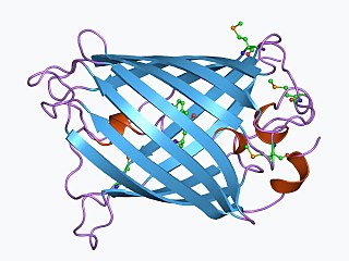
The green fluorescent protein (GFP) is a protein that exhibits green fluorescence when exposed to light in the blue to ultraviolet range. The label GFP traditionally refers to the protein first isolated from the jellyfish Aequorea victoria and is sometimes called avGFP. However, GFPs have been found in other organisms including corals, sea anemones, zoanithids, copepods and lancelets.

<span class="mw-page-title-main">Bioluminescence</span> Emission of light by a living organism

Bioluminescence is the production and emission of light by living organisms. It is a form of chemiluminescence. Bioluminescence occurs widely in marine vertebrates and invertebrates, as well as in some fungi, microorganisms including some bioluminescent bacteria, and terrestrial arthropods such as fireflies. In some animals, the light is bacteriogenic, produced by symbiotic bacteria such as those from the genus Vibrio; in others, it is autogenic, produced by the animals themselves.

<span class="mw-page-title-main">Luciferase</span> Enzyme family

Luciferase is a generic term for the class of oxidative enzymes that produce bioluminescence, and is usually distinguished from a photoprotein. The name was first used by Raphaël Dubois who invented the words luciferin and luciferase, for the substrate and enzyme, respectively. Both words are derived from the Latin word lucifer, meaning "lightbearer", which in turn is derived from the Latin words for "light" (lux) and "to bring or carry" (ferre).

<span class="mw-page-title-main">Luciferin</span> Class of light-emitting chemical compounds

Luciferin is a generic term for the light-emitting compound found in organisms that generate bioluminescence. Luciferins typically undergo an enzyme-catalyzed reaction with molecular oxygen. The resulting transformation, which usually involves breaking off a molecular fragment, produces an excited state intermediate that emits light upon decaying to its ground state. The term may refer to molecules that are substrates for both luciferases and photoproteins.

<i>Aequorea victoria</i> Species of hydrozoan

Aequorea victoria, also sometimes called the crystal jelly, is a bioluminescent hydrozoan jellyfish, or hydromedusa, that is found off the west coast of North America.

pGLO

The pGLO plasmid is an engineered plasmid used in biotechnology as a vector for creating genetically modified organisms. The plasmid contains several reporter genes, most notably the green fluorescent protein (GFP) and the ampicillin resistance gene. GFP was isolated from the jelly fish Aequorea victoria. Because it shares a bidirectional promoter with a gene for metabolizing arabinose, the GFP gene is expressed in the presence of arabinose, which makes the transgenic organism express its fluorescence under UV light. GFP can be induced in bacteria containing the pGLO plasmid by growing them on +arabinose plates. pGLO is made by Bio-Rad Laboratories.

<span class="mw-page-title-main">EF hand</span> Protein helix–loop–helix motif

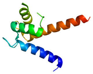
The EF hand is a helix–loop–helix structural domain or motif found in a large family of calcium-binding proteins.

<i>Aequorea forskalea</i> Species of hydrozoan

Aequorea forskalea is a species of hydrozoan in the family Aequoreidae. Discovered in 1810 by Péron and Lesueur, A. forskalea was initially found in coastal to offshore waters of the Mediterranean Sea. This species is commonly referred to as the many-ribbed jellyfish. The species is often mixed up with some other members of the genus due to some similarities including the capability of bioluminescence.

In enzymology, an Oplophorus-luciferin 2-monooxygenase, also known as Oplophorus luciferase is a luciferase, an enzyme, from the deep-sea shrimp Oplophorus gracilirostris [2], belonging to a group of coelenterazine luciferases. Unlike other luciferases, it has a broader substrate specificity [3,4,6] and can also bind to bisdeoxycoelenterazine efficiently [3,4]. It is the third example of a luciferase to be purified in lab [2]. The systematic name of this enzyme class is Oplophorus-luciferin:oxygen 2-oxidoreductase (decarboxylating). This enzyme is also called Oplophorus luciferase.

<span class="mw-page-title-main">Renilla-luciferin 2-monooxygenase</span>

Renilla-luciferin 2-monooxygenase, Renilla luciferase, or RLuc, is a bioluminescent enzyme found in Renilla reniformis, belonging to a group of coelenterazine luciferases. Of this group of enzymes, the luciferase from Renilla reniformis has been the most extensively studied, and due to its bioluminescence requiring only molecular oxygen, has a wide range of applications, with uses as a reporter gene probe in cell culture, in vivo imaging, and various other areas of biological research. Recently, chimeras of RLuc have been developed and demonstrated to be the brightest luminescent proteins to date, and have proved effective in both noninvasive single-cell and whole body imaging.

<span class="mw-page-title-main">S100P</span> Protein-coding gene in the species Homo sapiens

S100 calcium-binding protein P (S100P) is a protein that in humans is encoded by the S100P gene.

<span class="mw-page-title-main">Bioreporter</span> Genetically engineered microbial cells

Bioreporters are intact, living microbial cells that have been genetically engineered to produce a measurable signal in response to a specific chemical or physical agent in their environment. Bioreporters contain two essential genetic elements, a promoter gene and a reporter gene. The promoter gene is turned on (transcribed) when the target agent is present in the cell’s environment. The promoter gene in a normal bacterial cell is linked to other genes that are then likewise transcribed and then translated into proteins that help the cell in either combating or adapting to the agent to which it has been exposed. In the case of a bioreporter, these genes, or portions thereof, have been removed and replaced with a reporter gene. Consequently, turning on the promoter gene now causes the reporter gene to be turned on. Activation of the reporter gene leads to production of reporter proteins that ultimately generate some type of a detectable signal. Therefore, the presence of a signal indicates that the bioreporter has sensed a particular target agent in its environment.

Douglas C. Prasher is an American molecular biologist. He is known for his work to clone and sequence the genes for the photoprotein aequorin and green fluorescent protein (GFP) and for his proposal to use GFP as a tracer molecule. He communicated his pioneering work to Martin Chalfie and Roger Y. Tsien, but by 1991 he was unable to obtain further research funding, and left academia. Eventually, he had to abandon science. Chalfie and Tsien were awarded the 2008 Nobel Prize in Chemistry for work that they publicly acknowledged was substantially based on Prasher's work; through their efforts and those of others, he returned to scientific research in June 2010.

<span class="mw-page-title-main">Coelenterazine</span> Chemical compound

Coelenterazine is a luciferin, a molecule that emits light after reaction with oxygen, found in many aquatic organisms across eight phyla. It is the substrate of many luciferases such as Renilla reniformis luciferase (Rluc), Gaussia luciferase (Gluc), and photoproteins, including aequorin, and obelin. All these proteins catalyze the oxidation of this substance, a reaction catalogued EC 1.13.12.5.

<span class="mw-page-title-main">Vargulin</span> Chemical compound

Vargulin, also called Cypridinid luciferin, Cypridina luciferin, or Vargula luciferin, is the luciferin found in the ostracod Cypridina hilgendorfii, also named Vargula hilgendorfii. These bottom dwelling ostracods emit a light stream into water when disturbed presumably to deter predation. Vargulin is also used by the midshipman fish, Porichthys.

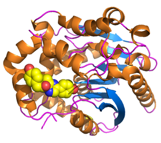
Photoproteins are a type of enzyme produced by bioluminescent organisms. They add to the function of the luciferins whose usual light-producing reaction is catalyzed by the enzyme luciferase.

<span class="mw-page-title-main">Coelenteramide</span> Chemical compound

Coelenteramide is the oxidized product, or oxyluciferin, of the bioluminescent reactions in many marine organisms that use coelenterazine. It was first isolated as a blue fluorescent protein from Aequorea victoria after the animals were stimulated to emit light. Under basic conditions, the compound will break down further into coelenteramine and 4-hydroxyphenylacetic acid.

<span class="mw-page-title-main">Coelenteramine</span> Chemical compound

Coelenteramine is a metabolic product of the bioluminescent reactions in organisms that utilize coelenterazine. It was first isolated from Aequorea victoria along with coelenteramide after coelenterates were stimulated to emit light.

Calcium imaging is a microscopy technique to optically measure the calcium (Ca2+) status of an isolated cell, tissue or medium. Calcium imaging takes advantage of calcium indicators, fluorescent molecules that respond to the binding of Ca2+ ions by fluorescence properties. Two main classes of calcium indicators exist: chemical indicators and genetically encoded calcium indicators (GECI). This technique has allowed studies of calcium signalling in a wide variety of cell types. In neurons, action potential generation is always accompanied by rapid influx of Ca2+ ions. Thus, calcium imaging can be used to monitor the electrical activity in hundreds of neurons in cell culture or in living animals, which has made it possible to observe the activity of neuronal circuits during ongoing behavior.

Aequorea macrodactyla is a species of hydrozoan in the family Aequoreidae. It was first described by Johann Friedrich von Brandt in 1835.

References

  1. 1 2 Shimomura O (1995). "A short story of aequorin". Biol. Bull. 189 (1). Biological Bulletin: 1–5. doi:10.2307/1542194. JSTOR   1542194. PMID   7654844.
  2. 1 2 Shimomura O, Johnson FH, Saiga Y (1962). "Extraction, purification and properties of aequorin, a bioluminescent protein from the luminous hydromedusan, Aequorea". J Cell Comp Physiol. 59 (3): 223–39. doi:10.1002/jcp.1030590302. PMID   13911999.
  3. Harvey EN (1921). "Studies on Bioluminescence. XIII. Luminescence in the Cœlenterate". Biological Bulletin. 41 (5): 280–287. doi:10.2307/1536528. JSTOR   1536528. S2CID   10826363.
  4. Morin JG, Hastings JW (1971). "Energy transfer in a bioluminescent system". J. Cell. Physiol. 77 (3): 313–318. doi:10.1002/jcp.1040770305. PMID   4397528. S2CID   42494355.
  5. Shimomura O (2005). "The discovery of aequorin and green fluorescent protein". J Microsc. 217 (Pt 1): 1–15. doi:10.1111/j.0022-2720.2005.01441.x. PMID   15655058. S2CID   36316988.
  6. Shimomura O, Johnson FH (1978). "Peroxidized coelenterazine, the active group in the photoprotein aequorin". PNAS USA. 75 (3): 2611–2615. Bibcode:1978PNAS...75.2611S. doi: 10.1073/pnas.75.6.2611 . PMC   392612 . PMID   275832.
  7. Charbonneau H, Walsh KA, McCann RO, Prendergast FG, Cormier MJ, Vanaman TC (1985). "Amino acid sequence of the calcium-dependent photoprotein aequorin". Biochemistry. 24 (24): 6762–6771. doi:10.1021/bi00345a006. PMID   2866797.
  8. Zhou Y, Yang W, Kirberger M, Lee HW, Ayalasomayajula G, Yang JJ (2006). "Prediction of EF-hand calcium-binding proteins and analysis of bacterial EF-hand proteins". Proteins. 65 (3): 643–655. doi:10.1002/prot.21139. PMID   16981205. S2CID   8904181.
  9. Head JF, Inouye S, Teranishi K, Shimomura O (2000). "The crystal structure of the photoprotein aequorin at 2.3 Å resolution". Nature. 405 (6784): 372–376. Bibcode:2000Natur.405..372H. doi:10.1038/35012659. PMID   10830969. S2CID   4425033.
  10. Shimomura O (1995). "Luminescence of aequorin is triggered by the binding of two calcium ions". Biochem. Biophys. Res. Commun. 211 (2): 359–363. doi:10.1006/bbrc.1995.1821. PMID   7794244.
  11. Shimomura O (1995). "Luminescence of aequorin is triggered by the binding of two calcium ions". Biochemical and Biophysical Research Communications. 211 (2): 359–363. doi:10.1006/bbrc.1995.1821. PMID   7794244.
  12. Deng L, Vysotski ES, Markova SV, Liu ZJ, Lee J, Rose J, Wang BC (2005). "All three Ca2+-binding loops of photoproteins bind calcium ions: the crystal structures of calcium-loaded apo-aequorin and apo-obelin". Protein Sci. 14 (3): 663–675. doi:10.1110/ps.041142905. PMC   2279293 . PMID   15689515.
  13. Shimomura O, Inouye S (1996). "Titration of recombinant aequorin with calcium chloride". Biochem. Biophys. Res. Commun. 221 (1): 77–81. doi: 10.1006/bbrc.1996.0548 . PMID   8660347.
  14. Ohmiya Y, Kurono S, Ohashi M, Fagan TF, Tsuji FI (1993). "Mass spectrometric evidence for a disulfide bond in aequorin regeneration". FEBS Lett. 332 (3): 226–228. doi: 10.1016/0014-5793(93)80637-a . PMID   8405461.
  15. Inouye S (2004). "Blue fluorescent protein from the calcium-sensitive photoprotein aequorin is a heat-resistant enzyme, catalyzing the oxidation of coelenterazine". FEBS Lett. 577 (1–2): 105–110. doi: 10.1016/j.febslet.2004.09.078 . PMID   15527769.
  16. Prasher D, McCann RO, Cormier MJ (1985). "Cloning and expression of the cDNA coding for aequorin, a bioluminescent calcium-binding protein". Biochem. Biophys. Res. Commun. 126 (3): 1259–68. doi:10.1016/0006-291X(85)90321-3. PMID   2579647.
  17. Inouye S, Noguchi M, Sakaki Y, Takagi Y, Miyata T, Iwanaga S, Miyata T, Tsuji FI (1985). "Cloning and sequence analysis of cDNA for the luminescent protein aequorin". Proc. Natl. Acad. Sci. U.S.A. 82 (10): 3154–58. Bibcode:1985PNAS...82.3154I. doi: 10.1073/pnas.82.10.3154 . PMC   397733 . PMID   3858813.
  18. Masuda H, Takenaka Y, Shikamoto Y, Kagawa M, Mizuno H, Tsuji FI (2003). "Chromatography of isoforms of recombinant apoaequorin and method for the preparation of aequorin". Protein Expr. Purif. 31 (2): 181–187. doi:10.1016/s1046-5928(03)00186-4. PMID   14550635.
  19. Harvey EN (1926). "Oxygen and Luminescence, with a Description of Methods for Removing Oxygen from Cells and Fluids". Biological Bulletin. 51 (2): 89–97. doi:10.2307/1536540. JSTOR   1536540.
  20. Harvey EN (1952). Bioluminescence. Academic Press.
  21. Shimomura O, Johnson FH (1975). "Regeneration of the photoprotein aequorin". Nature. 256 (5514): 236–238. Bibcode:1975Natur.256..236S. doi:10.1038/256236a0. PMID   239351. S2CID   4176627.
  22. Shimomura O, Johnson FH, Morise H (1974). "Mechanism of the luminescent intramolecular reaction of aequorin". Biochemistry. 13 (16): 3278–3286. doi:10.1021/bi00713a016. PMID   4152180.
  23. Shimomura O, Johnson FH (1970). "Calcium binding, quantum yield, and emitting molecule in aequorin bioluminescence". Nature. 227 (5265): 1356–1357. Bibcode:1970Natur.227.1356S. doi:10.1038/2271356a0. PMID   4393938. S2CID   4284185.
  24. Inouye S, Sasaki S (2006). "Blue fluorescent protein from the calcium-sensitive photoprotein aequorin: catalytic properties for the oxidation of coelenterazine as an oxygenase". FEBS Lett. 580 (8): 1977–1982. doi: 10.1016/j.febslet.2006.02.065 . PMID   16545379.
  25. Shimomura O, Johnson FH (1975). "Chemical nature of bioluminescence systems in coelenterates". Proceedings of the National Academy of Sciences . 72 (4): 1546–1549. Bibcode:1975PNAS...72.1546S. doi: 10.1073/pnas.72.4.1546 . PMC   432574 . PMID   236561.
  26. Shimomura O, Inouye S, Musicki B, Kishi Y (1990). "Recombinant aequorin and recombinant semi-synthetic aequorins. Cellular Ca2+ ion indicators". Biochem. J. 270 (2): 309–312. doi:10.1042/bj2700309. PMC   1131721 . PMID   2400391.
  27. Ridgway EB, Ashley CC (1967). "Calcium transients in single muscle fibers". Biochem. Biophys. Res. Commun. 29 (2): 229–234. doi:10.1016/0006-291x(67)90592-x. PMID   4383681.
  28. Cheung CY, Webb SE, Meng A, Miller AL (2006). "Transient expression of apoaequorin in zebrafish embryos: extending the ability to image calcium transients during later stages of development". Int. J. Dev. Biol. 50 (6): 561–569. doi: 10.1387/ijdb.062151cc . PMID   16741871.
  29. Rembold CM, Kendall JM, Campbell AK (January 1997). "Measurement of changes in sarcoplasmic reticulum [Ca2+] in rat tail artery with targeted apoaequorin delivered by an adenoviral vector". Cell Calcium. 21 (1): 69–79. doi:10.1016/s0143-4160(97)90098-1. PMID   9056079.
  30. Yamano K, Mori K, Nakano R, Kusunoki M, Inoue M, Satoh M (2007). "Identification of the functional expression of adenosine A3 receptor in pancreas using transgenic mice expressing jellyfish apoaequorin". Transgenic Res. 16 (4): 429–435. doi:10.1007/s11248-007-9084-0. PMID   17387626. S2CID   19339429.
  31. Sheu YA, Kricka LJ, Pritchett DB (1993). "Measurement of intracellular calcium using bioluminescent aequorin expressed in human cells". Anal. Biochem. 209 (2): 343–347. doi: 10.1006/abio.1993.1132 . PMID   8470808.
  32. Mithöfer A, Mazars C (2002). "Aequorin-based measurements of intracellular Ca2+ signatures in plant cells". Biol. Proced. Online. 4: 105–118. doi:10.1251/bpo40. PMC   145563 . PMID   12734562. Archived from the original on 2005-07-28.
  33. Blinks JR, Wier WG, Hess P, Prendergast FG (1982). "Measurement of Ca2+ concentrations in living cells". Prog Biophys Mol Biol. 40 (1–2): 1–114. doi: 10.1016/0079-6107(82)90011-6 . PMID   6758036.
  34. Montero M, Brini M, Marsault R, Alvarez J, Sitia R, Pozzan T, Rizzuto R (1995). "Monitoring dynamic changes in free Ca2+ concentration in the endoplasmic reticulum of intact cells". EMBO J. 14 (22): 5467–5475. doi:10.1002/j.1460-2075.1995.tb00233.x. PMC   394660 . PMID   8521803.
  35. Kendall JM, Badminton MN, Sala-Newby GB, Campbell AK, Rembold CM (1996). "Recombinant apoaequorin acting as a pseudo-luciferase reports micromolar changes in the endoplasmic reticulum free Ca2+ of intact cells". Biochem J. 318 (2): 383–387. doi:10.1042/bj3180383. PMC   1217633 . PMID   8809023.
  36. Miyawaki A, Llopis J, Heim R, McCaffery JM, Adams JA, Ikura M, Tsien RY (1997). "Fluorescent indicators for Ca2+ based on green fluorescent proteins and calmodulin". Nature. 388 (6645): 882–887. Bibcode:1997Natur.388..882M. doi: 10.1038/42264 . PMID   9278050. S2CID   13745050.
  37. Heim N, Griesbeck O (2004). "Genetically encoded indicators of cellular calcium dynamics based on troponin C and green fluorescent protein". J Biol Chem. 279 (14): 14280–14286. doi: 10.1074/jbc.M312751200 . PMID   14742421.
  38. Hamilton, Martha M. (September 11, 2021). "Does the supplement Prevagen improve memory? A court case is asking that question". The Washington Post . Retrieved September 11, 2021.
  39. Fox M (January 9, 2017). "Jellyfish Memory Supplement Prevagen Is a Hoax, FTC Says". NBC News . Retrieved January 9, 2017.
  40. Li DK (January 9, 2017). "Schneiderman slams Prevagen as a 'clear-cut fraud' in lawsuit". New York Post . Retrieved January 9, 2017.
  41. "Prevagen's Fishy Memory Claims Under Fire by Federal Regulators". Truth in Advertising. January 9, 2017. Retrieved January 9, 2017.
  42. University of California Berkeley School of Public Health, Health After 50, "Forget Jellyfish Protein", Winter, 2017–18, p. 6
  43. "Prevagen: How Can This Memory Supplement Flunk Its One Trial and Still Be Advertised as Effective?". Center for Science in the Public Interest. November 20, 2017. Retrieved September 20, 2018.
  44. "Quincy Bioscience's Prevagen Supplement October 2018", Truth in Advertising, October 2018. Retrieved November 14, 2018.
  45. FTC vs. Quincy Bioscience Holding Company, United States Court of Appeals for the Second Circuit, Case 17-3745, Document 257, February 21, 2019. Retrieved March 26, 2019.
  46. "Prevagen Is Going to the Dogs", Truth in Advertising, February 22, 2019. Retrieved March 26, 2019.
  47. 1 2 Mora, Michael A. (March 24, 2020). "Federal Magistrate Judge Recommends Certifying Nationwide Prevagen Class Action in Florida". Daily Business Review. law.com . Retrieved March 24, 2020.
  48. "Report and Recommendations on Plaintiff's Motion for Class Certification". Google Docs. United States District Court, Southern District of Florida, Miami Division. March 19, 2020. Retrieved October 24, 2020.
  49. "Prevagen Brain Health Supplement Class Action Settlement". Top Class Actions. September 21, 2020.
  50. Hall, Harriet (4 December 2018). "Reader's Digest Promotes Prevagen". Science-Based Medicine . Archived from the original on 5 December 2018. Retrieved 5 December 2018.
  51. Hume, Anne. "Apoaequorin for memory enhancement?". Pharmacist.com. Archived from the original on 5 December 2018. Retrieved 5 December 2018.