Alpha solenoid

Last updated
An example of an alpha solenoid structure composed of 15 HEAT repeats. The protein phosphatase 2A regulatory subunit is shown with the N-terminus in blue at bottom and the C-terminus in red at top. A single helix-turn-helix motif is shown in the center with the outer helix in pink, the inner helix in green, and the turn in white. From PDB: 2IAE . Alpha solenoid pp2a 2iae with single repeat center.png
An example of an alpha solenoid structure composed of 15 HEAT repeats. The protein phosphatase 2A regulatory subunit is shown with the N-terminus in blue at bottom and the C-terminus in red at top. A single helix-turn-helix motif is shown in the center with the outer helix in pink, the inner helix in green, and the turn in white. From PDB: 2IAE .

An alpha solenoid (sometimes also known as an alpha horseshoe or as stacked pairs of alpha helices, abbreviated SPAH) is a protein fold composed of repeating alpha helix subunits, commonly helix-turn-helix motifs, arranged in antiparallel fashion to form a superhelix. [2] Alpha solenoids are known for their flexibility and plasticity. [3] Like beta propellers, alpha solenoids are a form of solenoid protein domain commonly found in the proteins comprising the nuclear pore complex. [4] They are also common in membrane coat proteins known as coatomers, such as clathrin, and in regulatory proteins that form extensive protein-protein interactions with their binding partners. [2] [4] Examples of alpha solenoid structures binding RNA and lipids have also been described. [2]

Contents

Terminology and classification

The term "alpha solenoid" has been used somewhat inconsistently in the literature. [4] As originally defined, alpha solenoids were composed of helix-turn-helix motifs that stacked into an open superhelix. [5] However, protein structural classification systems have used varying terminology; the Structural Classification of Proteins (SCOP) database describes these proteins using the term "alpha alpha superhelix". The CATH database uses the term "alpha horseshoe" [6] for these proteins, and uses "alpha solenoid" for a somewhat different and more compact structure exemplified by the peridinin-chlorophyll binding protein. [4]

Structure

Alpha solenoid proteins are composed of repeating structural units containing at least two alpha helices arranged in an antiparallel orientation. Often the repeating unit is a helix-turn-helix motif, but it can be more elaborate, as in variants with an additional helix in the turn segment. [2] Alpha solenoids can be formed by several different types of helical tandem repeats, including HEAT repeats, Armadillo repeats, tetratricopeptide (TPR) repeats, leucine-rich repeats, and ankyrin repeats. [2] [4] [5]

Alpha solenoids have unusual elasticity and flexibility relative to globular proteins. [2] [3] They are sometimes considered to occupy an intermediate position between globular proteins and fibrous structural proteins, distinct from the latter in part due to the alpha solenoids' lack of need for intermolecular interactions to maintain their structure. [5] The extent of the curvature of an alpha solenoid superhelix varies considerably among the class, resulting in the ability of these proteins to form large, extended protein-protein interaction surfaces or to form deep concave areas for binding globular proteins. [2]

Because they are composed of repeating relatively short subunits, alpha solenoids can acquire additional subunits relatively easily, resulting in new interaction surface properties. [2] As a result, known alpha solenoid proteins vary substantially in length. [4]

Function

Nuclear pore complex components

Alpha solenoids feature prominently in the proteins making up the nuclear pore complex (NPC); alpha solenoid and beta propeller domains together account for up to half of the core NPC scaffold by mass. [4] A large number of the conserved nucleoporin proteins forming the NPC are either alpha solenoid proteins or consist of a beta propeller domain at the N-terminus and an alpha solenoid at the C-terminus. [7] [8] This latter domain architecture also occurs in clathrin and Sec31, and was thought to be unique to eukaryotes, [7] [9] though a few examples have been reported in planctomycetes. [10]

Vesicle coat proteins

The structure of the clathrin heavy chain leg segment showing helical repeats, with the N-terminus in blue at left and the C-terminus in red at right. 1b89 clathrin heavy chain leg.png
The structure of the clathrin heavy chain leg segment showing helical repeats, with the N-terminus in blue at left and the C-terminus in red at right.

Vesicle coat proteins frequently contain alpha solenoids and share common domain architecture with some NPC proteins. [7] Three major coat complexes involved in distinct cellular pathways all contain alpha solenoid proteins: the clathrin/adaptin complex, which buds vesicles from the plasma membrane and is involved in endocytosis; the COPI complex, which buds vesicles from the Golgi apparatus and is associated with retrograde transport; and the COPII complex, which buds vesicles from the endoplasmic reticulum and is associated with anterograde transport. [12]

Transport proteins

Due to their propensity for forming large interaction surfaces well-suited to protein-protein interactions, and their flexible surfaces permitting binding of various cargo molecules, alpha solenoid proteins commonly function as transport proteins, particularly in transport between the nucleus and the cytoplasm. [2] For example, the beta-karyopherin superfamily consists of alpha solenoid proteins formed from HEAT repeats; importin beta is a member of this family, and its adaptor protein importin alpha is an alpha solenoid formed from Armadillo repeats. [13] Transporters of other molecules, such as RNA, can also be of alpha solenoid architecture, as in exportin-5 [14] or pentatricopeptide-repeat-containing RNA-binding proteins, which are particularly common in plants. [15] [16]

Regulatory proteins

The assembled heterotrimer of protein phosphatase 2A. Subunit A, consisting of 15 HEAT repeats, is shown in rainbow color with the N-terminus in blue at bottom and the C-terminus in red at top. The regulatory subunit B, consisting of irregular pseudo-HEAT repeats, is shown in light blue. The catalytic subunit C is shown in tan. (All from PDB: 2IAE .) Superposed is the unbound form of the regulatory subunit B in gray (from PDB: 1B3U ), illustrating the flexibility of this alpha solenoid protein. Conformational changes in HEAT repeat 11 result in flexing the C-terminal end of the protein to accommodate binding of the catalytic subunit. Pp2a 2iae chainABC 1b3u chainA.png
The assembled heterotrimer of protein phosphatase 2A. Subunit A, consisting of 15 HEAT repeats, is shown in rainbow color with the N-terminus in blue at bottom and the C-terminus in red at top. The regulatory subunit B, consisting of irregular pseudo-HEAT repeats, is shown in light blue. The catalytic subunit C is shown in tan. (All from PDB: 2IAE .) Superposed is the unbound form of the regulatory subunit B in gray (from PDB: 1B3U ), illustrating the flexibility of this alpha solenoid protein. Conformational changes in HEAT repeat 11 result in flexing the C-terminal end of the protein to accommodate binding of the catalytic subunit.

The protein-protein interaction capacity of alpha solenoid proteins also makes them well suited to function as regulatory proteins. For example, regulatory subunit A (also known as PR65) of protein phosphatase 2A is a HEAT-repeat alpha solenoid whose conformational flexibility regulates access to the enzyme binding site. [18] [1]

Taxonomic distribution

Alpha solenoid proteins are found in all domains of life; however, their frequencies in different proteomes vary significantly. They are rare in viruses and bacteria, somewhat more common in archaea, and quite common in eukaryotes. Many of the eukaryotic alpha solenoid proteins have detectable homologs only in other eukaryotes and are often restricted even further, to the chordates. Prokaryotic alpha solenoid proteins are concentrated in particular taxa, notably the cyanobacteria and planctomycetes, which have unusually complex intracellular compartmentalization relative to most prokaryotes. [2]

Evolution

Evolutionary relationships between different alpha solenoid proteins are difficult to trace due to the low sequence homology of the repeats. Convergent evolution of similar protein structures from ancestrally unrelated proteins is thought to be significant in the evolutionary history of this fold class. [2]

Nuclear pore complexes and vesicle transport

The nuclear pore complex is an extremely large protein complex that mediates transit into and out of the cell nucleus. Homologous structures from which the NPC might have evolved have not been detected in prokaryotic transmembrane transport proteins; however, it has been suggested that the NPC components show distinct homology to vesicle coat proteins found in clathrin/adaptin, COPI, and COPII complexes. Most distinctively, a shared domain architecture consisting of an N-terminal beta propeller and a C-terminal alpha solenoid has been detected in both NPC and coat proteins, suggesting a possible common origin. [7] [8] An ancestral "protocoatomer" that diversified to acquire derived characteristics of all four modern complexes has been proposed. [4] [19] [20] [21]

Examination of the genome of Lokiarchaeum, thought to be among the closest archaeal relatives to eukaryotes, did not reveal any examples of the beta propeller/alpha solenoid domain architecture, although homologs of other proteins involved in eukaryotic membrane trafficking were identified. However, it is unclear whether this observation means that the propeller/solenoid architecture evolved later or was lost from modern lokiarchaea. [22]

Membrane coat proteins in prokaryotes

A survey of the sequenced genomes of complex prokaryotes from the PVC superphylum (Planctomycetota-Verrucomicrobiota-Chlamydiota) identified examples of proteins with homology to eukaryotic membrane trafficking proteins, including examples of the distinctive beta-propeller/alpha-solenoid domain architecture previously believed to be unique to eukaryotes. [10] The PVC superphylum is known for containing bacteria with unusually complex membrane morphology, and this discovery has been cited as evidence in favor of these organisms' status as an intermediate form between prokaryotes and eukaryotes. The planctomycete Gemmata obscuriglobus has exceptionally complex membrane architecture and has been a source of controversy in the literature regarding the possibility that it has a membrane-bound "nucleoid" compartment enclosing its DNA. [23] [24] [25] [26] [27] [28] The identification of proteins with sequence similarities to HEAT repeats in the G. obscuriglobus proteome has been interpreted as support for the membrane-bound nucleoid hypothesis; [29] however, this has been disputed. [24]

Bioinformatics

Low sequence similarity among alpha solenoid proteins of similar structure has impeded their identification using bioinformatics methods, since the repeats are often not well defined in sequence. A large number of different computational methods have been developed to identify candidate alpha solenoid proteins based on their amino acid sequence. [2] [30] [31]

Related Research Articles

<span class="mw-page-title-main">Endomembrane system</span> Membranes in the cytoplasm of a eukaryotic cell

The endomembrane system is composed of the different membranes (endomembranes) that are suspended in the cytoplasm within a eukaryotic cell. These membranes divide the cell into functional and structural compartments, or organelles. In eukaryotes the organelles of the endomembrane system include: the nuclear membrane, the endoplasmic reticulum, the Golgi apparatus, lysosomes, vesicles, endosomes, and plasma (cell) membrane among others. The system is defined more accurately as the set of membranes that forms a single functional and developmental unit, either being connected directly, or exchanging material through vesicle transport. Importantly, the endomembrane system does not include the membranes of plastids or mitochondria, but might have evolved partially from the actions of the latter.

<span class="mw-page-title-main">Endocytosis</span> Cellular process

Endocytosis is a cellular process in which substances are brought into the cell. The material to be internalized is surrounded by an area of cell membrane, which then buds off inside the cell to form a vesicle containing the ingested material. Endocytosis includes pinocytosis and phagocytosis. It is a form of active transport.

<span class="mw-page-title-main">Nuclear pore</span> Openings in nuclear envelope of eukaryotic cells

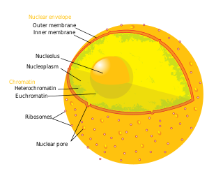
A nuclear pore is a channel as part of the nuclear pore complex (NPC), a large protein complex found in the nuclear envelope in eukaryotic cells, enveloping the cell nucleus containing DNA, which facilitates the selective membrane transport of various molecules across the membrane.

<span class="mw-page-title-main">Clathrin</span> Protein playing a major role in the formation of coated vesicles

Clathrin is a protein that plays a major role in the formation of coated vesicles. Clathrin was first isolated and named by Barbara Pearse in 1976. It forms a triskelion shape composed of three clathrin heavy chains and three light chains. When the triskelia interact they form a polyhedral lattice that surrounds the vesicle, hence the protein's name, which is derived from the Latin clathrum meaning lattice. Coat-proteins, like clathrin, are used to build small vesicles in order to transport molecules within cells. The endocytosis and exocytosis of vesicles allows cells to communicate, to transfer nutrients, to import signaling receptors, to mediate an immune response after sampling the extracellular world, and to clean up the cell debris left by tissue inflammation. The endocytic pathway can be hijacked by viruses and other pathogens in order to gain entry to the cell during infection.

The Coat Protein Complex II, or COPII, is a group of proteins that facilitate the formation of vesicles to transport proteins from the endoplasmic reticulum to the Golgi apparatus or endoplasmic-reticulum–Golgi intermediate compartment. This process is termed anterograde transport, in contrast to the retrograde transport associated with the COPI complex. COPII is assembled in two parts: first an inner layer of Sar1, Sec23, and Sec24 forms; then the inner coat is surrounded by an outer lattice of Sec13 and Sec31.

<span class="mw-page-title-main">COPI</span> Protein complex

COPI is a coatomer, a protein complex that coats vesicles transporting proteins from the cis end of the Golgi complex back to the rough endoplasmic reticulum (ER), where they were originally synthesized, and between Golgi compartments. This type of transport is retrograde transport, in contrast to the anterograde transport associated with the COPII protein. The name "COPI" refers to the specific coat protein complex that initiates the budding process on the cis-Golgi membrane. The coat consists of large protein subcomplexes that are made of seven different protein subunits, namely α, β, β', γ, δ, ε and ζ.

<span class="mw-page-title-main">Planctomycetota</span> Phylum of aquatic bacteria

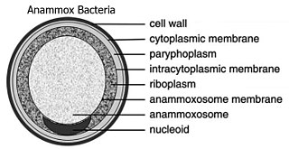
The Planctomycetota are a phylum of widely distributed bacteria, occurring in both aquatic and terrestrial habitats. They play a considerable role in global carbon and nitrogen cycles, with many species of this phylum capable of anaerobic ammonium oxidation, also known as anammox. Many Planctomycetota occur in relatively high abundance as biofilms, often associating with other organisms such as macroalgae and marine sponges.

<span class="mw-page-title-main">SNARE protein</span> Protein family

SNARE proteins – "SNAPREceptors" – are a large protein family consisting of at least 24 members in yeasts, more than 60 members in mammalian cells, and some numbers in plants. The primary role of SNARE proteins is to mediate the fusion of vesicles with the target membrane; this notably mediates exocytosis, but can also mediate the fusion of vesicles with membrane-bound compartments. The best studied SNAREs are those that mediate the release of synaptic vesicles containing neurotransmitters in neurons. These neuronal SNAREs are the targets of the neurotoxins responsible for botulism and tetanus produced by certain bacteria.

<span class="mw-page-title-main">Vesicular transport adaptor protein</span>

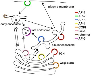
Vesicular transport adaptor proteins are proteins involved in forming complexes that function in the trafficking of molecules from one subcellular location to another. These complexes concentrate the correct cargo molecules in vesicles that bud or extrude off of one organelle and travel to another location, where the cargo is delivered. While some of the details of how these adaptor proteins achieve their trafficking specificity has been worked out, there is still much to be learned.

<span class="mw-page-title-main">ENTH domain</span> InterPro Domain

The epsin N-terminal homology (ENTH) domain is a structural domain that is found in proteins involved in endocytosis and cytoskeletal machinery.

<span class="mw-page-title-main">AP2 adaptor complex</span>

The AP2 adaptor complex is a multimeric protein that works on the cell membrane to internalize cargo in clathrin-mediated endocytosis. It is a stable complex of four adaptins which give rise to a structure that has a core domain and two appendage domains attached to the core domain by polypeptide linkers. These appendage domains are sometimes called 'ears'. The core domain binds to the membrane and to cargo destined for internalisation. The alpha and beta appendage domains bind to accessory proteins and to clathrin. Their interactions allow the temporal and spatial regulation of the assembly of clathrin-coated vesicles and their endocytosis.

<span class="mw-page-title-main">Nucleoporin</span> Family of proteins that form the nuclear pore complex

Nucleoporins are a family of proteins which are the constituent building blocks of the nuclear pore complex (NPC). The nuclear pore complex is a massive structure embedded in the nuclear envelope at sites where the inner and outer nuclear membranes fuse, forming a gateway that regulates the flow of macromolecules between the cell nucleus and the cytoplasm. Nuclear pores enable the passive and facilitated transport of molecules across the nuclear envelope. Nucleoporins, a family of around 30 proteins, are the main components of the nuclear pore complex in eukaryotic cells. Nucleoporin 62 is the most abundant member of this family. Nucleoporins are able to transport molecules across the nuclear envelope at a very high rate. A single NPC is able to transport 60,000 protein molecules across the nuclear envelope every minute.

<span class="mw-page-title-main">COPB1</span> Protein-coding gene in humans

Coatomer subunit beta is a protein that in humans is encoded by the COPB1 gene.

<span class="mw-page-title-main">Adaptor-related protein complex 2, alpha 1</span> Protein-coding gene in the species Homo sapiens

AP-2 complex subunit alpha-1 is a protein that in humans is encoded by the AP2A1 gene.

<span class="mw-page-title-main">AP1G1</span> Protein-coding gene in the species Homo sapiens

AP-1 complex subunit gamma-1 is a protein that in humans is encoded by the AP1G1 gene.

<span class="mw-page-title-main">CLTC</span> Protein-coding gene in the species Homo sapiens

Clathrin heavy chain 1 is a protein that in humans is encoded by the CLTC gene.

<span class="mw-page-title-main">AP1B1</span> Protein-coding gene in the species Homo sapiens

AP-1 complex subunit beta-1 is a protein that in humans is encoded by the AP1B1 gene.

Clathrin adaptor proteins, also known as adaptins, are vesicular transport adaptor proteins associated with clathrin. These proteins are synthesized in the ribosomes, processed in the endoplasmic reticulum and transported from the Golgi apparatus to the trans-Golgi network, and from there via small carrier vesicles to their final destination compartment. The association between adaptins and clathrin are important for vesicular cargo selection and transporting. Clathrin coats contain both clathrin and adaptor complexes that link clathrin to receptors in coated vesicles. Clathrin-associated protein complexes are believed to interact with the cytoplasmic tails of membrane proteins, leading to their selection and concentration. Therefore, adaptor proteins are responsible for the recruitment of cargo molecules into a growing clathrin-coated pits. The two major types of clathrin adaptor complexes are the heterotetrameric vesicular transport adaptor proteins (AP1-5), and the monomeric GGA adaptors. Adaptins are distantly related to the other main type of vesicular transport proteins, the coatomer subunits, sharing between 16% and 26% of their amino acid sequence.

<span class="mw-page-title-main">Beta2-adaptin C-terminal domain</span>

The C-terminal domain ofBeta2-adaptin is a protein domain is involved in cell trafficking by aiding import and export of substances in and out of the cell.

<i>Gemmata obscuriglobus</i> Species of bacteria

Gemmata obscuriglobus is a species of Gram-negative, aerobic, heterotrophic bacteria of the phylum Planctomycetota. G. obscuriglobus occur in freshwater habitats and was first described in 1984, and is the only described species in its genus.

References

  1. 1 2 3 Cho, Uhn Soo; Xu, Wenqing (1 November 2006). "Crystal structure of a protein phosphatase 2A heterotrimeric holoenzyme". Nature. 445 (7123): 53–57. doi:10.1038/nature05351. PMID   17086192. S2CID   4408160.
  2. 1 2 3 4 5 6 7 8 9 10 11 12 Fournier, David; Palidwor, Gareth A.; Shcherbinin, Sergey; Szengel, Angelika; Schaefer, Martin H.; Perez-Iratxeta, Carol; Andrade-Navarro, Miguel A.; E. Tosatto, Silvio C. (21 November 2013). "Functional and Genomic Analyses of Alpha-Solenoid Proteins". PLOS ONE. 8 (11): e79894. Bibcode:2013PLoSO...879894F. doi: 10.1371/journal.pone.0079894 . PMC   3837014 . PMID   24278209.
  3. 1 2 Kappel, Christian; Zachariae, Ulrich; Dölker, Nicole; Grubmüller, Helmut (September 2010). "An Unusual Hydrophobic Core Confers Extreme Flexibility to HEAT Repeat Proteins". Biophysical Journal. 99 (5): 1596–1603. Bibcode:2010BpJ....99.1596K. doi:10.1016/j.bpj.2010.06.032. PMC   2931736 . PMID   20816072.
  4. 1 2 3 4 5 6 7 8 Field, Mark C.; Sali, Andrej; Rout, Michael P. (13 June 2011). "On a bender—BARs, ESCRTs, COPs, and finally getting your coat". The Journal of Cell Biology. 193 (6): 963–972. doi:10.1083/jcb.201102042. PMC   3115789 . PMID   21670211.
  5. 1 2 3 Kobe, Bostjan; Kajava, Andrey V (October 2000). "When protein folding is simplified to protein coiling: the continuum of solenoid protein structures". Trends in Biochemical Sciences. 25 (10): 509–515. doi:10.1016/S0968-0004(00)01667-4. PMID   11050437.
  6. "CATH Topology "Alpha Horseshoe"".
  7. 1 2 3 4 Alber, Frank; Dokudovskaya, Svetlana; Veenhoff, Liesbeth M.; Zhang, Wenzhu; Kipper, Julia; Devos, Damien; Suprapto, Adisetyantari; Karni-Schmidt, Orit; Williams, Rosemary; Chait, Brian T.; Sali, Andrej; Rout, Michael P. (29 November 2007). "The molecular architecture of the nuclear pore complex". Nature. 450 (7170): 695–701. Bibcode:2007Natur.450..695A. doi:10.1038/nature06405. PMID   18046406. S2CID   4431057.
  8. 1 2 Devos, Damien; Dokudovskaya, Svetlana; Alber, Frank; Williams, Rosemary; Chait, Brian T; Sali, Andrej; Rout, Michael P; Greg Petsko (2 November 2004). "Components of Coated Vesicles and Nuclear Pore Complexes Share a Common Molecular Architecture". PLOS Biology. 2 (12): e380. doi: 10.1371/journal.pbio.0020380 . PMC   524472 . PMID   15523559.
  9. Antonin, Wolfram; Mattaj, Iain W. (January 2005). "Nuclear pore complexes: Round the bend?". Nature Cell Biology. 7 (1): 10–12. doi:10.1038/ncb0105-10. PMID   15632943. S2CID   9909704.
  10. 1 2 Santarella-Mellwig, Rachel; Franke, Josef; Jaedicke, Andreas; Gorjanacz, Matyas; Bauer, Ulrike; Budd, Aidan; Mattaj, Iain W.; Devos, Damien P.; Schmid, Sandra L. (19 January 2010). "The Compartmentalized Bacteria of the Planctomycetes-Verrucomicrobia-Chlamydiae Superphylum Have Membrane Coat-Like Proteins". PLOS Biology. 8 (1): e1000281. doi: 10.1371/journal.pbio.1000281 . PMC   2799638 . PMID   20087413.
  11. Ybe, Joel A.; Brodsky, Frances M.; Hofmann, Kay; Lin, Kai; Liu, Shu-Hui; Chen, Lin; Earnest, Thomas N.; Fletterick, Robert J.; Hwang, Peter K. (27 May 1999). "Clathrin self-assembly is mediated by a tandemly repeated superhelix". Nature. 399 (6734): 371–375. Bibcode:1999Natur.399..371Y. doi:10.1038/20708. PMID   10360576. S2CID   4406014.
  12. Lee, Changwook; Goldberg, Jonathan (July 2010). "Structure of Coatomer Cage Proteins and the Relationship among COPI, COPII, and Clathrin Vesicle Coats". Cell. 142 (1): 123–132. doi:10.1016/j.cell.2010.05.030. PMC   2943847 . PMID   20579721.
  13. Forwood, Jade K.; Lange, Allison; Zachariae, Ulrich; Marfori, Mary; Preast, Callie; Grubmüller, Helmut; Stewart, Murray; Corbett, Anita H.; Kobe, Bostjan (September 2010). "Quantitative Structural Analysis of Importin-β Flexibility: Paradigm for Solenoid Protein Structures". Structure. 18 (9): 1171–1183. doi:10.1016/j.str.2010.06.015. hdl: 11858/00-001M-0000-0027-C07B-1 . PMID   20826343.
  14. Katahira, Jun; Yoneda, Yoshihiro (November 2011). "Nucleocytoplasmic Transport of MicroRNAs and Related Small RNAs". Traffic. 12 (11): 1468–1474. doi:10.1111/j.1600-0854.2011.01211.x. PMID   21518166. S2CID   38777924.
  15. Barkan, Alice; Rojas, Margarita; Fujii, Sota; Yap, Aaron; Chong, Yee Seng; Bond, Charles S.; Small, Ian; Voytas, Dan (16 August 2012). "A Combinatorial Amino Acid Code for RNA Recognition by Pentatricopeptide Repeat Proteins". PLOS Genetics. 8 (8): e1002910. doi: 10.1371/journal.pgen.1002910 . PMC   3420917 . PMID   22916040.
  16. Barkan, Alice; Small, Ian (29 April 2014). "Pentatricopeptide Repeat Proteins in Plants". Annual Review of Plant Biology. 65 (1): 415–442. doi: 10.1146/annurev-arplant-050213-040159 . PMID   24471833.
  17. Groves, Matthew R.; Hanlon, Neil; Turowski, Patric; Hemmings, Brian A.; Barford, David (January 1999). "The Structure of the Protein Phosphatase 2A PR65/A Subunit Reveals the Conformation of Its 15 Tandemly Repeated HEAT Motifs". Cell. 96 (1): 99–110. doi: 10.1016/S0092-8674(00)80963-0 . PMID   9989501. S2CID   14465060.
  18. Grinthal, A.; Adamovic, I.; Weiner, B.; Karplus, M.; Kleckner, N. (25 January 2010). "PR65, the HEAT-repeat scaffold of phosphatase PP2A, is an elastic connector that links force and catalysis". Proceedings of the National Academy of Sciences. 107 (6): 2467–2472. Bibcode:2010PNAS..107.2467G. doi: 10.1073/pnas.0914073107 . PMC   2823866 . PMID   20133745.
  19. Field, Mark C; Dacks, Joel B (February 2009). "First and last ancestors: reconstructing evolution of the endomembrane system with ESCRTs, vesicle coat proteins, and nuclear pore complexes". Current Opinion in Cell Biology. 21 (1): 4–13. doi:10.1016/j.ceb.2008.12.004. PMID   19201590.
  20. Dacks, Joel B.; Field, Mark C.; Buick, Roger; Eme, Laura; Gribaldo, Simonetta; Roger, Andrew J.; Brochier-Armanet, Céline; Devos, Damien P. (26 September 2016). "The changing view of eukaryogenesis – fossils, cells, lineages and how they all come together". Journal of Cell Science. 129 (20): 3695–3703. doi: 10.1242/jcs.178566 . PMID   27672020.
  21. Promponas, Vasilis J.; Katsani, Katerina R.; Blencowe, Benjamin J.; Ouzounis, Christos A. (2 March 2016). "Sequence evidence for common ancestry of eukaryotic endomembrane coatomers". Scientific Reports. 6: 22311. Bibcode:2016NatSR...622311P. doi:10.1038/srep22311. PMC   4773986 . PMID   26931514.
  22. Klinger, Christen M.; Spang, Anja; Dacks, Joel B.; Ettema, Thijs J.G. (June 2016). "Tracing the Archaeal Origins of Eukaryotic Membrane-Trafficking System Building Blocks". Molecular Biology and Evolution. 33 (6): 1528–1541. doi: 10.1093/molbev/msw034 . PMID   26893300.
  23. Fuerst, John A. (2010). "Beyond Prokaryotes and Eukaryotes : Planctomycetes and Cell Organization". Nature Education. 3 (9): 44.
  24. 1 2 McInerney, JO; Martin, WF; Koonin, EV; Allen, JF; Galperin, MY; Lane, N; Archibald, JM; Embley, TM (November 2011). "Planctomycetes and eukaryotes: a case of analogy not homology". BioEssays. 33 (11): 810–7. doi:10.1002/bies.201100045. PMC   3795523 . PMID   21858844.
  25. Fuerst, JA (October 2013). "The PVC superphylum: exceptions to the bacterial definition?". Antonie van Leeuwenhoek. 104 (4): 451–66. doi:10.1007/s10482-013-9986-1. PMID   23912444. S2CID   14283647.
  26. Devos, Damien P. (September 2013). "Gemmata obscuriglobus". Current Biology. 23 (17): R705–R707. doi: 10.1016/j.cub.2013.07.013 . PMID   24028944.
  27. Devos, DP (February 2014). "Re-interpretation of the evidence for the PVC cell plan supports a Gram-negative origin". Antonie van Leeuwenhoek. 105 (2): 271–4. doi:10.1007/s10482-013-0087-y. hdl: 10261/129395 . PMID   24292377. S2CID   16557669.
  28. Devos, Damien P. (January 2014). "PVC bacteria: variation of, but not exception to, the Gram-negative cell plan". Trends in Microbiology. 22 (1): 14–20. doi:10.1016/j.tim.2013.10.008. hdl: 10261/129431 . PMID   24286661.
  29. Fuerst, John A.; Sagulenko, Evgeny (August 2014). "Towards understanding the molecular mechanism of the endocytosis-like process in the bacterium Gemmata obscuriglobus". Biochimica et Biophysica Acta (BBA) - Molecular Cell Research. 1843 (8): 1732–1738. doi: 10.1016/j.bbamcr.2013.10.002 . PMID   24144586.
  30. Di Domenico, Tomás; Potenza, Emilio; Walsh, Ian; Gonzalo Parra, R.; Giollo, Manuel; Minervini, Giovanni; Piovesan, Damiano; Ihsan, Awais; Ferrari, Carlo; Kajava, Andrey V.; Tosatto, Silvio C.E. (January 2014). "RepeatsDB: a database of tandem repeat protein structures". Nucleic Acids Research. 42 (D1): D352–D357. doi:10.1093/nar/gkt1175. PMC   3964956 . PMID   24311564.
  31. Pellegrini, Marco (24 September 2015). "Tandem Repeats in Proteins: Prediction Algorithms and Biological Role". Frontiers in Bioengineering and Biotechnology. 3: 143. doi: 10.3389/fbioe.2015.00143 . PMC   4585158 . PMID   26442257.