An electron microscope is a microscope that uses a beam of electrons as a source of illumination. They use electron optics that are analogous to the glass lenses of an optical light microscope. As the wavelength of an electron can be up to 100,000 times shorter than that of visible light, electron microscopes have a higher resolution of about 0.1 nm, which compares to about 200 nm for light microscopes. Electron microscope may refer to:

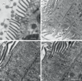
Transmission electron microscopy (TEM) is a microscopy technique in which a beam of electrons is transmitted through a specimen to form an image. The specimen is most often an ultrathin section less than 100 nm thick or a suspension on a grid. An image is formed from the interaction of the electrons with the sample as the beam is transmitted through the specimen. The image is then magnified and focused onto an imaging device, such as a fluorescent screen, a layer of photographic film, or a sensor such as a scintillator attached to a charge-coupled device.

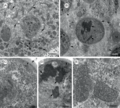
The opalines are a small group of peculiar heterokonts, currently assigned to the family Opalinidae, in the order Slopalinida. Their name is derived from the opalescent appearance of these microscopic organisms when illuminated with full sunlight. Most opalines live in the large intestine and cloaca of anurans, though they are sometimes found in fish, reptiles, molluscs and insects; whether they are parasitic is not certain. The unusual features of the opalines, first observed by Antonie van Leeuwenhoek in 1683, has led to much debate regarding their phylogenetic position among the protists.
Scanning probe microscopy (SPM) is a branch of microscopy that forms images of surfaces using a physical probe that scans the specimen. SPM was founded in 1981, with the invention of the scanning tunneling microscope, an instrument for imaging surfaces at the atomic level. The first successful scanning tunneling microscope experiment was done by Gerd Binnig and Heinrich Rohrer. The key to their success was using a feedback loop to regulate gap distance between the sample and the probe.
Electron crystallography is a method to determine the arrangement of atoms in solids using a transmission electron microscope (TEM). It can involve the use of high-resolution transmission electron microscopy images, electron diffraction patterns including convergent-beam electron diffraction or combinations of these. It has been successful in determining some bulk structures, and also surface structures. Two related methods are low-energy electron diffraction which has solved the structure of many surfaces, and reflection high-energy electron diffraction which is used to monitor surfaces often during growth.

A scanning transmission electron microscope (STEM) is a type of transmission electron microscope (TEM). Pronunciation is [stɛm] or [ɛsti:i:ɛm]. As with a conventional transmission electron microscope (CTEM), images are formed by electrons passing through a sufficiently thin specimen. However, unlike CTEM, in STEM the electron beam is focused to a fine spot which is then scanned over the sample in a raster illumination system constructed so that the sample is illuminated at each point with the beam parallel to the optical axis. The rastering of the beam across the sample makes STEM suitable for analytical techniques such as Z-contrast annular dark-field imaging, and spectroscopic mapping by energy dispersive X-ray (EDX) spectroscopy, or electron energy loss spectroscopy (EELS). These signals can be obtained simultaneously, allowing direct correlation of images and spectroscopic data.
This page deals with the electron affinity as a property of isolated atoms or molecules. Solid state electron affinities are not listed here.

Electron tomography (ET) is a tomography technique for obtaining detailed 3D structures of sub-cellular, macro-molecular, or materials specimens. Electron tomography is an extension of traditional transmission electron microscopy and uses a transmission electron microscope to collect the data. In the process, a beam of electrons is passed through the sample at incremental degrees of rotation around the center of the target sample. This information is collected and used to assemble a three-dimensional image of the target. For biological applications, the typical resolution of ET systems are in the 5–20 nm range, suitable for examining supra-molecular multi-protein structures, although not the secondary and tertiary structure of an individual protein or polypeptide. Recently, atomic resolution in 3D electron tomography reconstructions has been demonstrated.
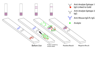
Malaria antigen detection tests are a group of commercially available rapid diagnostic tests of the rapid antigen test type that allow quick diagnosis of malaria by people who are not otherwise skilled in traditional laboratory techniques for diagnosing malaria or in situations where such equipment is not available. There are currently over 20 such tests commercially available. The first malaria antigen suitable as target for such a test was a soluble glycolytic enzyme Glutamate dehydrogenase. None of the rapid tests are currently as sensitive as a thick blood film, nor as cheap. A major drawback in the use of all current dipstick methods is that the result is essentially qualitative. In many endemic areas of tropical Africa, however, the quantitative assessment of parasitaemia is important, as a large percentage of the population will test positive in any qualitative assay.

Rafal Edward Dunin-Borkowski HonFRMS is a British experimental physicist. He is currently Director of the Institute for Microstructure Research (PGI-5) and the Ernst Ruska-Centre for Microscopy and Spectroscopy with Electrons (ER-C) in Forschungszentrum Jülich and Professor of Experimental Physics in RWTH Aachen University.

Piroplasmida is an order of parasites in the phylum Apicomplexa. They divide by binary fission and as sporozoan parasites they possess sexual and asexual phases. They include the tick parasites Babesia and Theileria.

Scanning thermal microscopy (SThM) is a type of scanning probe microscopy that maps the local temperature and thermal conductivity of an interface. The probe in a scanning thermal microscope is sensitive to local temperatures – providing a nano-scale thermometer. Thermal measurements at the nanometer scale are of both scientific and industrial interest. The technique was invented by Clayton C. Williams and H. Kumar Wickramasinghe in 1986.

Crystallographic image processing (CIP) is traditionally understood as being a set of key steps in the determination of the atomic structure of crystalline matter from high-resolution electron microscopy (HREM) images obtained in a transmission electron microscope (TEM) that is run in the parallel illumination mode. The term was created in the research group of Sven Hovmöller at Stockholm University during the early 1980s and became rapidly a label for the "3D crystal structure from 2D transmission/projection images" approach. Since the late 1990s, analogous and complementary image processing techniques that are directed towards the achieving of goals with are either complementary or entirely beyond the scope of the original inception of CIP have been developed independently by members of the computational symmetry/geometry, scanning transmission electron microscopy, scanning probe microscopy communities, and applied crystallography communities.

Ondrej L. Krivanek is a Czech/British physicist resident in the United States, and a leading developer of electron-optical instrumentation. He won the Kavli Prize for Nanoscience in 2020 for his substantial innovations in atomic resolution electron microscopy.

Setaria is a genus of parasitic roundworms that infect domesticated mammals such as pigs, camels, cattle and horses. Some species also infect wild mammals such as deer and antelope. The genus consists of about 43 species. Members of the genus are uniquely parasites in the abdominal cavity of the body. They are mostly large-sized roundworms, possessing an elaborate head (cephalic) region that is characterised by spines, presence of four lips, and well-guarded mouth. Little is known about their pathogenic effects, but some are known to affect nervous system and eye. The larval infective forms are transmitted from one animal to another by the bite of mosquitoes and flies. In addition Setaria marshalli can be transmitted from the womb to new-born calf.

Liquid-phase electron microscopy refers to a class of methods for imaging specimens in liquid with nanometer spatial resolution using electron microscopy. LP-EM overcomes the key limitation of electron microscopy: since the electron optics requires a high vacuum, the sample must be stable in a vacuum environment. Many types of specimens relevant to biology, materials science, chemistry, geology, and physics, however, change their properties when placed in a vacuum.

Jacques Dubochet is a retired Swiss biophysicist. He is a former researcher at the European Molecular Biology Laboratory in Heidelberg, Germany, and an honorary professor of biophysics at the University of Lausanne in Switzerland.

Cryogenic electron microscopy (cryo-EM) is a cryomicroscopy technique applied on samples cooled to cryogenic temperatures. For biological specimens, the structure is preserved by embedding in an environment of vitreous ice. An aqueous sample solution is applied to a grid-mesh and plunge-frozen in liquid ethane or a mixture of liquid ethane and propane. While development of the technique began in the 1970s, recent advances in detector technology and software algorithms have allowed for the determination of biomolecular structures at near-atomic resolution. This has attracted wide attention to the approach as an alternative to X-ray crystallography or NMR spectroscopy for macromolecular structure determination without the need for crystallization.

Cichlidogyrus philander is a species of Monopisthocotylean Monogenean, in the family Ancyrocephalidae. It is ectoparasitic on the gills of the fish Pseudocrenilabrus philander.
Joanne Etheridge is an Australian physicist. She is Director of the Monash Centre for Electron Microscopy and Professor in the Department of Materials Science and Engineering at Monash University.