Equine anatomy

Last updated

Points of a horse Points of a horse.jpg
Points of a horse

Equine anatomy encompasses the gross and microscopic anatomy of horses, ponies and other equids, including donkeys, mules and zebras. While all anatomical features of equids are described in the same terms as for other animals by the International Committee on Veterinary Gross Anatomical Nomenclature in the book Nomina Anatomica Veterinaria , there are many horse-specific colloquial terms used by equestrians.

Contents

External anatomy

Digestive system

Topography of Viscera of Horse Left Deep View.jpg
A dehydrated anatomical specimen Technique of dehydration applied in a foal.jpg
A dehydrated anatomical specimen

Horses and other equids evolved as grazing animals, adapted to eating small amounts of the same kind of food all day long. In the wild, the horse adapted to eating prairie grasses in semi-arid regions and traveling significant distances each day in order to obtain adequate nutrition. [13] Therefore, the digestive system of a horse is about 30 m (100 ft) long, and most of this is intestines.

Mouth

Digestion begins in the mouth, which is also called the "oral cavity." It is made up of the teeth, the hard palate, the soft palate, the tongue and related muscles, the cheeks and the lips. Horses also have three pairs of salivary glands, the parotoid (largest salivary gland and located near the poll), mandibular (located in the jaw), and sublingual (located under the tongue). Horses select pieces of forage and pick up finer foods, such as grain, with their sensitive, prehensile lips. The front teeth of the horse, called incisors, clip forage, and food is then pushed back in the mouth by the tongue, and ground up for swallowing by the premolars and molars. [14]

Esophagus

The esophagus is about 1.2 to 1.5 m (4 to 5 ft) in length, and carries food to the stomach. A muscular ring, called the cardiac sphincter, connects the stomach to the esophagus. This sphincter is very well developed in horses. This and the oblique angle at which the esophagus connects to the stomach explains why horses cannot vomit. [14] The esophagus is also the area of the digestive tract where horses may suffer from choke.

Stomach

Equine stomach. Equine stomach-FMVZ USP-25.jpeg
Equine stomach.

Horses have a relatively small stomach for their size, and this limits the amount of feed a horse can take in at one time. The average sized horse (360 to 540 kg [800 to 1,200 lb]) has a stomach with a capacity of around 19 L (5 US gal), and works best when it contains about 7.6 L (2 US gal). Because the stomach empties when 23 full, whether stomach enzymes have completed their processing of the food or not, and doing so prevents full digestion and proper utilization of feed, continuous foraging or several small feedings per day are preferable to one or two large ones. [14] The horse stomach consists of a non-glandular proximal region (saccus cecus), divided by a distinct border, the margo plicatus, from the glandular distal stomach. [15]

In the stomach, assorted acids and the enzyme pepsin break down food. Pepsin allows for the further breakdown of proteins into amino acid chains. [14] Other enzymes include resin and lipase. Additionally, the stomach absorbs some water, as well as ions and lipid-soluble compounds.

Small intestine

The horse's small intestine is 15 to 21 m (50 to 70 ft) long and holds 38 to 45 L (10 to 12 US gal). This is the major digestive organ, and where most nutrients are absorbed. [16] It has three parts, the duodenum, jejunum and ileum. The majority of digestion occurs in the duodenum while the majority of absorption occurs in the jejunum. Bile from the liver aids in digesting fats in the duodenum combined with enzymes from the pancreas and small intestine. Horses, in common with mammals such as camels, do not have a gall bladder, meaning bile flows constantly. [14] Most food is digested and absorbed into the bloodstream from the small intestine, including proteins, simple carbohydrate, fats, and vitamins A, D, and E. Any remaining liquids and roughage move into the large intestine.

Large intestine

Equine colon Equine colon.jpg
Equine colon

The large intestine, also known as the hindgut consists of the cecum, large colon, small colon and rectum, terminating in the anus.

Cecum

The cecum (called the "water gut" in old textbooks) is the first section of the large intestine, analagous to the appendix in humans. It is a cul-de-sac pouch, [16] about 1.2 m (4 ft) long that holds 26 to 30 L (7 to 8 US gal). It contains bacteria and other microbes that break down cellulose and other indigestible plant fiber through fermentation into volatile fatty acids. [17] These microbes feed upon the portion of chyme not absorbed by the small intestine, and produce vitamin K and B complex vitamins. [17]

Large colon

The large colon is 3.0 to 3.7 m (10 to 12 ft) long and holds up to 76 L (20 US gal) of semi-liquid matter. It is made up of the right ventral (lower) colon, the left ventral colon, the left dorsal (upper) colon, the right dorsal colon, and the transverse colon, in that order. [14] Three flexures are also named: the sternal flexure, between right and left ventral colon; the pelvic flexure, between left ventral and left dorsal colon; the diaphragmatic flexure, between left dorsal and right dorsal colon. Besides the transverse colon, these sections are all analogous to the ascending colon in humans. The large colon continues the fermentation process, and absorbs volatile fatty acids as an energy source. Due to its many twists and turns, the large colon is a common place for certain forms of colic including impaction, displacement and volvulus. [16] [17]

Small colon

The small colon is 3.0 to 3.7 m (10 to 12 ft) in length and holds only 19 L (5 US gal) of material. It is the area where the majority of water in the horse's diet is absorbed, and is the place where fecal balls are formed. [14] This section is analagous to the descending colon in humans.

Rectum and anus

The rectum is about 30 cm (1 ft) long, and acts as a holding chamber for waste matter, which is then expelled from the body via the anus. [14]

Reproductive system

Mare

The mare's reproductive system is responsible for controlling gestation, birth, and lactation, as well as her estrous cycle and mating behavior. It lies ventral to the 4th or 5th lumbar vertebrae, although its position within the mare can vary depending on the movement of the intestines and distention of the bladder.

The mare has two ovaries, usually 7 to 8 cm (2.8 to 3.1 in) in length and 3 to 4 cm (1.2 to 1.6 in) thick, that generally tend to decrease in size as the mare ages. In equine ovaries, unlike in humans, the vascular tissue is cortical to follicular tissue, so ovulation can only occur at an ovulation fossa near the infundibulum. The ovaries connect to the fallopian tubes (oviducts), which serve to move the ovum from the ovary to the uterus. To do so, the oviducts are lined with a layer of cilia, which produce a current that flows toward the uterus. Each oviduct attaches to one of the two horns of the uterus, which are approximately 20 to 25 cm (7.9 to 9.8 in) in length. These horns attach to the body of the uterus (18 to 20 cm [7.1 to 7.9 in] long). The equine uterus is bipartite, meaning the two uterine horns fuse into a relatively large uterine body (resembling a shortened bicornuate uterus or a stretched simplex uterus). Caudal to the uterus is the cervix, about 5 to 7 cm (2.0 to 2.8 in) long, which separates the uterus from the vagina. Usually 3.5 to 4 cm (1.4 to 1.6 in) in diameter with longitudinal folds on the interior surface, it can expand to allow the passage of the foal. The vagina of the mare is 15 to 20 cm (5.9 to 7.9 in) long, and is quite elastic, allowing it to expand.[ citation needed ] The vulva is the external opening of the vagina, and consists of the clitoris and two labia. [18] It lies ventral to the rectum. [19] [20] The mare has two mammary glands, which are smaller in maiden mares. They have two ducts each, which open externally.[ citation needed ]

Stallion

Secondary characteristics of a stallion include heavier muscling for a given breed than is seen in mares or geldings, often with considerable development along the crest of the neck, as shown in this image. RCMP Farm Hannoverian2.jpg
Secondary characteristics of a stallion include heavier muscling for a given breed than is seen in mares or geldings, often with considerable development along the crest of the neck, as shown in this image.

The stallion's reproductive system is responsible for his sexual behavior and secondary sex characteristics (such as a large crest). The external genitalia include the urethra; the testes, which average 8 to 12 cm (3.1 to 4.7 in) long; the penis, which, when housed within the prepuce, is 50 cm (20 in) long and 2.5 to 6 cm (0.98 to 2.36 in) in diameter with the distal end 15 to 20 cm (5.9 to 7.9 in) and when erect, increases by 3 to 4 times. The internal genitalia accessory sex glands are the vesicular glands, prostate gland, and bulbourethral glands, which contribute fluid to the semen at ejaculation, but are not strictly necessary for fertility. [21]

Teeth

A horse's teeth include incisors, premolars, molars, and sometimes canine teeth. A horse's incisors, premolars, and molars, once fully developed, continue to erupt throughout its lifetime as the grinding surface is worn down through chewing. Because of this pattern of wear, a rough estimate of a horse's age can be made from an examination of the teeth. Abnormal wear of the teeth, caused by conformational defects, abnormal behaviors, or improper diets, can cause serious health issues and can even result in the death of the horse.

Feet/hooves

1- Heel perioplium, 2-Bulb, 3-Frog, 4-Frog cleft, 5-Lateral groove, 6-Heel, 7-Bar, 8-Seat-of-corn, 9-Pigmented walls 10-Water line, 11-White line, 12-Apex of the frog, 13-Sole, 14-Toe, 15-How to measure hoof width (blue dotted line), 16-Quarter, 17-How to measure length (blue dotted line) Hoof quoted.jpg
1- Heel perioplium, 2-Bulb, 3-Frog, 4-Frog cleft, 5-Lateral groove, 6-Heel, 7-Bar, 8-Seat-of-corn, 9-Pigmented walls 10-Water line, 11-White line, 12-Apex of the frog, 13-Sole, 14-Toe, 15-How to measure hoof width (blue dotted line), 16-Quarter, 17-How to measure length (blue dotted line)

The hoof of the horse encases part of the second and all of the third phalanx of the lower limbs, analogous to the fingertip or toe tip of a human. In essence, a horse travels on its "tiptoes". The hoof wall is a much larger, thicker and stronger version of the human fingernail or toenail, made up of similar materials, primarily keratin, a very strong protein molecule. The horse's hoof contains a high proportion of sulfur-containing amino acids which contribute to its resilience and toughness. Vascular fold-like structures called laminae suspend the distal phalanx from the hoof wall.

Skeletal system

A horse's skeleton Skeleton with outline of a horse.png
A horse's skeleton

The skeleton of the horse has three major functions in the body. It protects vital organs, provides framework, and supports soft parts of the body. Horses have 205 bones, which are divided into the appendicular skeleton (the legs) and the axial skeleton (the skull, vertebral column, sternum, and ribs). Both pelvic and thoracic limbs contain the same number of bones, 20 bones per limb. Bones are connected to muscles via tendons and other bones via ligaments. Bones are also used to store minerals, and are the site of red blood cell formation.

The bones of the horse are the same as those of other domestic species, but the third metacarpal and metatarsal are much more developed and the second and fourth are undeveloped, having the first and fifth metacarpal and metatarsal. [22]

Horse skeleton bones
Spine54
Ribs36
Sternum01
Head (including ear )34
Thoracic region40
Pelvic region40

Ligaments and tendons

Ligaments

Ligaments attach bone to bone or bone to tendon, and are vital in stabilizing joints as well as supporting structures. They are made up of fibrous material that is generally quite strong. Due to their relatively poor blood supply, ligament injuries generally take a long time to heal.

Tendons

Tendons are cords of connective tissue attaching muscle to bone, cartilage or other tendons. They are a major contributor to shock absorption, are necessary for support of the horse's body, and translate the force generated by muscles into movement. Tendons are classified as flexors (flex a joint) or extensors (extend a joint). However, some tendons will flex multiple joints while extending another (the flexor tendons of the hind limb, for example, will flex the fetlock, pastern, and coffin joint, but extend the hock joint). In this case, the tendons (and associated muscles) are named for their most distal action (digital flexion).

Tendons form in the embryo from fibroblasts which become more tightly packed as the tendon grows. As tendons develop they lay down collagen, which is the main structural protein of connective tissue. As tendons pass near bony prominences, they are protected by a fluid filled synovial structure, either a tendon sheath or a sac called a bursa.

Tendons are easily damaged if placed under too much strain, which can result in a painful, and possibly career-ending, injury. Tendinitis is most commonly seen in high performance horses that gallop or jump. When a tendon is damaged the healing process is slow because tendons have a poor blood supply, reducing the availability of nutrients and oxygen to the tendon. Once a tendon is damaged the tendon will always be weaker, because the collagen fibres tend to line up in random arrangements instead of the stronger linear pattern. Scar tissue within the tendon decreases the overall elasticity in the damaged section of the tendon as well, causing an increase in strain on adjacent uninjured tissue.

Muscular system

Leisering's Atlas of the Anatomy of the Horse Muscles.jpg

When a muscle contracts, it pulls a tendon, which acts on the horse's bones to move them. Muscles are commonly arranged in pairs so that they oppose each other (they are "antagonists"), with one flexing the joint (a flexor muscle) and the other extending it (extensor muscle). Therefore, one muscle of the pair must be relaxed in order for the other muscle in the pair to contract and bend the joint properly. A muscle is made up of several muscle bundles, which in turn are made up of muscle fibers. Muscle fibers have myofibrils, which are able to contract due to actin and myosin. A muscle together with its tendon and bony attachments form an extensor or flexor unit.

Respiratory system and smell

The horse's respiratory system consists of the nostrils, pharynx, larynx, trachea, diaphragm, and lungs. Additionally, the nasolacrimal duct and sinuses are connected to the nasal passage. The horse's respiratory system not only allows the animal to breathe, but also is important in the horse's sense of smell (olfactory ability) as well as in communicating. The soft palate blocks off the pharynx from the mouth (oral cavity) of the horse, except when swallowing. This helps prevent the horse from inhaling food, but also means that a horse cannot use its mouth to breathe when in respiratory distress—a horse can only breathe through its nostrils, also called obligate nasal breathing. [23] For this same reason, horses also cannot pant as a method of thermoregulation. The genus Equus also has a unique part of the respiratory system called the guttural pouch, which is thought to equalize air pressure on the tympanic membrane. Located between the mandibles but below the occiput, it fills with air when the horse swallows or exhales.

Circulatory system

The horse's circulatory system includes the four-chambered heart, averaging 3.9 kg (8.5 lb) in weight, as well as the blood and blood vessels. Its main purpose is to circulate blood throughout the body to deliver oxygen and nutrients to tissues, and to remove waste from these tissues. The hoof (including the frog - the V-shaped part on the bottom of the horses hoof) is a very important part of the circulatory system. As the horse puts weight onto the hoof, the hoof wall is pushed outwards and the frog compressed, driving blood out of the frog, the digital pad, and the laminae of the hoof. When weight is removed from the hoof, the release of pressure pulls blood back down into the foot again. This effectively creates an auxiliary blood-pumping system at the end of each leg. Some of this effect may be lost when a horse is shod (eliminating the expansion and contraction of the hoof wall and raising the frog higher from the ground). [24]

The eye

A horse's eye HorseEye.jpg
A horse's eye

The horse has one of the largest eyes of all land mammals. [25] Eye size in mammals is significantly correlated to maximum running speed as well as to body size, in accordance with Leuckart's law; animals capable of fast locomotion require large eyes. [26] The eye of the horse is set to the side of its skull, consistent with that of a prey animal. [25] The horse has a wide field of monocular vision, as well as good visual acuity. Horses have two-color, or dichromatic vision, which is somewhat like red-green color blindness in humans. [27] Because the horse's vision is closely tied to behavior, the horse's visual abilities are often taken into account when handling and training the animal.

Hearing

The pinna of a horse's ears can rotate in any direction to pick up sounds SuffolkHead.jpg
The pinna of a horse's ears can rotate in any direction to pick up sounds

The hearing of horses is good, [28] superior to that of humans, and the pinna of each ear can rotate up to 180°, giving the potential for 360° hearing without having to move the head. [29] Often, the eye of the horse is looking in the same direction as the ear is directed.

See also

Related Research Articles

<span class="mw-page-title-main">Gastrointestinal tract</span> Organ system within humans and other animals

The gastrointestinal tract is the tract or passageway of the digestive system that leads from the mouth to the anus. The GI tract contains all the major organs of the digestive system, in humans and other animals, including the esophagus, stomach, and intestines. Food taken in through the mouth is digested to extract nutrients and absorb energy, and the waste expelled at the anus as faeces. Gastrointestinal is an adjective meaning of or pertaining to the stomach and intestines.

Articles related to anatomy include:

<span class="mw-page-title-main">Navicular bone</span> Small bone found in the feet of most mammals

The navicular bone is a small bone found in the feet of most mammals.

<span class="mw-page-title-main">Bowed tendon</span>

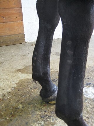
Tendinitis/tendonitis is inflammation of a tendon, often involving torn collagen fibers. A bowed tendon is a horseman's term for a tendon after a horse has sustained an injury that causes swelling in one or more tendons creating a "bowed" appearance.

<span class="mw-page-title-main">Equine conformation</span> Evaluation of a horses bone and muscle structure

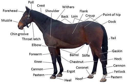
Equine conformation evaluates a horse's bone structure, musculature, and its body proportions in relation to each other. Undesirable conformation can limit the ability to perform a specific task. Although there are several faults with universal disadvantages, a horse's conformation is usually judged by what its intended use may be. Thus "form to function" is one of the first set of traits considered in judging conformation. A horse with poor form for a Grand Prix show jumper could have excellent conformation for a World Champion cutting horse, or to be a champion draft horse. Every horse has good and bad points of its conformation and many horses excel even with conformation faults.

<span class="mw-page-title-main">Abdomen</span> Part of the body between the chest and pelvis

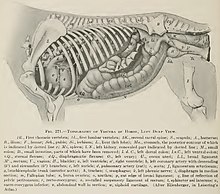
The abdomen is the part of the body between the thorax (chest) and pelvis, in humans and in other vertebrates. The abdomen is the front part of the abdominal segment of the torso. The area occupied by the abdomen is called the abdominal cavity. In arthropods, it is the posterior tagma of the body; it follows the thorax or cephalothorax.

<span class="mw-page-title-main">Fetlock</span>

Fetlock is the common name in horses, large animals, and sometimes dogs for the metacarpophalangeal and metatarsophalangeal joints.

<span class="mw-page-title-main">Metacarpophalangeal joint</span> Bodily joint at the base of each finger

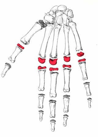
The metacarpophalangeal joints (MCP) are situated between the metacarpal bones and the proximal phalanges of the fingers. These joints are of the condyloid kind, formed by the reception of the rounded heads of the metacarpal bones into shallow cavities on the proximal ends of the proximal phalanges. Being condyloid, they allow the movements of flexion, extension, abduction, adduction and circumduction at the joint.

<span class="mw-page-title-main">Pastern</span> Part of the leg of a horse

The pastern is a part of the leg of a horse between the fetlock and the top of the hoof. It incorporates the long pastern bone and the short pastern bone, which are held together by two sets of paired ligaments to form the pastern joint. Anatomically homologous to the two largest bones found in the human finger, the pastern was famously mis-defined by Samuel Johnson in his dictionary as "the knee of a horse". When a lady asked Johnson how this had happened, he gave the much-quoted reply: "Ignorance, madam, pure ignorance."

Osselet is arthritis in the fetlock joint of a horse, caused by trauma. Osselets usually occur in the front legs of the horse, because there is more strain and concussion on the fetlock there than in the hind legs. The arthritis will occur at the joint between the cannon bone and large pastern bone, at the front of the fetlock.

A flexion test is a preliminary veterinary procedure performed on a horse, generally during a prepurchase or a lameness exam. The purpose is to accentuate any pain that may be associated with a joint or soft-tissue structure, allowing the practitioner to localize a lameness to a specific area, or to alert a practitioner to the presence of sub-clinical disease that may be present during a pre-purchase exam.

<span class="mw-page-title-main">Skeletal system of the horse</span> The skeletal system is made of many interconnected tissues including bone, cartilage, and tendons

The skeletal system of the horse is a skeletal system of a horse that has three major functions in the body. It protects vital organs, provides framework, and supports soft parts of the body. Horses typically have 205 bones. The pelvic limb typically contains 19 bones, while the thoracic limb contains 20 bones.

<span class="mw-page-title-main">Outline of human anatomy</span> Overview of and topical guide to human anatomy

The following outline is provided as an overview of and topical guide to human anatomy:

Lameness is an abnormal gait or stance of an animal that is the result of dysfunction of the locomotor system. In the horse, it is most commonly caused by pain, but can be due to neurologic or mechanical dysfunction. Lameness is a common veterinary problem in racehorses, sport horses, and pleasure horses. It is one of the most costly health problems for the equine industry, both monetarily for the cost of diagnosis and treatment, and for the cost of time off resulting in loss-of-use.

Racehorse injuries and fatalities are a side effect of the training and competition of horse racing. Racehorse injuries are considered especially difficult to treat, as they frequently result in the death of the horse. A 2005 study by the United States Department of Agriculture found that injuries are the second leading cause of death in horses, second only to old age.

<span class="mw-page-title-main">Limbs of the horse</span> Structures made of bones, joints, muscles, tendons, and ligaments

The limbs of the horse are structures made of dozens of bones, joints, muscles, tendons, and ligaments that support the weight of the equine body. They include two apparatuses: the suspensory apparatus, which carries much of the weight, prevents overextension of the joint and absorbs shock, and the stay apparatus, which locks major joints in the limbs, allowing horses to remain standing while relaxed or asleep. The limbs play a major part in the movement of the horse, with the legs performing the functions of absorbing impact, bearing weight, and providing thrust. In general, the majority of the weight is borne by the front legs, while the rear legs provide propulsion. The hooves are also important structures, providing support, traction and shock absorption, and containing structures that provide blood flow through the lower leg. As the horse developed as a cursorial animal, with a primary defense mechanism of running over hard ground, its legs evolved to the long, sturdy, light-weight, one-toed form seen today.

<span class="mw-page-title-main">Anatomical terminology</span> Scientific terminology used by anatomists, zoologists, and health professionals

Anatomical terminology is a form of scientific terminology used by anatomists, zoologists, and health professionals such as doctors, physicians, and pharmacists.

<span class="mw-page-title-main">Stay apparatus</span>

The stay apparatus is an arrangement of muscles, tendons and ligaments that work together so that an animal can remain standing with virtually no muscular effort. It is best known as the mechanism by which horses can enter a light sleep while still standing up. The effect is that an animal can distribute its weight on three limbs while resting a fourth in a flexed, non-weight bearing position. The animal can periodically shift its weight to rest a different leg and thus all limbs are able to be individually rested, reducing overall wear and tear. The relatively slim legs of certain large mammals such as horses and cows would be subject to dangerous levels of fatigue if not for the stay apparatus.

<span class="mw-page-title-main">Glossary of medicine</span> List of definitions of terms and concepts commonly used in the study of medicine

This glossary of medical terms is a list of definitions about medicine, its sub-disciplines, and related fields.

References

  1. Goody, John (2000). Horse Anatomy (2nd ed.). J A Allen. ISBN   0851317693.
  2. Pavord, Tony; Pavord, Marcy (2007). Complete Equine Veterinary Manual. David & Charles. ISBN   978-0715318836.
  3. 1 2 3 4 Brander, Michael (1971). The Complete Guide to Horsemanship. London: A & C Black. p. 444. ISBN   0-7136-1701-2. p.38
  4. Getty (1975)"Equine Osteology" in Sisson and Grossman's The Anatomy of the Domestic Animals Volume 1", Sunders, ISBN   0-7216-4102-4
  5. Interactive points of the horse chart Archived 29 February 2012 at the Wayback Machine
  6. "OSU Extension Catalog - Oregon State University" (PDF). extension.oregonstate.edu. Archived (PDF) from the original on 11 April 2013. Retrieved 1 May 2018.
  7. Whittington, Beverly. "Body Parts of the Horse". Gaited Horses. Archived from the original on 30 March 2018. Retrieved 1 May 2018.
  8. "Skeleton of the horse" (PDF). Archived from the original (PDF) on 29 September 2011. Retrieved 25 August 2011.
  9. "Parts of the Horse" (PDF). Archived from the original (PDF) on 22 August 2011. Retrieved 25 August 2011.
  10. "American Quarter Horse". Archived from the original on 29 August 2011. Retrieved 25 August 2011.
  11. "Points - the Horse". HorseData.co.uk. Archived from the original on 26 September 2011.
  12. "Definition of THROATLATCH". www.merriam-webster.com. Retrieved 1 May 2018.
  13. Budiansky, Stephen. The Nature of Horses. Free Press, 1997. ISBN   0-684-82768-9
  14. 1 2 3 4 5 6 7 8 Giffen, James M.; Gore, Tom (1998) [1989]. Horse Owner's Veterinary Handbook (2nd ed.). New York: Howell Book House. ISBN   0-87605-606-0.
  15. Andrews, F. M.; Buchanan, B. R.; Elliot, S. B.; Clariday, N. A.; Edwards, L. H. (2005). "Gastric ulcers in horses". J. Anim. Sci. 83 (13): E18–E21. doi:10.2527/2005.8313_supplE18x. Archived from the original on 23 February 2013.
  16. 1 2 3 "Horse Nutrition - The Horse's Digestive System." Bulletin 762-00, Ohio State University. Web site accessed 9 February 2007.
  17. 1 2 3 Williams, Carey A. (April 2004). "The Basics of Equine Nutrition". Equine Science Center. Rutgers University. FS #038.
  18. Budras, Klaus-Dieter; Sack, W. O.; Rock, Sabine (2003). Anatomy of the Horse: An Illustrated Text. Schlütersche. ISBN   978-3-89993-003-0.
  19. Mina C G Davies Morel (5 June 2015). Equine Reproductive Physiology, Breeding and Stud Management, 4th Edition. CABI. ISBN   978-1-78064-442-4.
  20. Juan C. Samper (1 January 2009). Equine Breeding Management and Artificial Insemination. Elsevier Health Sciences. ISBN   978-1-4160-5234-0.
  21. "The Stallion: Breeding Soundness Examination & Reproductive Anatomy". University of Wisconsin-Madison. Archived from the original on 16 July 2007. Retrieved 7 July 2007.
  22. Riegal, Ronald J. DVM, and Susan E. Hakola RN. Illustrated Atlas of Clinical Equine Anatomy and Common Disorders of the Horse Vol. II. Equistar Publication, Limited. Marysville, OH. Copyright 2000.
  23. Susan J. Holcombe (1998). "Neuromuscular Regulation of the Larynx and Nasopharynx in the Horse" (PDF). Proceedings of the Annual Convention of the AAEP. 44: 26.
  24. Cook FRCVS PhD, Robert (2008), Shoeing your horse is like foot binding your daughter, Veterinary Times, p. 8, archived from the original (PDF) on 13 August 2017, retrieved 23 August 2011
  25. 1 2 Hartley, C; Grundon, RA (2016). "Chapter 5: Diseases and surgery of the globe and orbit". In Gilger, BC (ed.). Equine Ophthalmology (3rd ed.). John Wiley & Sons. p. 151. ISBN   9781119047742.
  26. Heard-Booth, A. N.; Kirk, E. C. (2012). "The Influence of Maximum Running Speed on Eye Size: A Test of Leuckart's Law in Mammals". The Anatomical Record. 295 (6): 1053–62. doi:10.1002/ar.22480. PMID   22539450. S2CID   26726269.
  27. McDonnell, Sue. "In Living Color." The Horse, Online edition, 1 June 2007. Web site accessed 27 July 2007 at "The Horse – Your Guide to Equine Health Care". Archived from the original on 27 September 2007. Retrieved 27 July 2007.
  28. Ensminger, M.E. Horses and Horsemanship, p. 309
  29. Myers Horse Safe p.7