The bird skeleton is highly adapted for flight. It is extremely lightweight but strong enough to withstand the stresses of taking off, flying, and landing. One key adaptation is the fusing of bones into single ossifications, such as the pygostyle. Because of this, birds usually have a smaller number of bones than other terrestrial vertebrates. Birds also lack teeth or even a true jaw and instead have a beak, which is far more lightweight. The beaks of many baby birds have a projection called an egg tooth, which facilitates their exit from the amniotic egg. It falls off once the egg has been penetrated.
Collage of bird anatomical illustrations with the different vertebral sections color-coded across various species. The species included are as follows: Top row (left to right)Struthio camelus and Sagittarius serpentarius (formerly Gypogeranus serpentarius) Bottom row (left to right)Megascops choliba decussatus (formerly known as Strix decussata) and Falco rusticolus islandus (formerly Falco islandus).
Sections of the vertebral column in anatomical bird diagrams
Color
Vertebral section
Pink
Cervical vertebrae
Orange
Thoracic/dorsal vertebrae
Yellow
Synsacrum
Green
Caudal vertebrae
Blue
Pygostyle
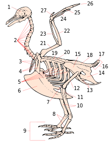
Vertebral column
The vertebral column is divided into five sections of vertebrae:
Cervical vertebrae
The cervical vertebrae provide structural support to the neck and number between 8 and as many as 25 vertebrae in certain swan species (Cygninae) and other long-necked birds. All cervical vertebrae have transverse processes attached except the first one. This vertebra (C1) is called the atlas which articulates with the occipital condyles of the skull and lacks the foramen typical of most vertebrae.[7] The neck of a bird is composed of many cervical vertebrae enabling birds to have increased flexibility. A flexible neck allows many birds with immobile eyes to move their head more productively and center their sight on objects that are close or far in distance.[8] Most birds have about three times as many neck vertebrae as humans, which allows for increased stability during fast movements such as flying, landing, and taking-off.[9] The neck plays a role in head-bobbing which is present in at least 8 out of 27 orders of birds, including Columbiformes, Galliformes, and Gruiformes.[10] Head-bobbing is an optokinetic response which stabilizes a bird's surroundings as it alternates between a thrust phase and a hold phase.[11] Head-bobbing is synchronous with the feet as the head moves in accordance with the rest of the body.[11] Data from various studies suggest that the main reason for head-bobbing in some birds is for the stabilization of their surroundings, although it is uncertain why some but not all bird orders show head-bob.[12]
Thoracic vertebrae
The thoracic vertebrae number between 5 and 10, and the first thoracic vertebra is distinguishable due to the fusion of its attached rib to the sternum while the ribs of cervical vertebrae are free.[7] Anterior thoracic vertebrae are fused in many birds and articulate with the notarium of the pectoral girdle.[13]
Diagram of a general bird pelvic girdle skeleton including the lower vertebral column sections. Note that the caudal vertebrae (5–10) are not fused in this diagram but can be in certain species.
Synsacrum
The synsacrum consists of one thoracic, six lumbar, two sacral, and five sacro-caudal vertebrae fused into one ossified structure that then fuse with the ilium.[14] When not in flight, this structure provides the main support for the rest of the body.[7] Similar to the sacrum of mammals, the synsacrum lacks the distinct disc shape of cervical and thoracic vertebrae.[15]
Caudal vertebrae
The free vertebrae immediately following the fused sacro-caudal vertebrae of the synsacrum are known as the caudal vertebrae. Birds have between 5 and 8 free caudal vertebrae.[7] The caudal vertebrae provide structure to the tails of vertebrates and are homologous to the coccyx found in mammals lacking tails.[16]
Pygostyle
In birds, the last 5 to 6 caudal vertebrae are fused to form the pygostyle.[14] Some sources note that up to 10 caudal vertebrae may make up this fused structure. This structure provides an attachment point for tail feathers that aid in control of flight.[7]
Highlighted in red is an intact keeled sternum of a dissected pigeon. In flying birds the sternum is enlarged for increased muscle attachment.
Scapular girdle
Birds are the only living vertebrates to have fused collarbones and a keeled breastbone.[17] The keeled sternum serves as an attachment site for the muscles used in flying or swimming.[17] Flightless birds, such as ostriches, lack a keeled sternum and have denser and heavier bones compared to birds that fly.[18] Swimming birds have a wide sternum, walking birds have a long sternum, and flying birds have a sternum that is nearly equal in width and height.[19] The chest consists of the furcula (wishbone) and coracoid (collar bone) which, together with the scapula, form the pectoral girdle; the side of the chest is formed by the ribs, which meet at the sternum (mid-line of the chest).[7]
Ribs
Birds have uncinate processes on the ribs. These are hooked extensions of bone which help to strengthen the rib cage by overlapping with the rib behind them. This feature is also found in the tuatara (Sphenodon).
Skull
The typical cranial anatomy of a bird. Pmx= premaxilla, M= maxilla, D= dentary, V= vomer, Pal= palatine, Pt= Pterygoid, Lc= Lacrimal
The skull consists of five major bones: the frontal (top of head), parietal (back of head), premaxillary and nasal (top beak), and the mandible (bottom beak). The skull of a normal bird usually weighs about 1% of the bird's total body weight. The eye occupies a considerable amount of the skull and is surrounded by a sclerotic eye-ring, a ring of tiny bones. This characteristic is also seen in their reptile cousins.
Broadly speaking, avian skulls consist of many small, non-overlapping bones. Pedomorphosis, maintenance of the ancestral state in adults, is thought to have facilitated the evolution of the avian skull. In essence, adult bird skulls will resemble the juvenile form of their theropod dinosaur ancestors.[20] As the avian lineage has progressed and as pedomorphosis has occurred, they have lost the postorbital bone behind the eye, the ectopterygoid at the back of the palate, and teeth.[21][22] The palate structures have also become greatly altered with changes, mostly reductions, seen in the ptyergoid, palatine, and jugal bones. A reduction in the adductor chambers has also occurred. [22] These are all conditions seen in the juvenile form of their ancestors. The premaxillary bone has also hypertrophied to form the beak while the maxilla has become diminished, as suggested by both developmental[20] and paleontological [23] studies. This expansion into the beak has occurred in tandem with the loss of a functional hand and the developmental of a point at the front of the beak that resembles a "finger".[22] The premaxilla is also known to play a large role in feeding behaviours in fish.[24][25]
The structure of the avian skull has important implications for their feeding behaviours. Birds show independent movement of the skull bones known as cranial kinesis. Cranial kinesis in birds occurs in several forms, but all of the different varieties are all made possible by the anatomy of the skull. Animals with large, overlapping bones (including the ancestors of modern birds)[26][27][28] have akinetic (non-kinetic) skulls.[29][30] For this reason it has been argued that the pedomorphic bird beak can be seen as an evolutionary innovation.[22]
Birds have a diapsid skull, as in reptiles, with a pre-lachrymal fossa (present in some reptiles). The skull has a single occipital condyle.[31]
The shoulder consists of the scapula (shoulder blade), coracoid, and humerus (upper arm). The humerus joins the radius and ulna (forearm) to form the elbow. The carpus and metacarpus form the "wrist" and "hand" of the bird, and the digits are fused together. The bones in the wing are extremely light so that the bird can fly more easily.
The hips consist of the pelvis, which includes three major bones: the ilium (top of the hip), ischium (sides of hip), and pubis (front of the hip). These are fused into one (the innominate bone). Innominate bones are evolutionary significant in that they allow birds to lay eggs. They meet at the acetabulum (hip socket) and articulate with the femur, which is the first bone of the hind limb.
The upper leg consists of the femur. At the knee joint, the femur connects to the tibiotarsus (shin) and fibula (side of lower leg). The tarsometatarsus forms the upper part of the foot, digits make up the toes. The leg bones of birds are the heaviest, contributing to a low center of gravity, which aids in flight. A bird's skeleton accounts for only about 5% of its total body weight.
They have a greatly elongate tetradiate pelvis, similar to some reptiles. The hind limb has an intra-tarsal joint found also in some reptiles. There is extensive fusion of the trunk vertebrae as well as fusion with the pectoral girdle.
Syndactyly, as it occurs in birds, is like anisodactyly, except that the second and third toes (the inner and middle forward-pointing toes), or three toes, are fused together, as in the belted kingfisherCeryle alcyon. This is characteristic of Coraciiformes (kingfishers, bee-eaters, rollers, etc.).
Zygodactyl (from Greek ζυγον, a yoke) feet have two toes facing forward (digits two and three) and two back (digits one and four). This arrangement is most common in arboreal species, particularly those that climb tree trunks or clamber through foliage. Zygodactyly occurs in the parrots, woodpeckers (including flickers), cuckoos (including roadrunners), and some owls. Zygodactyl tracks have been found dating to 120–110 Ma (early Cretaceous), 50 million years before the first identified zygodactyl fossils.[33]
Heterodactyly is like zygodactyly, except that digits three and four point forward and digits one and two point back. This is found only in trogons, while pamprodactyl is an arrangement in which all four toes may point forward, or birds may rotate the outer two toes backward. It is a characteristic of swifts (Apodidae).
Evolution
Hind limb change
A significant similarity in the structure of the hind limbs of birds and other dinosaurs is associated with their ability to walk on two legs, or bipedalism.[34] In the 20th century, the prevailing opinion was that the transition to bipedalism occurred due to the transformation of the forelimbs into wings. Modern scientists believe that, on the contrary, it was a necessary condition for the occurrence of flight.[35]
The transition to the use of only the hind limbs for movement was accompanied by an increase in the rigidity of the lumbar and sacral regions. The pubic bones of birds and some other bipedal dinosaurs are turned backward. Scientists associate this with a shift in the center of gravity of the body backward. The reason for this shift is called the transition to bipedality or the development of powerful forelimbs, as in Archaeopteryx.[36][37] The large and heavy tail of two-legged dinosaurs may have been an additional support. Partial tail reduction and subsequent formation of pygostyle occurred due to the backward deviation of the first toe of the hind limb; in dinosaurs with a long rigid tail, the development of the foot proceeded differently. This process, apparently, took place in parallel in birds and some other dinosaurs. In general, the anisodactyl foot, which also has a better grasping ability and allows confident movement both on the ground and along branches, is ancestral for birds. Against this background, pterosaurs stand out, which, in the process of unsuccessful evolutionary changes, could not fully move on two legs, but instead developed a physical means of flight[further explanation needed] that was fundamentally different from birds.[37]
Forelimb changes
Changes in the hindlimbs did not affect the location of the forelimbs, which in birds remained laterally spaced, and in non-avian dinosaurs they switched to a parasagittal orientation.[36] At the same time, the forelimbs, freed from the support function, had ample opportunities for evolutionary changes. Proponents of the running hypothesis believe that flight was formed through fast running, bouncing, and then gliding. The forelimbs could be used for grasping after a jump or as "insect trapping nets", animals could wave them, helping themselves during the jump. According to the arboreal hypothesis, the ancestors of birds climbed trees with the help of their forelimbs, and from there they planned, after which they proceeded to flight.[38]
Muscular system
The supracoracoideus works using a pulley-like system to lift the wing while the pectorals provide the powerful downstroke
Most birds have approximately 175 different muscles, mainly controlling the wings, skin, and legs. Overall, the muscle mass of birds is concentrated ventrally. The largest muscles in the bird are the pectorals, or the pectoralis major, which control the wings and make up about 15–25% of a flighted bird's body weight. They provide the powerful wing stroke essential for flight. The muscle deep to (underneath) the pectorals is the supracoracoideus, or the pectoralis minor. It raises the wing between wingbeats. Both muscle groups attach to the keel of the sternum. This is remarkable, because other vertebrates have the muscles to raise the upper limbs generally attached to areas on the back of the spine. The supracoracoideus and the pectorals together make up about 25–40% of the bird's full body weight.[39] Caudal to the pectorals and supracoracoideus are the internal and external obliques which compress the abdomen. Additionally, there are other abdominal muscles present that expand and contract the chest, and hold the ribcage. The muscles of the wing, as seen in the labelled images, function mainly in extending or flexing the elbow, moving the wing as a whole or in extending or flexing particular digits. These muscles work to adjust the wings for flight and all other actions.[39] Muscle composition does vary between species and even within families.[40]
Labelled ventral musculature of a pigeon wing
Birds have unique necks which are elongated with complex musculature as it must allow for the head to perform functions other animals may utilize pectoral limbs for.[39]
Labelled dorsal musculature of a pigeon wing
The skin muscles help a bird in its flight by adjusting the feathers, which are attached to the skin muscle and help the bird in its flight maneuvers as well as aiding in mating rituals.
There are only a few muscles in the trunk and the tail, but they are very strong and are essential for the bird. These include the lateralis caudae and the levator caudae which control movement of the tail and the spreading of rectrices, giving the tail a larger surface area which helps keep the bird in the air as well as aiding in turning.[39]
Muscle composition and adaptation differ by theories of muscle adaptation in whether evolution of flight came from flapping or gliding first.[41]
The scales of birds are composed of keratin, like beaks, claws, and spurs. They are found mainly on the toes and tarsi (lower leg of birds), usually up to the tibio-tarsal joint, but may be found further up the legs in some birds. In many of the eagles and owls the legs are feathered down to (but not including) their toes.[42][43][44] Most bird scales do not overlap significantly, except in the cases of kingfishers and woodpeckers. The scales and scutes of birds were originally thought to be homologous to those of reptiles;[45] however, more recent research suggests that scales in birds re-evolved after the evolution of feathers.[46][47][48]
Bird embryos begin development with smooth skin. On the feet, the corneum, or outermost layer, of this skin may keratinize, thicken and form scales. These scales can be organized into;
Cancella – minute scales which are really just a thickening and hardening of the skin, crisscrossed with shallow grooves.
Scutella – scales that are not quite as large as scutes, such as those found on the caudal, or hind part, of the chicken metatarsus.
Scutes – the largest scales, usually on the anterior surface of the metatarsus and dorsal surface of the toes.
The rows of scutes on the anterior of the metatarsus can be called an "acrometatarsium" or "acrotarsium".
Reticula are located on the lateral and medial surfaces (sides) of the foot and were originally thought to be separate scales. However, histological and evolutionary developmental work in this area revealed that these structures lack beta-keratin (a hallmark of reptilian scales) and are entirely composed of alpha-keratin.[47][49] This, along with their unique structure, has led to the suggestion that these are actually feather buds that were arrested early in development.[47]
Collectively, the scaly covering present on the foot of the birds is called podotheca.
Herbst corpuscles and lore
The bills of many waders have Herbst corpuscles which help them find prey hidden under wet sand, by detecting minute pressure differences in the water.[50] All extant birds can move the parts of the upper jaw relative to the brain case. However, this is more prominent in some birds and can be readily detected in parrots.[51]
The region between the eye and bill on the side of a bird's head is called the lore. This region is sometimes featherless, and the skin may be tinted, as in many species of the cormorant family.
The beak, bill, or rostrum is an external anatomical structure of birds which is used for eating and for preening, manipulating objects, killing prey, fighting, probing for food, courtship and feeding young. Although beaks vary significantly in size, shape and color, they share a similar underlying structure. Two bony projections—the upper and lower mandibles—covered with a thin keratinized layer of epidermis known as the rhamphotheca. In most species, two holes known as nares lead to the respiratory system.
The arrangement of the air sacs and lungs in birdsThe anatomy of bird's respiratory system, showing the relationships of the trachea, primary and intra-pulmonary bronchi, the dorso- and ventro-bronchi, with the parabronchi running between the two. The posterior and anterior air sacs are also indicated, but not to scale.Inhalation–exhalation cycle in birds.
Due to the high metabolic rate required for flight, birds have a high oxygen demand. Their highly effective respiratory system helps them meet that demand.
Although birds have lungs, theirs are fairly rigid structures that do not expand and contract as they do in mammals, reptiles and many amphibians. Instead, the structures that act as the bellows that ventilate the lungs are the air sacs, which are distributed throughout much of the birds' bodies.[52] The airsacs move air unidirectionally through the parabronchi of the rigid lungs.[53][54] The primary mechanism of unidirectional flows in bird lungs is flow irreversibility at high Reynolds number manifested in asymmetric junctions and their loop-forming connectivity.[55]
Although bird lungs are smaller than those of mammals of comparable size, the air sacs account for 15% of the total body volume, whereas in mammals, the alveoli, which act as the bellows, constitute only 7% of the total body volume.[56] The walls of the air sacs do not have a good blood supply and so do not play a direct role in gas exchange.
Birds lack a diaphragm, and therefore use their intercostal and abdominal muscles to expand and contract their entire thoraco-abdominal cavities, thus rhythmically changing the volumes of all their air sacs in unison (illustration on the right). The active phase of respiration in birds is exhalation, requiring contraction of their muscles of respiration.[54] Relaxation of these muscles causes inhalation.
Three distinct sets of organs perform respiration — the anterior air sacs (interclavicular, cervicals, and anterior thoracics), the lungs, and the posterior air sacs (posterior thoracics and abdominals). Typically there are nine air sacs within the system;[54] however, that number can range between seven and twelve, depending on the species of bird. Passerines possess seven air sacs, as the clavicular air sacs may interconnect or be fused with the anterior thoracic sacs.
During inhalation, environmental air initially enters the bird through the nostrils from where it is heated, humidified, and filtered in the nasal passages and upper parts of the trachea.[56] From there, the air enters the lower trachea and continues to just beyond the syrinx, at which point the trachea branches into two primary bronchi, going to the two lungs. The primary bronchi enter the lungs to become the intrapulmonary bronchi, which give off a set of parallel branches called ventrobronchi and, a little further on, an equivalent set of dorsobronchi.[57] The ends of the intrapulmonary bronchi discharge air into the posterior air sacs at the caudal end of the bird. Each pair of dorso-ventrobronchi is connected by a large number of parallel microscopic air capillaries (or parabronchi) where gas exchange occurs.[57] As the bird inhales, tracheal air flows through the intrapulmonary bronchi into the posterior air sacs, as well as into the dorsobronchi (but not into the ventrobronchi whose openings into the intrapulmonary bronchi were previously believed to be tightly closed during inhalation.[57] However, more recent studies have shown that the aerodynamics of the bronchial architecture directs the inhaled air away from the openings of the ventrobronchi, into the continuation of the intrapulmonary bronchus towards the dorsobronchi and posterior air sacs[53][58]). From the dorsobronchi the air flows through the parabronchi (and therefore the gas exchanger) to the ventrobronchi from where the air can only escape into the expanding anterior air sacs. So, during inhalation, both the posterior and anterior air sacs expand,[57] the posterior air sacs filling with fresh inhaled air, while the anterior air sacs fill with "spent" (oxygen-poor) air that has just passed through the lungs.
During exhalation the intrapulmonary bronchi were believed to be tightly constricted between the region where the ventrobronchi branch off and the region where the dorsobronchi branch off.[57] But it is now believed that more intricate aerodynamic features have the same effect.[53][58] The contracting posterior air sacs can therefore only empty into the dorsobronchi. From there the fresh air from the posterior air sacs flows through the parabronchi (in the same direction as occurred during inhalation) into ventrobronchi. The air passages connecting the ventrobronchi and anterior air sacs to the intrapulmonary bronchi open up during exhalation, thus allowing oxygen-poor air from these two organs to escape via the trachea to the exterior.[57] Oxygenated air therefore flows constantly (during the entire breathing cycle) in a single direction through the parabronchi.[1]
The cross-current respiratory gas exchanger in the lungs of birds. Air is forced from the air sacs unidirectionally (from right to left in the diagram) through the parabronchi. The pulmonary capillaries surround the parabronchi in the manner shown (blood flowing from below the parabronchus to above it in the diagram). Blood or air with a high oxygen content is shown in red; oxygen-poor air or blood is shown in various shades of purple-blue.
The blood flow through the bird lung is at right angles to the flow of air through the parabronchi, forming a cross-current flow exchange system (see illustration on the left).[57][59] The partial pressure of oxygen in the parabronchi declines along their lengths as O2 diffuses into the blood. The blood capillaries leaving the exchanger near the entrance of airflow take up more O2 than do the capillaries leaving near the exit end of the parabronchi. When the contents of all capillaries mix, the final partial pressure of oxygen of the mixed pulmonary venous blood is higher than that of the exhaled air,[57][59] but is nevertheless less than half that of the inhaled air,[57] thus achieving roughly the same systemic arterial blood partial pressure of oxygen as mammals do with their bellows-type lungs.[57]
The trachea is an area of dead space: the oxygen-poor air it contains at the end of exhalation is the first air to re-enter the posterior air sacs and lungs. In comparison to the mammalian respiratory tract, the dead space volume in a bird is, on average, 4.5times greater than it is in mammals of the same size.[57][56] Birds with long necks will inevitably have long tracheae, and must therefore take deeper breaths than mammals do to make allowances for their greater dead space volumes. In some birds (e.g. the whooper swan, Cygnus cygnus, the white spoonbill, Platalea leucorodia, the whooping crane, Grus americana, and the helmeted curassow, Pauxi pauxi) the trachea, which some cranes can be 1.5m long,[57] is coiled back and forth within the body, drastically increasing the dead space ventilation.[57] The purpose of this extraordinary feature is unknown.
Air passes unidirectionally through the lungs during both exhalation and inspiration, causing, except for the oxygen-poor dead space air left in the trachea after exhalation and breathed in at the beginning of inhalation, little to no mixing of new oxygen-rich air with spent oxygen-poor air (as occurs in mammalian lungs), changing only (from oxygen-rich to oxygen-poor) as it moves (unidirectionally) through the parabronchi.
Avian lungs do not have alveoli as mammalian lungs do. Instead they contain millions of narrow passages known as parabronchi, connecting the dorsobronchi to the ventrobronchi at either ends of the lungs. Air flows anteriorly (caudal to cranial) through the parallel parabronchi. These parabronchi have honeycombed walls. The cells of the honeycomb are dead-end air vesicles, called atria, which project radially from the parabronchi. The atria are the site of gas exchange by simple diffusion.[60] The blood flow around the parabronchi (and their atria), forms a cross-current gas exchanger (see diagram on the left).[57][59]
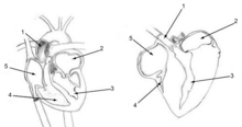
The human heart (left) and chicken heart (right) share many similar characteristics. Avian hearts pump faster than mammalian hearts. Due to the faster heart rate, the muscles surrounding the ventricles of the chicken heart are thicker. Both hearts are labeled with the following parts: 1. Ascending Aorta 2. Left Atrium 3. Left Ventricle 4. Right Ventricle 5. Right Atrium. In chickens and others birds, the superior cava is double.
All species of birds with the exception of the penguin, have a small region of their lungs devoted to "neopulmonic parabronchi". This unorganized network of microscopic tubes branches off from the posterior air sacs, and open haphazardly into both the dorso- and ventrobronchi, as well as directly into the intrapulmonary bronchi. Unlike the parabronchi, in which the air moves unidirectionally, the air flow in the neopulmonic parabronchi is bidirectional. The neopulmonic parabronchi never make up more than 25% of the total gas exchange surface of birds.[56]
Vocal Bird anatomy: Birds produce sounds through the air that passes through the Syrinx, which is shown close up in the bottom right.
In order for birds to produce sound, they use an organ located above the lungs called the syrinx, which is composed of tracheal rings, syringeal muscles, Tympaniform membrane, and internal bony structures that contribute to the production of sound. Air then passes through this organ, resulting in the vocalization of birds. Sound can then be produced through the movement of the Tympaniform membrane. Pitch can also be changed by opening and closing of the Tympaniform membrane, allowing for higher and lower production of sound.[61]
Birds have a four-chambered heart,[62] in common with mammals, and some reptiles (mainly the crocodilia). This adaptation allows for an efficient nutrient and oxygen transport throughout the body, providing birds with energy to fly and maintain high levels of activity. A ruby-throated hummingbird's heart beats up to 1200 times per minute (about 20 beats per second).[63]
Digestive system
Pigeon crop containing ingested food particles is highlighted in yellow. The crop is an out-pouching of the esophagus and the wall of the esophagus is shown in blue.Simplified depiction of avian digestive system.
Many birds possess a muscular pouch along the esophagus called a crop. The crop functions to both soften food and regulate its flow through the system by storing it temporarily. The size and shape of the crop is quite variable among the birds.[64] Members of the family Columbidae, such as pigeons, produce a nutritious crop milk which is fed to their young by regurgitation.[65]
The gizzard is composed of four muscular bands that rotate and crush food by shifting the food from one area to the next within the gizzard. The gizzard of some species of herbivorous birds, like turkey and quails,[64] contains small pieces of grit or stone called gastroliths that are swallowed by the bird to aid in the grinding process, serving the function of teeth. The use of gizzard stones is a similarity found between birds and dinosaurs, which left gastroliths as trace fossils.[65]
Intestines
The partially digested and pulverized gizzard contents, now called a bolus, are passed into the intestine, where pancreatic and intestinal enzymes complete the digestion of the digestible food. The digestion products are then absorbed through the intestinal mucosa into the blood. The intestine ends via the large intestine in the vent or cloaca which serves as the common exit for renal and intestinal excrements as well as for the laying of eggs.[69] However, unlike mammals, many birds do not excrete the bulky portions (roughage) of their undigested food (e.g. feathers, fur, bone fragments, and seed husks) via the cloaca, but regurgitate them as food pellets.[70][71]
Drinking behaviour
There are three general ways in which birds drink: using gravity itself, sucking, and by using the tongue. Fluid is also obtained from food.
Most birds are unable to swallow by the "sucking" or "pumping" action of peristalsis in their esophagus (as humans do), and drink by repeatedly raising their heads after filling their mouths to allow the liquid to flow by gravity, a method usually described as "sipping" or "tipping up".[72] The notable exception is the family of pigeons and doves, the Columbidae; in fact, according to Konrad Lorenz in 1939:
one recognizes the order by the single behavioral characteristic, namely that in drinking the water is pumped up by peristalsis of the esophagus which occurs without exception within the order. The only other group, however, which shows the same behavior, the Pteroclidae, is placed near the doves just by this doubtlessly very old characteristic.[73]
Although this general rule still stands, since that time, observations have been made of a few exceptions in both directions.[72][74]
In addition, specialized nectar feeders like sunbirds (Nectariniidae) and hummingbirds (Trochilidae) drink by using protrusible grooved or trough-like tongues, and parrots (Psittacidae) lap up water.[72]
Many seabirds have glands near the eyes that allow them to drink seawater. Excess salt is eliminated from the nostrils. Many desert birds get the water that they need entirely from their food. The elimination of nitrogenous wastes as uric acid reduces the physiological demand for water,[75] as uric acid is not very toxic and thus does not need to be diluted in as much water.[76]
Reproductive and urogenital systems
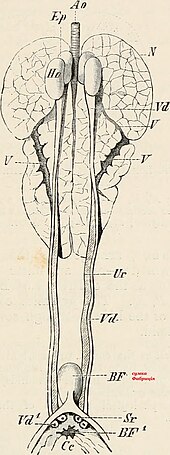
Seen here is a diagram of a female chicken reproduction system. A.Mature ovum, B.Infundibulum, C.Magnum, D.Isthmus, E.Uterus, F.Vagina, G.Cloaca, H.Large intestine, I.rudiment of right oviductFledgling
Male birds have two testes which become hundreds of times larger during the breeding season to produce sperm.[77] The testes in birds are generally asymmetric with most birds having a larger left testis.[78] Female birds in most families have only one functional ovary (the left one), connected to an oviduct — although two ovaries are present in the embryonic stage of each female bird. Some species of birds have two functional ovaries, and the kiwis always retain both.[79][80] Birds do not have male accessory glands.[81]
Most male birds have no phallus. In the males of species without a phallus, sperm is stored in the seminal glomera within the cloacal protuberance prior to copulation. During copulation, the female moves her tail to the side and the male either mounts the female from behind or in front (as in the stitchbird), or moves very close to her. The cloacae then touch, so that the sperm can enter the female's reproductive tract. This can happen very fast, sometimes in less than half a second.[82]
The sperm is stored in the female's sperm storage tubules for a period varying from a week to more than 100 days,[83] depending on the species. Then, eggs will be fertilized individually as they leave the ovaries, before the shell is calcified in the oviduct. After the egg is laid by the female, the embryo continues to develop in the egg outside the female body.
Many waterfowl and some other birds, such as the ostrich and turkey, possess a phallus.[84] This appears to be the ancestral condition among birds; most birds have lost the phallus.[85] The length is thought to be related to sperm competition in species that usually mate many times in a breeding season; sperm deposited closer to the ovaries is more likely to achieve fertilization.[86][87] The longer and more complicated phalli tend to occur in waterfowl whose females have unusual anatomical features of the vagina (such as dead end sacs and clockwise coils). These vaginal structures may be used to prevent penetration by the male phallus (which coils counter-clockwise). In these species, copulation is often violent and female co-operation is not required; the female ability to prevent fertilization may allow the female to choose the father for her offspring.[87][88][89][90] When not copulating, the phallus is hidden within the proctodeum compartment within the cloaca, just inside the vent.
After the eggs hatch, parents provide varying degrees of care in terms of food and protection. Precocial birds can care for themselves independently within minutes of hatching; altricial hatchlings are helpless, blind, and naked, and require extended parental care. The chicks of many ground-nesting birds such as partridges and waders are often able to run virtually immediately after hatching; such birds are referred to as nidifugous. The young of hole-nesters, though, are often totally incapable of unassisted survival. The process whereby a chick acquires feathers until it can fly is called "fledging".
Some birds, such as pigeons, geese, and red-crowned cranes, remain with their mates for life and may produce offspring on a regular basis.
Kidney
Avian kidneys function in almost the same way as the more extensively studied mammalian kidney, but with a few important adaptations; while much of the anatomy remains unchanged in design, some important modifications have occurred during their evolution.
The three-sectioned kidneys are placed on the bilateral side of the vertebral column, and there are connected to the lower gastrointestinal tract.[91] Depending on the bird species, the cortex makes up around 71–80% of the kidney's mass, while the medulla is much smaller at about 5–15% of the mass. Blood vessels and other tubes make up the remaining mass.
Unique to birds is the presence of two different types of nephrons (the functional unit of the kidney): both reptilian-like nephrons located in the cortex; and mammalian-like nephrons located in the medulla. Reptilian nephrons are more abundant but lack the distinctive loops of Henle seen in mammals. Because of the absence of the loop of Henle in birds, their ability to concentrate water does not depend heavily on it. Water reabsorption depends entirely on the coprodeum and the rectum.[20]
The urine collected by the kidney is emptied into the cloaca through the ureters and then to the colon by reverse peristalsis.
A Roseate spoonbill excreting urine in flightSuperior (towards the top) is the chicken's head, inferior (towards the bottom) is the chicken's feet. Chicken's kidneys are visualized at the bottom of the abdomen cavity, along the medial spine of the chicken. Testes are labeled as they sit above the kidneys.
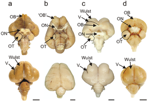
Brains of an emu, a kiwi, a barn owl, and a pigeon, with visual processing areas labelled
The avian brain is the central organ of the nervous system in birds. Birds possess large, complex brains, which process, integrate, and coordinate information received from the environment and make decisions on how to respond with the rest of the body. Like in all chordates, the avian brain is contained within the skull bones of the head.
The bird brain is divided into a number of sections, each with a different function. The cerebrum or telencephalon is divided into two hemispheres, and controls higher functions. The telencephalon is dominated by a large pallium, which corresponds to the mammaliancerebral cortex and is responsible for the cognitive functions of birds. The pallium is made up of several major structures: the hyperpallium, a dorsal bulge of the pallium found only in birds, as well as the nidopallium, mesopallium, and archipallium. The bird telencephalon nuclear structure, wherein neurons are distributed in three-dimensionally arranged clusters, with no large-scale separation of white matter and grey matter, though there exist layer-like and column-like connections. Structures in the pallium are associated with perception, learning, and cognition. Beneath the pallium are the two components of the subpallium, the striatum and pallidum. The subpallium connects different parts of the telencephalon and plays major roles in a number of critical behaviours. To the rear of the telencephalon are the thalamus, midbrain, and cerebellum. The hindbrain connects the rest of the brain to the spinal cord.
The size and structure of the avian brain enables prominent behaviours of birds such as flight and vocalization. Dedicated structures and pathways integrate the auditory and visual senses, strong in most species of birds, as well as the typically weaker olfactory and tactile senses. Social behaviour, widespread among birds, depends on the organisation and functions of the brain. Some birds exhibit strong abilities of cognition, enabled by the unique structure and physiology of the avian brain.
Birds have a large brain to body mass ratio. This is reflected in the advanced and complex bird intelligence.
Birds have acute eyesight—raptors (birds of prey) have vision eight times sharper than humans—thanks to higher densities of photoreceptors in the retina (up to 1,000,000 per square mm in Buteos, compared to 200,000 for humans), a high number of neurons in the optic nerves, a second set of eye muscles not found in other animals, and, in some cases, an indented fovea which magnifies the central part of the visual field. Many species, including hummingbirds and albatrosses, have two foveas in each eye. Many birds can detect polarised light.
Hearing
The avian ear is adapted to pick up on slight and rapid changes of pitch found in bird song. General avian tympanic membrane form is ovular and slightly conical. Morphological differences in the middle ear are observed between species. Ossicles within green finches, blackbirds, song thrushes, and house sparrows are proportionately shorter to those found in pheasants, Mallard ducks, and sea birds. In song birds, a syrinx allows the respective possessors to create intricate melodies and tones. The middle avian ear is made up of three semicircular canals, each ending in an ampulla and joining to connect with the macula sacculus and lagena, of which the cochlea, a straight short tube to the external ear, branches from.[92]
Taste
Birds evolved from an ancestor that had lost the taste bud type called T1R2, which allows other animals, like alligators, to taste sweet. After many birds adapted to a diet with high sugar content, they modified their umami taste receptors (T1R1-T1R3) to also sense sweet tastes. TR2, used to detect bitter tastes, is reduced in birds.[93][94]
The immune system of birds resembles that of other jawed vertebrates. Birds have both innate and adaptive immune systems. Birds are susceptible to tumours, immune deficiency and autoimmune diseases.
Bursa of fabricius
Internal view of the location of bursa of fabricius
The bursa of fabricius is a circular pouch connected to the superior dorsal side of the cloaca . The bursa is composed of many folds, known as plica, which are lined by more than 10,000 follicles encompassed by connective tissue and surrounded by mesenchyme. Each follicle consists of a cortex that surrounds a medulla. The cortex houses the highly compacted B lymphocytes, whereas the medulla houses lymphocytes loosely.[95] The medulla is separated from the lumen by the epithelium and this aids in the transport of epithelial cells into the lumen of the bursa. There are 150,000 B lymphocytes located around each follicle.[96]
Pterosaurs are an extinct clade of flying reptiles in the order Pterosauria. They existed during most of the Mesozoic: from the Late Triassic to the end of the Cretaceous. Pterosaurs are the earliest vertebrates known to have evolved powered flight. Their wings were formed by a membrane of skin, muscle, and other tissues stretching from the ankles to a dramatically lengthened fourth finger.
The lungs are the central organs of the respiratory system in humans and most other animals, including some snails and a small number of fish. In mammals and most other vertebrates, two lungs are located near the backbone on either side of the heart. Their function in the respiratory system is to extract oxygen from the air and transfer it into the bloodstream, and to release carbon dioxide from the bloodstream into the atmosphere, in a process of gas exchange. The pleurae, which are thin, smooth, and moist, serve to reduce friction between the lungs and chest wall during breathing, allowing for easy and effortless movements of the lungs.
The respiratory system is a biological system consisting of specific organs and structures used for gas exchange in animals and plants. The anatomy and physiology that make this happen varies greatly, depending on the size of the organism, the environment in which it lives and its evolutionary history. In land animals, the respiratory surface is internalized as linings of the lungs. Gas exchange in the lungs occurs in millions of small air sacs; in mammals and reptiles, these are called alveoli, and in birds, they are known as atria. These microscopic air sacs have a very rich blood supply, thus bringing the air into close contact with the blood. These air sacs communicate with the external environment via a system of airways, or hollow tubes, of which the largest is the trachea, which branches in the middle of the chest into the two main bronchi. These enter the lungs where they branch into progressively narrower secondary and tertiary bronchi that branch into numerous smaller tubes, the bronchioles. In birds, the bronchioles are termed parabronchi. It is the bronchioles, or parabronchi that generally open into the microscopic alveoli in mammals and atria in birds. Air has to be pumped from the environment into the alveoli or atria by the process of breathing which involves the muscles of respiration.
Aquatic respiration is the process whereby an aquatic organism exchanges respiratory gases with water, obtaining oxygen from oxygen dissolved in water and excreting carbon dioxide and some other metabolic waste products into the water.
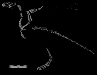
Gallimimus is a genus of theropod dinosaur that lived in what is now Mongolia during the Late Cretaceous period, about seventy million years ago (mya). Several fossils in various stages of growth were discovered by Polish-Mongolian expeditions in the Gobi Desert of Mongolia during the 1960s; a large skeleton discovered in this region was made the holotype specimen of the new genus and species Gallimimus bullatus in 1972. The generic name means "chicken mimic", referring to the similarities between its neck vertebrae and those of the Galliformes. The specific name is derived from bulla, a golden capsule worn by Roman youth, in reference to a bulbous structure at the base of the skull of Gallimimus. At the time it was named, the fossils of Gallimimus represented the most complete and best preserved ornithomimid material yet discovered, and the genus remains one of the best known members of the group.
Protoavis is a problematic taxon known from fragmentary remains from Late Triassic Norian stage deposits near Post, Texas. The animal's true classification has been the subject of much controversy, and there are many different interpretations of what the taxon actually is. When it was first described, the fossils were described as being from a primitive bird which, if the identification is valid, would push back avian origins some 60-75 million years.
Confuciusornis is a genus of basal crow-sized avialan from the Early Cretaceous Period of the Yixian and Jiufotang Formations of China, dating from 125 to 120 million years ago. Like modern birds, Confuciusornis had a toothless beak, but closer and later relatives of modern birds such as Hesperornis and Ichthyornis were toothed, indicating that the loss of teeth occurred convergently in Confuciusornis and living birds. It was thought to be the oldest known bird to have a beak, though this title now belongs to an earlier relative Eoconfuciusornis. It was named after the Chinese moral philosopher Confucius. Confuciusornis is one of the most abundant vertebrates found in the Yixian Formation, and several hundred complete specimens have been found.
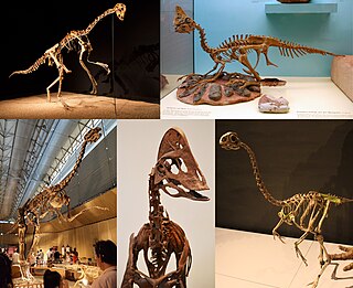
Oviraptorosaurs are a group of feathered maniraptoran dinosaurs from the Cretaceous Period of what are now Asia and North America. They are distinct for their characteristically short, beaked, parrot-like skulls, with or without bony crests atop the head. They ranged in size from Caudipteryx, which was the size of a turkey, to the 8-meter-long, 1.4-ton Gigantoraptor. The group is close to the ancestry of birds. Some researchers such as Maryanska et al (2002) and Osmólska et al. (2004) have proposed that they may represent primitive flightless birds. The most complete oviraptorosaur specimens have been found in Asia. The North American oviraptorosaur record is sparse.
The syrinx is the vocal organ of birds. Located at the base of a bird's trachea, it produces sounds without the vocal folds of mammals. The sound is produced by vibrations of some or all of the membrana tympaniformis and the pessulus, caused by air flowing through the syrinx. This sets up a self-oscillating system that modulates the airflow creating the sound. The muscles modulate the sound shape by changing the tension of the membranes and the bronchial openings. The syrinx enables some species of birds to mimic human speech.
Garudimimus is a genus of ornithomimosaur that lived in Asia during the Late Cretaceous. The genus is known from a single specimen found in 1981 by a Soviet-Mongolian paleontological expedition in the Bayan Shireh Formation and formally described in the same year by Rinchen Barsbold; the only species is Garudimimus brevipes. Several interpretations about the anatomical traits of Garudimimus were made in posterior examinations of the specimen, but most of them were criticized during its comprehensive redescription in 2005. Extensive undescribed ornithomimosaur remains at the type locality of Garudimimus may represent additional specimens of the genus.
Pelecanimimus is an extinct genus of basal ("primitive") ornithomimosaurian dinosaur from the Early Cretaceous of Spain. It is notable for possessing more teeth than any other member of the Ornithomimosauria, most of which were toothless.
Air sacs are spaces within an organism where there is the constant presence of air. Among modern animals, birds possess the most air sacs (9–11), with their extinct dinosaurian relatives showing a great increase in the pneumatization in their bones. Birds use air sacs for respiration as well as a number of other things. Theropods, like Aerosteon, have many air sacs in the body that are not just in bones, and they can be identified as the more primitive form of modern bird airways. Sauropods are well known for the large number of air pockets in their bones, although one theropod, Deinocheirus, shows a rivalling number of air pockets.
Majungasaurus is a genus of abelisaurid theropod dinosaur that lived in Madagascar from 70 to 66 million years ago, at the end of the Cretaceous Period, making it one of the last-known non-avian dinosaurs that went extinct during the Cretaceous–Paleogene extinction event. The genus contains a single species, Majungasaurus crenatissimus. This dinosaur is also called Majungatholus, a name which is considered a junior synonym of Majungasaurus.
The physiology of dinosaurs has historically been a controversial subject, particularly their thermoregulation. Recently, many new lines of evidence have been brought to bear on dinosaur physiology generally, including not only metabolic systems and thermoregulation, but on respiratory and cardiovascular systems as well.
Confuciusornithidae is an extinct family of pygostylian avialans known from the Early Cretaceous, found in northern China. They are commonly placed as a sister group to Ornithothoraces, a group that contains all extant birds along with their closest extinct relatives. Confuciusornithidae contains four genera, possessing both shafted and non-shafted (downy) feathers. They are also noted for their distinctive pair of ribbon-like tail feathers of disputed function.
Zhongornis is a genus of primitive maniraptoran dinosaurs that lived during the Early Cretaceous. It was found in rocks of the Yixian Formation in Lingyuan City (China), and described by Gao et al. in 2008.
Aerosteon is a genus of megaraptoran dinosaur from the Late Cretaceous period of Argentina. Its remains were discovered in 1996 in the Anacleto Formation, which is from the late Campanian. The type and only known species is A. riocoloradensis. Its specific name indicates that its remains were found 1 km north of the Río Colorado, in Mendoza Province, Argentina.
Skeletal pneumaticity is the presence of air spaces within bones. It is generally produced during development by excavation of bone by pneumatic diverticula from an air-filled space, such as the lungs or nasal cavity. Pneumatization is highly variable between individuals, and bones not normally pneumatized can become pneumatized in pathological development.
The vertebral column, also known as the spinal column, spine or backbone, is the core part of the axial skeleton in vertebrate animals. The vertebral column is the defining and eponymous characteristic of the vertebrate endoskeleton, where the notochord found in all chordates has been replaced by a segmented series of mineralized irregular bones called vertebrae, separated by fibrocartilaginous intervertebral discs. The dorsal portion of the vertebral column houses the spinal canal, an elongated cavity formed by alignment of the vertebral neural arches that encloses and protects the spinal cord, with spinal nerves exiting via the intervertebral foramina to innervate each body segments.
This glossary explains technical terms commonly employed in the description of dinosaur body fossils. Besides dinosaur-specific terms, it covers terms with wider usage, when these are of central importance in the study of dinosaurs or when their discussion in the context of dinosaurs is beneficial. The glossary does not cover ichnological and bone histological terms, nor does it cover measurements.
↑ Louchart, Antoine; Viriot, Laurent (2011). "From snout to beak: the loss of teeth in birds". Trends in Ecology & Evolution. 26 (12): 663–673. doi:10.1016/j.tree.2011.09.004. PMID21978465.
↑ Simonetta, Alberto M. (1960-09-01). "On the Mechanical Implications of the Avian Skull and Their Bearing on the Evolution and Classification of Birds". The Quarterly Review of Biology. 35 (3): 206–220. doi:10.1086/403106. ISSN0033-5770. S2CID85091693.
↑ Lingham-Soliar, Theagarten (1995-01-30). "Anatomy and functional morphology of the largest marine reptile known, Mosasaurus hoffmanni (Mosasauridae, Reptilia) from the Upper Cretaceous, Upper Maastrichtian of The Netherlands". Phil. Trans. R. Soc. Lond. B. 347 (1320): 155–180. Bibcode:1995RSPTB.347..155L. doi:10.1098/rstb.1995.0019. ISSN0962-8436.
↑ Holliday, Casey M.; Witmer, Lawrence M. (2008). "Cranial kinesis in dinosaurs: intracranial joints, protractor muscles, and their significance for cranial evolution and function in diapsids". Journal of Vertebrate Paleontology. 28 (4): 1073–1088. Bibcode:2008JVPal..28.1073H. doi:10.1671/0272-4634-28.4.1073. S2CID15142387.
1 2 Kurochkin, E. N. (1995). "Synopsis of Mesozoic Birds and Early evolution of Class Aves". Archaeopteryx: Jahreszeitschrift der Freunde des Jura-Museums Eichstätt. 13: 47–66. ISSN0933-288X. OCLC85132179.
1 2 3 4 Proctor, Noble S., Lynch, Patrick J. (1993). Manual of Ornithology. New Haven and London: Yale University Press. pp.149–170. ISBN978-0-300-07619-6.{{cite book}}: CS1 maint: multiple names: authors list (link)
↑ Oberprieler, Ulrich; Cillie, Burger (2002). Raptor Identification Guide for Southern Africa. Parklands: Random House. p.8. ISBN978-0-9584195-7-4.
↑ Lucas, Alfred M. (1972). Avian Anatomy – integument. East Lansing, Michigan, USA: USDA Avian Anatomy Project, Michigan State University. pp.67, 344, 394–601.
↑ Sawyer, R.H.; Knapp, L.W. (2003). "Avian Skin Development and the Evolutionary Origin of Feathers". Journal of Experimental Zoology Part B: Molecular and Developmental Evolution. 298 (1): 57–72. doi:10.1002/jez.b.26. PMID12949769.
↑ Zusi, R L (1984). "A Functional and Evolutionary Analysis of Rhynchokinesis in Birds". Smithsonian Contributions to Zoology. 395 (395): 1–40. doi:10.5479/si.00810282.395. hdl:10088/5187.
↑ Maclean, Gordon L. (1996). The Ecophysiology of Desert Birds. Springer. ISBN3-540-59269-5.
↑ Elphick, Jonathan (2016). Birds: A Complete Guide to their Biology and Behavior. Buffalo, New York: Firefly Books. pp.53–54. ISBN978-1-77085-762-9.
↑ A study of the seasonal changes in avian testesArchived 2009-03-16 at the Wayback Machine Alexander Watson, J. Physiol. 1919;53;86–91, 'greenfinch (Carduelis chloris)', "In early summer (May and June) they are as big as a whole pea and in early winter (November) they are no bigger than a pin head"
↑ Lake, PE (1981). "Male genital organs". In King AS; McLelland J (eds.). Form and function in birds. Vol.2. New York: Academic. pp.1–61. ISBN978-0-12-407502-3.
↑ Kinsky, FC (1971). "The consistent presence of paired ovaries in the Kiwi(Apteryx) with some discussion of this condition in other birds". Journal of Ornithology. 112 (3): 334–357. doi:10.1007/BF01640692. S2CID28261057.
↑ Lynch, Wayne; Lynch, photographs by Wayne (2007). Owls of the United States and Canada: a complete guide to their biology and behavior. Baltimore: Johns Hopkins University Press. p.151. ISBN978-0-8018-8687-4.
↑ Birkhead, TR; A. P. Moller (1993). "Sexual selection and the temporal separation of reproductive events: sperm storage data from reptiles, birds and mammals". Biological Journal of the Linnean Society. 50 (4): 295–311. doi:10.1111/j.1095-8312.1993.tb00933.x.
1 2 Arnqvist, G.; I. Danielsson (1999). "Copulatory Behavior, Genital Morphology, and Male Fertilization Success in Water Striders". Evolution. 53 (1): 147–156. doi:10.2307/2640927. JSTOR2640927. PMID28565197.
This page is based on this Wikipedia article Text is available under the CC BY-SA 4.0 license; additional terms may apply. Images, videos and audio are available under their respective licenses.