
Brain abscess is an abscess caused by inflammation and collection of infected material, coming from local or remote infectious sources, within the brain tissue. The infection may also be introduced through a skull fracture following a head trauma or surgical procedures. Brain abscess is usually associated with congenital heart disease in young children. It may occur at any age but is most frequent in the third decade of life.

Histoplasmosis is a fungal infection caused by Histoplasma capsulatum. Symptoms of this infection vary greatly, but the disease affects primarily the lungs. Occasionally, other organs are affected; called disseminated histoplasmosis, it can be fatal if left untreated.
Nontuberculous mycobacteria (NTM), also known as environmental mycobacteria, atypical mycobacteria and mycobacteria other than tuberculosis (MOTT), are mycobacteria which do not cause tuberculosis or leprosy. NTM do cause pulmonary diseases that resemble tuberculosis. Mycobacteriosis is any of these illnesses, usually meant to exclude tuberculosis. They occur in many animals, including humans and are commonly found in soil and water.
Radiology (X-rays) is used in the diagnosis of tuberculosis. Abnormalities on chest radiographs may be suggestive of, but are never diagnostic of TB, but can be used to rule out pulmonary TB.
Mycobacterium xenopi is a slow-growing scotochromogenic species of Mycobacterium. It was first reported by Schwabacher in 1959, having been isolated in lesions found on a Xenopus laevis, but the possibility of human infection was not confirmed until 1965. It has been cultured from hot and cold water taps, hospital hot water generators and storage tanks, and other environmental sources.

Caseous necrosis or caseous degeneration is a unique form of cell death in which the tissue maintains a cheese-like appearance. It is also a distinctive form of coagulative necrosis. The dead tissue appears as a soft and white proteinaceous dead cell mass.

Miliary tuberculosis is a form of tuberculosis that is characterized by a wide dissemination into the human body and by the tiny size of the lesions (1–5 mm). Its name comes from a distinctive pattern seen on a chest radiograph of many tiny spots distributed throughout the lung fields with the appearance similar to millet seeds—thus the term "miliary" tuberculosis. Miliary TB may infect any number of organs, including the lungs, liver, and spleen. Miliary tuberculosis is present in about 2% of all reported cases of tuberculosis and accounts for up to 20% of all extra-pulmonary tuberculosis cases.
AIDS-defining clinical conditions is the list of diseases published by the Centers for Disease Control and Prevention (CDC) that are associated with AIDS, and used worldwide as a guideline for AIDS diagnosis. CDC exclusively uses the term AIDS-defining clinical conditions, but the other terms remain in common use.
The CDC Classification System for HIV Infection is the medical classification system used by the United States Centers for Disease Control and Prevention (CDC) to classify HIV disease and infection. The system is used to allow the government to handle epidemic statistics and define who receives US government assistance.
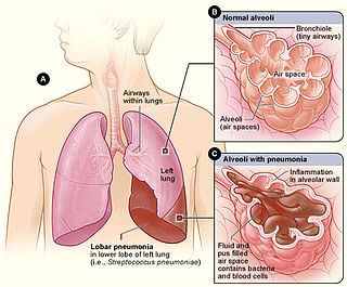
Lobar pneumonia is a form of pneumonia characterized by inflammatory exudate within the intra-alveolar space resulting in consolidation that affects a large and continuous area of the lobe of a lung.

Mycobacterium avium-intracellulare infection (MAI) is an atypical mycobacterial infection, i.e. one with nontuberculous mycobacteria or NTM, caused by Mycobacterium avium complex (MAC), which is made of two Mycobacterium species, M. avium and M. intracellulare. This infection causes respiratory illness in birds, pigs, and humans, especially in immunocompromised people. In the later stages of AIDS, it can be very severe. It usually first presents as a persistent cough. It is typically treated with a series of three antibiotics for a period of at least six months.

A Ghon focus is a primary lesion usually subpleural, often in the mid to lower zones, caused by Mycobacterium bacilli (tuberculosis) developed in the lung of a nonimmune host. It is named for Anton Ghon (1866–1936), an Austrian pathologist.

Tuberculous meningitis, also known as TB meningitis or tubercular meningitis, is a specific type of bacterial meningitis caused by the Mycobacterium tuberculosis infection of the meninges—the system of membranes which envelop the central nervous system.
Mycobacterium avium complex is a group of mycobacteria comprising Mycobacterium intracellulare and Mycobacterium avium that are commonly grouped because they infect humans together; this group, in turn, is part of the group of nontuberculous mycobacteria. These bacteria cause disease in humans called Mycobacterium avium-intracellulare infection or Mycobacterium avium complex infection. These bacteria are common and are found in fresh and salt water, in household dust and in soil. MAC bacteria usually cause infection in those who are immunocompromised or those with severe lung disease.
Mycobacterium canettii, a novel pathogenic taxon of the Mycobacterium tuberculosis complex (MTBC), was first reported in 1969 by the French microbiologist Georges Canetti, for whom the organism has been named. It formed smooth and shiny colonies, which is highly exceptional for the MTBC. It was described in detail in 1997 on the isolation of a new strain from a 2-year-old Somali patient with lymphadenitis. It did not differ from Mycobacterium tuberculosis in the biochemical tests and in its 16S rRNA sequence. It had shorter generation time than clinical isolates of M. tuberculosis and presented a unique, characteristic phenolic glycolipid and lipo-oligosaccharide. In 1998, Pfyffer described abdominal lymphatic TB in a 56-year-old Swiss man with HIV infection who lived in Kenya. Tuberculosis caused by M. canettii appears to be an emerging disease in the Horn of Africa. A history of a stay to the region should induce the clinician to consider this organism promptly even if the clinical features of TB caused by M. canettii are not specific. The natural reservoir, host range, and mode of transmission of the organism are still unknown.
Anton Ghon was an Austrian pathologist and bacteriologist who was a native of Villach. He is best known for his research on tuberculosis.
A Simon focus is a tuberculosis (TB) nodule that can form in the apex of the lung when a primary TB infection elsewhere in the body spreads to the lung apex via the bloodstream. Simon focus nodules are often calcified.
Karl Ernst Ranke was a German internist, pediatrician and pulmonologist known for his research of tuberculosis. He was the son of anthropologist Johannes Ranke (1836–1916).

A lung cavity or pulmonary cavity is an abnormal, thick-walled, air-filled space within the lung. Cavities in the lung can be caused by infections, cancer, autoimmune conditions, trauma, congenital defects, or pulmonary embolism. The most common cause of a single lung cavity is lung cancer. Bacterial, mycobacterial, and fungal infections are common causes of lung cavities. Globally, tuberculosis is likely the most common infectious cause of lung cavities. Less commonly, parasitic infections can cause cavities. Viral infections almost never cause cavities. The terms cavity and cyst are frequently used interchangeably; however, a cavity is thick walled, while a cyst is thin walled. The distinction is important because cystic lesions are unlikely to be cancer, while cavitary lesions are often caused by cancer.

Abdominal tuberculosis is a type of extrapulmonary tuberculosis which involves the abdominal organs such as intestines, peritoneum and abdominal lymph nodes. It can either occur in isolation or along with a primary focus in patients with disseminated tuberculosis.
