Related Research Articles
Panniculitis is a group of diseases whose hallmark is inflammation of subcutaneous adipose tissue. Symptoms include tender skin nodules, and systemic signs such as weight loss and fatigue.

A skin condition, also known as cutaneous condition, is any medical condition that affects the integumentary system—the organ system that encloses the body and includes skin, hair, nails, and related muscle and glands. The major function of this system is as a barrier against the external environment.

Paronychia is an inflammation of the skin around the nail, which can occur suddenly, when it is usually due to the bacteria Staph. aureus, or gradually when it is commonly caused by Candida albicans. The term is from Greek: παρωνυχία from para, "around", onyx, "nail" and the abstract noun suffix -ia.

Tuberculosis verrucosa cutis is a rash of small, red papular nodules in the skin that may appear 2–4 weeks after inoculation by Mycobacterium tuberculosis in a previously infected and immunocompetent individual.
Chrysiasis is a dermatological condition induced by the parenteral administration of gold salts, usually for the treatment of rheumatoid arthritis. Such treatment has been superseded as the best practice for treating the disease because of "numerous side effects and monitoring requirements, their limited efficacy, and very slow onset of action".

Scrofuloderma is a skin condition caused by tuberculous involvement of the skin by direct extension, usually from underlying tuberculous lymphadenitis.
Keratoderma is a hornlike skin condition.

Lupus vulgaris are painful cutaneous tuberculosis skin lesions with nodular appearance, most often on the face around the nose, eyelids, lips, cheeks, ears and neck. It is the most common Mycobacterium tuberculosis skin infection. The lesions may ultimately develop into disfiguring skin ulcers if left untreated.

The Koebner phenomenon or Köbner phenomenon, also called the Koebner response or the isomorphic response, attributed to Heinrich Köbner, is the appearance of skin lesions on lines of trauma. The Koebner phenomenon may result from either a linear exposure or irritation. Conditions demonstrating linear lesions after a linear exposure to a causative agent include: molluscum contagiosum, warts and toxicodendron dermatitis. Warts and molluscum contagiosum lesions can be spread in linear patterns by self-scratching ("auto-inoculation"). Toxicodendron dermatitis lesions are often linear from brushing up against the plant. Causes of the Koebner phenomenon that are secondary to scratching rather than an infective or chemical cause include vitiligo, psoriasis, lichen planus, lichen nitidus, pityriasis rubra pilaris, and keratosis follicularis.

Tinea nigra, also known as superficial phaeohyphomycosis and Tinea nigra palmaris et plantaris, is a superficial fungal infection that causes dark brown to black, painless patches called macules on the palms of the hands and the soles of the feet of otherwise healthy individuals. The macules occasionally extend to the fingers, toes, and nails, and may be reported on the chest, neck, or genital area. Tinea nigra infections can present with multiple macules that can be mottled or velvety in appearance, and may be oval or irregular in shape. The macules can be anywhere from a few mm to several cm in size.
Prurigo gestationis is an eruption consisting of pruritic, excoriated papules of the proximal limbs and upper trunk, most often occurring between the 20th and 34th week of gestation.
Tuberculosis cutis orificialis is a form of cutaneous tuberculosis that occurs at the mucocutaneous borders of the nose, mouth, anus, urinary meatus, and vagina, and on the mucous membrane of the mouth or tongue.
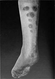
Papulonecrotic tuberculid is usually an asymptomatic, chronic skin disorder, presenting in successive crops, skin lesions symmetrically distributed on the extensor extremities.
Aquarium granuloma is a skin condition caused by Mycobacterium marinum, characterized by a skin lesion that presents roughly three weeks after exposure.
Primary cutaneous coccidioidomycosis is a skin condition caused by Coccidioides immitis following a definite history of inoculation or a colonized splinter found in the skin lesion.
Primary gonococcal dermatitis is a rare infection of the skin that occurs after primary inoculation of the skin from an infected focus.

Progressive vaccinia is a rare cutaneous condition caused by the vaccinia virus, characterized by painless, but progressive, necrosis and ulceration.

Cutaneous lymphoid hyperplasia refers to a groups of benign cutaneous disorders characterized by collections of lymphocytes, macrophages, and dendritic cells in the skin. Conditions included in this groups are:

Trichoblastomas are a cutaneous condition characterized by benign neoplasms of follicular germinative cells. Trichoblastic fibroma is a designation used to characterize small nodular trichoblastomas with conspicuous fibrocytic stroma, sometimes constituting over 50% of the lesion.

Tuberculous gumma is a skin condition characterized histologically by massive necrosis. Restated, this is a skin condition that results from hematogenous dissemination of mycobacteria from a primary focus, resulting in firm, nontender erythematous nodules that soften, ulcerate, and form sinuses.
References
- 1 2 Rapini, Ronald P.; Bolognia, Jean L.; Jorizzo, Joseph L. (2007). Dermatology: 2-Volume Set. St. Louis: Mosby. pp. 1114, 1116. ISBN 978-1-4160-2999-1.
- 1 2 James, William D.; Berger, Timothy G.; et al. (2006). Andrews' Diseases of the Skin: clinical Dermatology. Saunders Elsevier. ISBN 978-0-7216-2921-6.