Tuberculosis (TB), also known colloquially as the "white death", or historically as consumption, is an infectious disease usually caused by Mycobacterium tuberculosis (MTB) bacteria. Tuberculosis generally affects the lungs, but it can also affect other parts of the body. Most infections show no symptoms, in which case it is known as latent tuberculosis. Around 10% of latent infections progress to active disease which, if left untreated, kill about half of those affected. Typical symptoms of active TB are chronic cough with blood-containing mucus, fever, night sweats, and weight loss. Infection of other organs can cause a wide range of symptoms.
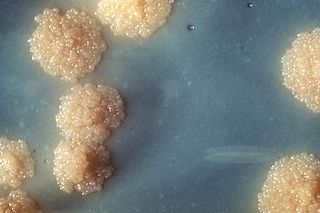
Mycobacterium tuberculosis, also known as Koch's bacillus, is a species of pathogenic bacteria in the family Mycobacteriaceae and the causative agent of tuberculosis. First discovered in 1882 by Robert Koch, M. tuberculosis has an unusual, waxy coating on its cell surface primarily due to the presence of mycolic acid. This coating makes the cells impervious to Gram staining, and as a result, M. tuberculosis can appear weakly Gram-positive. Acid-fast stains such as Ziehl–Neelsen, or fluorescent stains such as auramine are used instead to identify M. tuberculosis with a microscope. The physiology of M. tuberculosis is highly aerobic and requires high levels of oxygen. Primarily a pathogen of the mammalian respiratory system, it infects the lungs. The most frequently used diagnostic methods for tuberculosis are the tuberculin skin test, acid-fast stain, culture, and polymerase chain reaction.

Mycobacterium is a genus of over 190 species in the phylum Actinomycetota, assigned its own family, Mycobacteriaceae. This genus includes pathogens known to cause serious diseases in mammals, including tuberculosis and leprosy in humans. The Greek prefix myco- means 'fungus', alluding to this genus' mold-like colony surfaces. Since this genus has cell walls with a waxy lipid-rich outer layer that contains high concentrations of mycolic acid, acid-fast staining is used to emphasize their resistance to acids, compared to other cell types.
Nontuberculous mycobacteria (NTM), also known as environmental mycobacteria, atypical mycobacteria and mycobacteria other than tuberculosis (MOTT), are mycobacteria which do not cause tuberculosis or leprosy/Hansen's disease. NTM are able to cause pulmonary diseases that resemble tuberculosis. Mycobacteriosis is any of these illnesses, usually meant to exclude tuberculosis. They occur in many animals, including humans and are commonly found in soil and water.
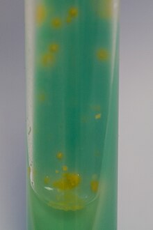
Mycobacterium xenopi is a slow-growing scotochromogenic species of Mycobacterium. It was first reported by Schwabacher in 1959, having been isolated in lesions found on a Xenopus laevis, but the possibility of human infection was not confirmed until 1965. It has been cultured from hot and cold water taps, hospital hot water generators and storage tanks, and other environmental sources.

Ethionamide is an antibiotic used to treat tuberculosis. Specifically it is used, along with other antituberculosis medications, to treat active multidrug-resistant tuberculosis. It is no longer recommended for leprosy. It is taken by mouth.

A mycobacteriophage is a member of a group of bacteriophages known to have mycobacteria as host bacterial species. While originally isolated from the bacterial species Mycobacterium smegmatis and Mycobacterium tuberculosis, the causative agent of tuberculosis, more than 4,200 mycobacteriophage have since been isolated from various environmental and clinical sources. 2,042 have been completely sequenced. Mycobacteriophages have served as examples of viral lysogeny and of the divergent morphology and genetic arrangement characteristic of many phage types.
The Timpe and Runyon classification of nontuberculous mycobacteria based on the rate of growth, production of yellow pigment and whether this pigment was produced in the dark or only after exposure to light.
Lipoarabinomannan, also called LAM, is a glycolipid, and a virulence factor associated with Mycobacterium tuberculosis, the bacteria responsible for tuberculosis. Its primary function is to inactivate macrophages and scavenge oxidative radicals.

Mycobacteroides abscessus is a species of rapidly growing, multidrug-resistant, nontuberculous mycobacteria (NTM) that is a common soil and water contaminant. Although M. abscessus most commonly causes chronic lung infection and skin and soft tissue infection (SSTI), it can also cause infection in almost all human organs, mostly in patients with suppressed immune systems. Amongst NTM species responsible for disease, infection caused by M. abscessus complex are more difficult to treat due to antimicrobial drug resistance.

Mycolicibacter arupensis is a slowly growing mycobacterium first isolated from soil and human sputum samples in Spain. Etymology: arupense, pertaining to the ARUP Institute for Clinical and Experimental Pathology, where the type strain was characterized.

Mycobacterium fortuitum is a nontuberculous species of the phylum Actinomycetota, belonging to the genus Mycobacterium.
Mycobacterium gastri is a species of the phylum Actinomycetota, belonging to the genus Mycobacterium.
Mycobacterium gordonae is a species of Mycobacterium named for Ruth E. Gordon. It is a species of the phylum Actinomycetota, belonging to the genus Mycobacterium.
Mycobacterium hassiacum is a rapid-growing thermophilic mycobacterium that was isolated in human urine in 1997 by researchers at the German University of Regensburg. It's a species of the phylum Actinomycetota, belonging to the genus Mycobacterium.
Mycobacterium avium complex is a group of mycobacteria comprising Mycobacterium intracellulare and Mycobacterium avium that are commonly grouped because they infect humans together; this group, in turn, is part of the group of nontuberculous mycobacteria. These bacteria cause Mycobacterium avium-intracellulare infections or Mycobacterium avium complex infections in humans. These bacteria are common and are found in fresh and salt water, in household dust and in soil. MAC bacteria usually cause infection in those who are immunocompromised or those with severe lung disease.
Mycobacterium malmoense is a Gram-positive bacterium from the genus Mycobacterium.
Mycobacterium canettii, a novel pathogenic taxon of the Mycobacterium tuberculosis complex (MTBC), was first reported in 1969 by the French microbiologist Georges Canetti, for whom the organism has been named. It formed smooth and shiny colonies, which is highly exceptional for the MTBC. It was described in detail in 1997 on the isolation of a new strain from a 2-year-old Somali patient with lymphadenitis. It did not differ from Mycobacterium tuberculosis in the biochemical tests and in its 16S rRNA sequence. It had shorter generation time than clinical isolates of M. tuberculosis and presented a unique, characteristic phenolic glycolipid and lipo-oligosaccharide. In 1998, Pfyffer described abdominal lymphatic TB in a 56-year-old Swiss man with HIV infection who lived in Kenya. Tuberculosis caused by M. canettii appears to be an emerging disease in the Horn of Africa. A history of a stay to the region should induce the clinician to consider this organism promptly even if the clinical features of TB caused by M. canettii are not specific. The natural reservoir, host range, and mode of transmission of the organism are still unknown.
Mycobacterium wolinskyi is a rapidly growing mycobacterium most commonly seen in post-traumatic wound infections, especially those following open fractures and with associated osteomyelitis. Mycobacterium wolinskyi is clearly clinically significant, and occurs in the same settings as Mycobacterium smegmatis and members of the Mycobacterium fortuitum complex; they differ from members of the Mycobacterium fortuitum complex in the type of chronic lung disease they produce, with essentially all cases occurring in the setting of chronic lipoid pneumonia, either secondary to chronic oil ingestion or chronic aspiration. Etymology: Wolinsky, named after Emanuel Wolinsky in honour of, and in recognition for, significant contributions to the study of the non-tuberculous mycobacteria.
Mycobacteria that form colonies clearly visible to the naked eye in more than 7 days on subculture are termed slow growers.