Glutaminase

Last updated
glutaminase
Glutaminase.png
Crystallographic structure of dimeric protein-glutaminase from Chryseobacterium proteolyticum . [1]
Identifiers
EC no. 3.5.1.2
CAS no. 9001-47-2
Databases
IntEnz IntEnz view
BRENDA BRENDA entry
ExPASy NiceZyme view
KEGG KEGG entry
MetaCyc metabolic pathway
PRIAM profile
PDB structures RCSB PDB PDBe PDBsum
Gene Ontology AmiGO / QuickGO
Search
PMC articles
PubMed articles
NCBI proteins
Glutaminase
PDB 2osu EBI.jpg
probable glutaminase from bacillus subtilis complexed with 6-diazo-5-oxo-ʟ-norleucine
Identifiers
SymbolGlutaminase
Pfam PF04960
Pfam clan CL0013
InterPro IPR015868
SCOP2 1mki / SCOPe / SUPFAM
Available protein structures:
Pfam   structures / ECOD  
PDB RCSB PDB; PDBe; PDBj
PDBsum structure summary

Glutaminase (EC 3.5.1.2, glutaminase I, L-glutaminase, glutamine aminohydrolase) is an amidohydrolase enzyme that generates glutamate from glutamine. Glutaminase has tissue-specific isoenzymes. Glutaminase has an important role in glial cells.

Contents

Glutaminase catalyzes the following reaction:

Glutamine + H2Oglutamate + NH+4

Tissue distribution

Glutaminase is expressed and active in periportal hepatocytes, where it generates ammonium for urea synthesis, as does glutamate dehydrogenase. [2] Glutaminase is also expressed in the epithelial cells of the renal tubules, where the produced ammonia is excreted as ammonium ions. This excretion of ammonium ions is an important mechanism of renal acid-base regulation. During chronic acidosis, glutaminase is induced in the kidney, which leads to an increase in the amount of ammonium ions excreted. Glutaminase can also be found in the intestines, whereby hepatic portal ammonia can reach as high as 0.26 mM (compared to an arterial blood ammonia of 0.02 mM).

One of the most important roles of glutaminase is found in the axonal terminals of neurons in the central nervous system. Glutamate is the most abundantly used excitatory neurotransmitter in the CNS. After being released into the synapse for neurotransmission, glutamate is rapidly taken up by nearby astrocytes, which convert it to glutamine. This glutamine is then supplied to the presynaptic terminals of the neurons, where glutaminases convert it back to glutamate for loading into synaptic vesicles. Although both "kidney-type" (GLS1) and "liver-type" (GLS2) glutaminases are expressed in brain, GLS2 has been reported to exist only in cellular nuclei in CNS neurons. [3]

Regulation

ADP is the strongest adenine nucleotide activator of glutaminase. Studies have also suggested ADP lowered the Km for glutamine and increased the Vmax. They found that these effects were increased even more when ATP was present. [4]

The end product of the glutaminase reaction, glutamate, is a strong inhibitor of the reaction. Changes in glutamate dehydrogenase, which converts glutamate to 2-oxoglutarate and thereby decreases intramitochondrial glutamate levels, are thereby an important regulatory mechanism of glutaminase activity.

Phosphate-activated mitochondrial glutaminase (GLS1) is suggested to be linked with elevated metabolism, decreased intracellular reactive oxygen species (ROS) levels, and overall decreased DNA oxidation in both normal and stressed cells. It is suggested that GLS2's control of ROS levels facilitates “the ability of p53 to protect cells from accumulation of genomic damage and allows cells to survive after mild and repairable genotoxic stress.” [5]

Structure

The structure of glutaminase has been determined using X-ray diffraction to a resolution of up to 1.73 Å. There are 2 chains containing 305 residues that make up the length of this dimeric protein. On each strand, 23% of the amino acid content, or 71 residues, are found in the 8 helices. Twenty-one percent, or 95 residues, make up the 23 beta sheet strands. [1]

Isozymes

Humans express 4 isoforms of glutaminase. GLS encodes 2 types of kidney-type glutaminase with a high activity and low Km. GLS2 encodes 2 forms of liver-type glutaminase with a low activity and allosteric regulation. [2]

glutaminase (kidney, mitochondrial) [6]
Identifiers
SymbolGLS
NCBI gene 2744
HGNC 4331
OMIM 138280
RefSeq NM_014905
UniProt O94925
Other data
EC number 3.5.1.2
Locus Chr. 2 q32-q34
Search for
Structures Swiss-model
Domains InterPro
glutaminase 2
(liver)
Identifiers
SymbolGLS2
NCBI gene 27165
HGNC 29570
OMIM 606365
RefSeq NM_013267
UniProt Q9UI32
Other data
EC number 3.5.1.2
Locus Chr. 12 q13
Search for
Structures Swiss-model
Domains InterPro

Glutaminases belong to a larger family that includes serine-dependent beta-lactamases and penicillin-binding proteins. Many bacteria have two isozymes. This model is based on selected known glutaminases and their homologs within prokaryotes, with the exclusion of highly derived (long-branch) and architecturally varied homologs, so as to achieve conservative assignments. A sharp drop in scores occurs below 250, and cutoffs are set accordingly. The enzyme converts glutamine to glutamate, with the release of ammonia. Members tend to be described as glutaminase A (glsA), where B (glsB) is unknown and may not be homologous (as in Rhizobium etli; some species have two isozymes that may both be designated A (GlsA1 and GlsA2).

Clinical significance

Many cancers rely on glutaminase thus glutaminase inhibitors have been proposed as a cancer treatment. [7] [8] Some glutaminase inhibitors such as JHU-083 [9] are in clinical trials. In 2021, it was reported that a GLS1 inhibitor eliminated senescent cells from various organs and tissues in aged mice, ameliorating age-associated tissue dysfunction. Results suggest that senescent cells rely on glutaminolysis, and inhibition of glutaminase 1 may offer a promising strategy for inducing senolysis in vivo. [10]

Related Research Articles

α-Ketoglutaric acid Chemical compound

α-Ketoglutaric acid is a keto acid.

<span class="mw-page-title-main">Metabolic pathway</span> Linked series of chemical reactions occurring within a cell

In biochemistry, a metabolic pathway is a linked series of chemical reactions occurring within a cell. The reactants, products, and intermediates of an enzymatic reaction are known as metabolites, which are modified by a sequence of chemical reactions catalyzed by enzymes. In most cases of a metabolic pathway, the product of one enzyme acts as the substrate for the next. However, side products are considered waste and removed from the cell. These enzymes often require dietary minerals, vitamins, and other cofactors to function.

<span class="mw-page-title-main">Neurotoxin</span> Toxin harmful to nervous tissue

Neurotoxins are toxins that are destructive to nerve tissue. Neurotoxins are an extensive class of exogenous chemical neurological insults that can adversely affect function in both developing and mature nervous tissue. The term can also be used to classify endogenous compounds, which, when abnormally contacted, can prove neurologically toxic. Though neurotoxins are often neurologically destructive, their ability to specifically target neural components is important in the study of nervous systems. Common examples of neurotoxins include lead, ethanol, glutamate, nitric oxide, botulinum toxin, tetanus toxin, and tetrodotoxin. Some substances such as nitric oxide and glutamate are in fact essential for proper function of the body and only exert neurotoxic effects at excessive concentrations.

<span class="mw-page-title-main">Phosphofructokinase 1</span> Class of enzymes

Phosphofructokinase-1 (PFK-1) is one of the most important regulatory enzymes of glycolysis. It is an allosteric enzyme made of 4 subunits and controlled by many activators and inhibitors. PFK-1 catalyzes the important "committed" step of glycolysis, the conversion of fructose 6-phosphate and ATP to fructose 1,6-bisphosphate and ADP. Glycolysis is the foundation for respiration, both anaerobic and aerobic. Because phosphofructokinase (PFK) catalyzes the ATP-dependent phosphorylation to convert fructose-6-phosphate into fructose 1,6-bisphosphate and ADP, it is one of the key regulatory steps of glycolysis. PFK is able to regulate glycolysis through allosteric inhibition, and in this way, the cell can increase or decrease the rate of glycolysis in response to the cell's energy requirements. For example, a high ratio of ATP to ADP will inhibit PFK and glycolysis. The key difference between the regulation of PFK in eukaryotes and prokaryotes is that in eukaryotes PFK is activated by fructose 2,6-bisphosphate. The purpose of fructose 2,6-bisphosphate is to supersede ATP inhibition, thus allowing eukaryotes to have greater sensitivity to regulation by hormones like glucagon and insulin.

<span class="mw-page-title-main">Pyridoxal phosphate</span> Active form of vitamin B6

Pyridoxal phosphate (PLP, pyridoxal 5'-phosphate, P5P), the active form of vitamin B6, is a coenzyme in a variety of enzymatic reactions. The International Union of Biochemistry and Molecular Biology has catalogued more than 140 PLP-dependent activities, corresponding to ~4% of all classified activities. The versatility of PLP arises from its ability to covalently bind the substrate, and then to act as an electrophilic catalyst, thereby stabilizing different types of carbanionic reaction intermediates.

<span class="mw-page-title-main">Glutamate dehydrogenase</span> Hexameric enzyme

Glutamate dehydrogenase is an enzyme observed in both prokaryotes and eukaryotic mitochondria. The aforementioned reaction also yields ammonia, which in eukaryotes is canonically processed as a substrate in the urea cycle. Typically, the α-ketoglutarate to glutamate reaction does not occur in mammals, as glutamate dehydrogenase equilibrium favours the production of ammonia and α-ketoglutarate. Glutamate dehydrogenase also has a very low affinity for ammonia, and therefore toxic levels of ammonia would have to be present in the body for the reverse reaction to proceed. However, in brain, the NAD+/NADH ratio in brain mitochondria encourages oxidative deamination. In bacteria, the ammonia is assimilated to amino acids via glutamate and aminotransferases. In plants, the enzyme can work in either direction depending on environment and stress. Transgenic plants expressing microbial GLDHs are improved in tolerance to herbicide, water deficit, and pathogen infections. They are more nutritionally valuable.

<span class="mw-page-title-main">Glutamine synthetase</span> Class of enzymes

Glutamine synthetase (GS) is an enzyme that plays an essential role in the metabolism of nitrogen by catalyzing the condensation of glutamate and ammonia to form glutamine:

<span class="mw-page-title-main">Glutamate dehydrogenase 1</span> Enzyme

GLUD1 is a mitochondrial matrix enzyme, one of the family of glutamate dehydrogenases that are ubiquitous in life, with a key role in nitrogen and glutamate (Glu) metabolism and energy homeostasis. This dehydrogenase is expressed at high levels in liver, brain, pancreas and kidney, but not in muscle. In the pancreatic cells, GLUD1 is thought to be involved in insulin secretion mechanisms. In nervous tissue, where glutamate is present in concentrations higher than in the other tissues, GLUD1 appears to function in both the synthesis and the catabolism of glutamate and perhaps in ammonia detoxification.

Carbamoyl phosphate synthetase I (CPS I) is a ligase enzyme located in the mitochondria involved in the production of urea. Carbamoyl phosphate synthetase I (CPS1 or CPSI) transfers an ammonia molecule to a molecule of bicarbonate that has been phosphorylated by a molecule of ATP. The resulting carbamate is then phosphorylated with another molecule of ATP. The resulting molecule of carbamoyl phosphate leaves the enzyme.

<span class="mw-page-title-main">Amino acid synthesis</span> The set of biochemical processes by which amino acids are produced

Amino acid synthesis is the set of biochemical processes by which the amino acids are produced. The substrates for these processes are various compounds in the organism's diet or growth media. Not all organisms are able to synthesize all amino acids. For example, humans can synthesize 11 of the 20 standard amino acids. These 11 are called the non-essential amino acids).

<span class="mw-page-title-main">Carbamoyl phosphate synthetase</span> Class of enzymes

Carbamoyl phosphate synthetase catalyzes the ATP-dependent synthesis of carbamoyl phosphate from glutamine or ammonia and bicarbonate. This enzyme catalyzes the reaction of ATP and bicarbonate to produce carboxy phosphate and ADP. Carboxy phosphate reacts with ammonia to give carbamic acid. In turn, carbamic acid reacts with a second ATP to give carbamoyl phosphate plus ADP.

Carbamoyl phosphate synthetase (glutamine-hydrolysing) is an enzyme that catalyzes the reactions that produce carbamoyl phosphate in the cytosol. Its systemic name is hydrogen-carbonate:L-glutamine amido-ligase .

<span class="mw-page-title-main">Amidophosphoribosyltransferase</span> Mammalian protein found in Homo sapiens

Amidophosphoribosyltransferase (ATase), also known as glutamine phosphoribosylpyrophosphate amidotransferase (GPAT), is an enzyme responsible for catalyzing the conversion of 5-phosphoribosyl-1-pyrophosphate (PRPP) into 5-phosphoribosyl-1-amine (PRA), using the amine group from a glutamine side-chain. This is the committing step in de novo purine synthesis. In humans it is encoded by the PPAT gene. ATase is a member of the purine/pyrimidine phosphoribosyltransferase family.

<span class="mw-page-title-main">GLS2</span> Protein

Glutaminase 2 is a protein that in humans is encoded by the GLS2 gene.

<span class="mw-page-title-main">PFKFB3</span> Protein-coding gene in the species Homo sapiens

PFKFB3 is a gene that encodes the 6-phosphofructo-2-kinase/fructose-2,6-biphosphatase 3 enzyme in humans. It is one of 4 tissue-specific PFKFB isoenzymes identified currently (PFKFB1-4).

Glutaminolysis (glutamine + -lysis) is a series of biochemical reactions by which the amino acid glutamine is lysed to glutamate, aspartate, CO2, pyruvate, lactate, alanine and citrate.

In biochemistry, the glutamate–glutamine cycle is a cyclic metabolic pathway which maintains an adequate supply of the neurotransmitter glutamate in the central nervous system. Neurons are unable to synthesize either the excitatory neurotransmitter glutamate, or the inhibitory GABA from glucose. Discoveries of glutamate and glutamine pools within intercellular compartments led to suggestions of the glutamate–glutamine cycle working between neurons and astrocytes. The glutamate/GABA–glutamine cycle is a metabolic pathway that describes the release of either glutamate or GABA from neurons which is then taken up into astrocytes. In return, astrocytes release glutamine to be taken up into neurons for use as a precursor to the synthesis of either glutamate or GABA.

<span class="mw-page-title-main">Purine nucleotide cycle</span>

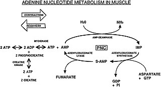
The Purine Nucleotide Cycle is a metabolic pathway in protein metabolism requiring the amino acids aspartate and glutamate. The cycle is used to regulate the levels of adenine nucleotides, in which ammonia and fumarate are generated. AMP converts into IMP and the byproduct ammonia. IMP converts to S-AMP (adenylosuccinate), which then converts to AMP and the byproduct fumarate. The fumarate goes on to produce ATP (energy) via oxidative phosphorylation as it enters the Krebs cycle and then the electron transport chain. Lowenstein first described this pathway and outlined its importance in processes including amino acid catabolism and regulation of flux through glycolysis and the Krebs cycle.

Carbamoyl phosphate synthetase III is one of the three isoforms of the carbamoyl phosphate synthetase, an enzyme that catalyzes the active production of carbamoyl phosphate in many organisms.

<span class="mw-page-title-main">JHU-083</span> Experimental Drug

JHU-083 is an experimental drug which acts as a glutaminase inhibitor. It is a prodrug which is cleaved in vivo to the active form 6-diazo-5-oxo-L-norleucine. It has been researched for the treatment of various neurological conditions such as depression, Alzheimer's disease, and cerebral malaria, as well as multiple sclerosis, atherosclerosis, hepatitis, and some forms of cancer in which it was found to target senescent cells.

References

  1. 1 2 PDB: 3A56 ; Hashizume R, Mizutani K, Takahashi N, Matsubara H, Matsunaga A, Yamaguchi S, Mikami B (2010). "Crystal structure of protein-glutaminase". doi:10.2210/pdb3a56/pdb.{{cite journal}}: Cite journal requires |journal= (help)
  2. 1 2 Botman D, Tigchelaar W, Van Noorden CJ (November 2014). "Determination of phosphate-activated glutaminase activity and its kinetics in mouse tissues using metabolic mapping (quantitative enzyme histochemistry)". The Journal of Histochemistry and Cytochemistry. 62 (11): 813–26. doi:10.1369/0022155414551177. PMC   4230542 . PMID   25163927.
  3. Olalla L, Gutiérrez A, Campos JA, Khan ZU, Alonso FJ, Segura JA, et al. (October 2002). "Nuclear localization of L-type glutaminase in mammalian brain". The Journal of Biological Chemistry. 277 (41): 38939–44. doi: 10.1074/jbc.C200373200 . PMID   12163477.
  4. Masola B, Ngubane NP (December 2010). "The activity of phosphate-dependent glutaminase from the rat small intestine is modulated by ADP and is dependent on integrity of mitochondria". Archives of Biochemistry and Biophysics. 504 (2): 197–203. doi:10.1016/j.abb.2010.09.002. PMID   20831857.
  5. Suzuki S, Tanaka T, Poyurovsky MV, Nagano H, Mayama T, Ohkubo S, et al. (April 2010). "Phosphate-activated glutaminase (GLS2), a p53-inducible regulator of glutamine metabolism and reactive oxygen species". Proceedings of the National Academy of Sciences of the United States of America. 107 (16): 7461–6. Bibcode:2010PNAS..107.7461S. doi: 10.1073/pnas.1002459107 . PMC   2867754 . PMID   20351271.
  6. DeBerardinis RJ, Cheng T. Q's next: the diverse functions of glutamine in metabolism, cell biology and cancer. Oncogene. 2010 Jan 21;29(3):313-24. doi: 10.1038/onc.2009.358. Epub 2009 Nov 2. PMID 19881548; PMCID: PMC2809806.
  7. Chen L, Cui H (September 2015). "Targeting Glutamine Induces Apoptosis: A Cancer Therapy Approach". International Journal of Molecular Sciences. 16 (9): 22830–55. doi: 10.3390/ijms160922830 . PMC   4613338 . PMID   26402672.
  8. Sheikh TN, Patwardhan PP, Cremers S, Schwartz GK (November 2017). "Targeted inhibition of glutaminase as a potential new approach for the treatment of NF1 associated soft tissue malignancies". Oncotarget. 8 (55): 94054–94068. doi:10.18632/oncotarget.21573. PMC   5706855 . PMID   29212209.
  9. Yamashita AS, da Costa Rosa M, Stumpo V, Rais R, Slusher BS, Riggins GJ. The glutamine antagonist prodrug JHU-083 slows malignant glioma growth and disrupts mTOR signaling. Neurooncol Adv. 2020 Oct 29;3(1):vdaa149. doi : 10.1093/noajnl/vdaa149 PMID   33681764
  10. Johmura Y, Yamanaka T, Omori S, Wang TW, Sugiura Y, Matsumoto M, et al. (January 2021). "Senolysis by glutaminolysis inhibition ameliorates various age-associated disorders". Science. 371 (6526): 265–270. Bibcode:2021Sci...371..265J. doi:10.1126/science.abb5916. PMID   33446552. S2CID   231606800.