Glutathione peroxidase

Last updated
Glutathione peroxidase
GlutPeroxidase-1GP1.png
Crystallographic structure of bovine glutathione peroxidase 1. [1]
Identifiers
EC no. 1.11.1.9
CAS no. 9013-66-5
Databases
IntEnz IntEnz view
BRENDA BRENDA entry
ExPASy NiceZyme view
KEGG KEGG entry
MetaCyc metabolic pathway
PRIAM profile
PDB structures RCSB PDB PDBe PDBsum
Gene Ontology AmiGO / QuickGO
Search
PMC articles
PubMed articles
NCBI proteins
Glutathione peroxidase
Identifiers
SymbolGSHPx
Pfam PF00255
InterPro IPR000889
PROSITE PDOC00396
SCOP2 1gp1 / SCOPe / SUPFAM
Available protein structures:
Pfam   structures / ECOD  
PDB RCSB PDB; PDBe; PDBj
PDBsum structure summary
PDB 2f8a B:14-128 1gp1 A:19-133

Glutathione peroxidase (GPx) (EC 1.11.1.9) is the general name of an enzyme family with peroxidase activity whose main biological role is to protect the organism from oxidative damage. [2] The biochemical function of glutathione peroxidase is to reduce lipid hydroperoxides to their corresponding alcohols and to reduce free hydrogen peroxide to water. [3]

Contents

Isozymes

Several isozymes are encoded by different genes, which vary in cellular location and substrate specificity. Glutathione peroxidase 1 (GPx1) is the most abundant version, found in the cytoplasm of nearly all mammalian tissues, whose preferred substrate is hydrogen peroxide. Glutathione peroxidase 4 (GPx4) has a high preference for lipid hydroperoxides; it is expressed in nearly every mammalian cell, though at much lower levels. Glutathione peroxidase 2 is an intestinal and extracellular enzyme, while glutathione peroxidase 3 is extracellular, especially abundant in plasma. [4] So far, eight different isoforms of glutathione peroxidase (GPx1-8) have been identified in humans.

GeneLocusEnzyme
GPX1 Chr. 3 p21.3glutathione peroxidase 1
GPX2 Chr. 14 q24.1glutathione peroxidase 2 (gastrointestinal)
GPX3 Chr. 5 q23glutathione peroxidase 3 (plasma)
GPX4 Chr. 19 p13.3glutathione peroxidase 4 (phospholipid hydroperoxidase)
GPX5 Chr. 6 p21.32glutathione peroxidase 5 (epididymal androgen-related protein)
GPX6 Chr. 6 p21glutathione peroxidase 6 (olfactory)
GPX7 Chr. 1 p32glutathione peroxidase 7
GPX8 Chr. 5 q11.2glutathione peroxidase 8 (putative)

Reaction

The main reaction that glutathione peroxidase catalyzes is:

2GSH + H2O2 → GS–SG + 2H2O

where GSH represents reduced monomeric glutathione, and GS–SG represents glutathione disulfide. The mechanism involves oxidation of the selenol of a selenocysteine residue by hydrogen peroxide. This process gives the derivative with a selenenic acid (RSeOH) group. The selenenic acid is then converted back to the selenol by a two step process that begins with reaction with GSH to form the GS-SeR and water. A second GSH molecule reduces the GS-SeR intermediate back to the selenol, releasing GS-SG as the by-product. A simplified representation is shown below: [5]

RSeH + H2O2 → RSeOH + H2O
RSeOH + GSH → GS-SeR + H2O
GS-SeR + GSH → GS-SG + RSeH

Glutathione reductase then reduces the oxidized glutathione to complete the cycle:

GS–SG + NADPH + H+ → 2 GSH + NADP+.

Structure

Mammalian GPx1, GPx2, GPx3, and GPx4 have been shown to be selenium-containing enzymes, whereas GPx6 is a selenoprotein in humans with cysteine-containing homologues in rodents. GPx1, GPx2, and GPx3 are homotetrameric proteins, whereas GPx4 has a monomeric structure. As the integrity of the cellular and subcellular membranes depends heavily on glutathione peroxidase, its antioxidative protective system itself depends heavily on the presence of selenium.

Animal models

Mice genetically engineered to lack glutathione peroxidase 1 (Gpx1−/− mice) are grossly phenotypically normal and have normal lifespans, indicating this enzyme is not critical for life. However, Gpx1−/− mice develop cataracts at an early age and exhibit defects in muscle satellite cell proliferation. [4] Gpx1 −/− mice showed up to 16 dB higher auditory brainstem response (ABR) thresholds than control mice. After 110 dB noise exposure for one hour, Gpx1 −/− mice had up to 15 dB greater noise-induced hearing loss compared with control mice. [6] "

Mice with knockouts for GPX3 (GPX3−/−) or GPX2 (GPX2−/−) also develop normally [7] [8]

However, glutathione peroxidase 4 knockout mice die during early embryonic development. [4] Some evidence, though, indicates reduced levels of glutathione peroxidase 4 can increase life expectancy in mice. [9]

The bovine erythrocyte enzyme has a molecular weight of 84 kDa.

Discovery

Glutathione peroxidase was discovered in 1957 by Gordon C. Mills. [10]

Methods for determining glutathione peroxidase activity

Activity of glutathione peroxidase is measured spectrophotometrically using several methods. A direct assay by linking the peroxidase reaction with glutathione reductase with measurement of the conversion of NADPH to NADP is widely used. [11] The other approach is measuring residual GSH in the reaction with Ellman's reagent. Based on this, several procedures for measuring glutathione peroxidase activity were developed using various hydroperoxides as substrates for reduction, e.g. cumene hydroperoxide, [12] tert-butyl hydroperoxide [13] and hydrogen peroxide. [14]

The other methods include the use of CUPRAC reagent with spectrophotometric detection of the reaction product [15] or o-phtalaldehyde as a fluorescent reagent. [16]

Clinical significance

It has been shown that low levels of glutathione peroxidase as measured in the serum may be a contributing factor to vitiligo. [17] Lower plasma glutathione peroxide levels were also observed in patients with type 2 diabetes with macroalbuminuria and this was correlated to the stage of diabetic nephropathy.[ citation needed ] In one study, the activity of glutathione peroxidase along with other antioxidant enzymes such as superoxide dismutase and catalase was not associated with coronary heart disease risk in women. [18] Glutathione peroxidase activity was found to be much lower in patients with relapsing-remitting multiple sclerosis. [19] One study has suggested that glutathione peroxidase and superoxide dismutase polymorphisms play a role in the development of celiac disease. [20]

The activity of this enzyme has been reported to be decreased in case of copper deficiency in the liver and plasma. [21]

See also

Related Research Articles

Antioxidants are compounds that inhibit oxidation, a chemical reaction that can produce free radicals. Autoxidation leads to degradation of organic compounds, including living matter. Antioxidants are frequently added to industrial products, such as polymers, fuels, and lubricants, to extend their usable lifetimes. Food are also treated with antioxidants to forestall spoilage, in particular the rancidification of oils and fats. In cells, antioxidants such as glutathione, mycothiol or bacillithiol, and enzyme systems like superoxide dismutase, can prevent damage from oxidative stress.

<span class="mw-page-title-main">Superoxide dismutase</span> Class of enzymes

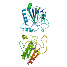
Superoxide dismutase (SOD, EC 1.15.1.1) is an enzyme that alternately catalyzes the dismutation (or partitioning) of the superoxide (O
2
) radical into ordinary molecular oxygen (O2) and hydrogen peroxide (H
2
O
2
). Superoxide is produced as a by-product of oxygen metabolism and, if not regulated, causes many types of cell damage. Hydrogen peroxide is also damaging and is degraded by other enzymes such as catalase. Thus, SOD is an important antioxidant defense in nearly all living cells exposed to oxygen. One exception is Lactobacillus plantarum and related lactobacilli, which use a different mechanism to prevent damage from reactive O
2
.

<span class="mw-page-title-main">Catalase</span> Biocatalyst decomposing hydrogen peroxide

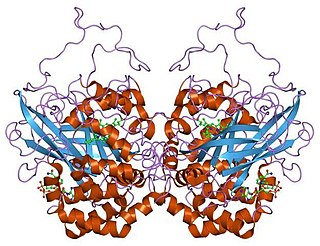
Catalase is a common enzyme found in nearly all living organisms exposed to oxygen which catalyzes the decomposition of hydrogen peroxide to water and oxygen. It is a very important enzyme in protecting the cell from oxidative damage by reactive oxygen species (ROS). Catalase has one of the highest turnover numbers of all enzymes; one catalase molecule can convert millions of hydrogen peroxide molecules to water and oxygen each second.

<span class="mw-page-title-main">Peroxidase</span> Peroxide-decomposing enzyme

Peroxidases or peroxide reductases are a large group of enzymes which play a role in various biological processes. They are named after the fact that they commonly break up peroxides.

<span class="mw-page-title-main">Reactive oxygen species</span> Highly reactive molecules formed from diatomic oxygen (O₂)

In chemistry, reactive oxygen species (ROS) are highly reactive chemicals formed from diatomic oxygen. Examples of ROS include peroxides, superoxide, hydroxyl radical, singlet oxygen, and alpha-oxygen.

<span class="mw-page-title-main">Lipid peroxidation</span> Reaction(s) leading to production of (phospho)lipid peroxides

Lipid peroxidation is the chain of reactions of oxidative degradation of lipids. It is the process in which free radicals "steal" electrons from the lipids in cell membranes, resulting in cell damage. This process proceeds by a free radical chain reaction mechanism. It most often affects polyunsaturated fatty acids, because they contain multiple double bonds in between which lie methylene bridges (-CH2-) that possess especially reactive hydrogen atoms. As with any radical reaction, the reaction consists of three major steps: initiation, propagation, and termination. The chemical products of this oxidation are known as lipid peroxides or lipid oxidation products (LOPs).

Organoselenium chemistry is the science exploring the properties and reactivity of organoselenium compounds, chemical compounds containing carbon-to-selenium chemical bonds. Selenium belongs with oxygen and sulfur to the group 16 elements or chalcogens, and similarities in chemistry are to be expected. Organoselenium compounds are found at trace levels in ambient waters, soils and sediments.

<span class="mw-page-title-main">Glutathione reductase</span> Enzyme

Glutathione reductase (GR) also known as glutathione-disulfide reductase (GSR) is an enzyme that in humans is encoded by the GSR gene. Glutathione reductase catalyzes the reduction of glutathione disulfide (GSSG) to the sulfhydryl form glutathione (GSH), which is a critical molecule in resisting oxidative stress and maintaining the reducing environment of the cell. Glutathione reductase functions as dimeric disulfide oxidoreductase and utilizes an FAD prosthetic group and NADPH to reduce one molar equivalent of GSSG to two molar equivalents of GSH:

<span class="mw-page-title-main">Selenol</span> Class of chemical compounds

Selenols are organic compounds that contain the functional group with the connectivity C–Se–H. Selenols are sometimes also called selenomercaptans and selenothiols. Selenols are one of the principal classes of organoselenium compounds. A well-known selenol is the amino acid selenocysteine.

<span class="mw-page-title-main">Peroxiredoxin</span> Family of antioxidant enzymes

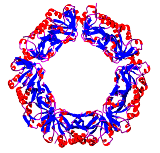
Peroxiredoxins are a ubiquitous family of antioxidant enzymes that also control cytokine-induced peroxide levels and thereby mediate signal transduction in mammalian cells. The family members in humans are PRDX1, PRDX2, PRDX3, PRDX4, PRDX5, and PRDX6. The physiological importance of peroxiredoxins is indicated by their relative abundance. Their function is the reduction of peroxides, specifically hydrogen peroxide, alkyl hydroperoxides, and peroxynitrite.

<span class="mw-page-title-main">GPX1</span> Protein-coding gene in the species Homo sapiens

Glutathione peroxidase 1, also known as GPx1, is an enzyme that in humans is encoded by the GPX1 gene on chromosome 3. This gene encodes a member of the glutathione peroxidase family. Glutathione peroxidase functions in the detoxification of hydrogen peroxide, and is one of the most important antioxidant enzymes in humans.

<span class="mw-page-title-main">NADH peroxidase</span>

In enzymology, a NADH peroxidase (EC 1.11.1.1) is an enzyme that catalyzes the chemical reaction

In enzymology, a phospholipid-hydroperoxide glutathione peroxidase (EC 1.11.1.12) is an enzyme that catalyzes the chemical reaction

<span class="mw-page-title-main">GPX4</span> Mammalian protein found in Homo sapiens

Glutathione peroxidase 4, also known as GPX4, is an enzyme that in humans is encoded by the GPX4 gene. GPX4 is a phospholipid hydroperoxidase that protects cells against membrane lipid peroxidation.

<span class="mw-page-title-main">GPX2 (gene)</span> Protein-coding gene in the species Homo sapiens

Glutathione peroxidase 2 is an enzyme that in humans is encoded by the GPX2 gene.

<span class="mw-page-title-main">GSTZ1</span> Protein-coding gene in the species Homo sapiens

Glutathione S-transferase Zeta 1 is an enzyme that in humans is encoded by the GSTZ1 gene on chromosome 14.

<span class="mw-page-title-main">GPX3</span> Enzyme in humans

Glutathione peroxidase 3 (GPx-3), also known as plasma glutathione peroxidase (GPx-P) or extracellular glutathione peroxidase is an enzyme that in humans is encoded by the GPX3 gene.

<span class="mw-page-title-main">Selenenic acid</span> Class of chemical compounds

A selenenic acid is an organoselenium compound and an oxoacid with the general formula RSeOH, where R ≠ H. It is the first member of the family of organoselenium oxoacids, which also include seleninic acids and selenonic acids, which are RSeO2H and RSeO3H, respectively. Selenenic acids derived from selenoenzymes are thought to be responsible for the antioxidant activity of these enzymes. This functional group is sometimes called SeO-selenoperoxol.

Oxidation response is stimulated by a disturbance in the balance between the production of reactive oxygen species and antioxidant responses, known as oxidative stress. Active species of oxygen naturally occur in aerobic cells and have both intracellular and extracellular sources. These species, if not controlled, damage all components of the cell, including proteins, lipids and DNA. Hence cells need to maintain a strong defense against the damage. The following table gives an idea of the antioxidant defense system in bacterial system.

<span class="mw-page-title-main">Superoxide dismutase mimetics</span> Synthetic compounds

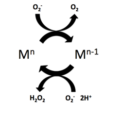
Superoxide dismutase (SOD) mimetics are synthetic compounds that mimic the native superoxide dismutase enzyme. SOD mimetics effectively convert the superoxide anion, a reactive oxygen species, into hydrogen peroxide, which is further converted into water by catalase. Reactive oxygen species are natural byproducts of cellular respiration and cause oxidative stress and cell damage, which has been linked to causing cancers, neurodegeneration, age-related declines in health, and inflammatory diseases. SOD mimetics are a prime interest in therapeutic treatment of oxidative stress because of their smaller size, longer half-life, and similarity in function to the native enzyme.

References

  1. PDB: 1GP1 ; Epp O, Ladenstein R, Wendel A (June 1983). "The refined structure of the selenoenzyme glutathione peroxidase at 0.2-nm resolution". European Journal of Biochemistry. 133 (1): 51–69. doi:10.1111/j.1432-1033.1983.tb07429.x. PMID   6852035.
  2. Muthukumar K, Nachiappan V (December 2010). "Cadmium-induced oxidative stress in Saccharomyces cerevisiae". Indian Journal of Biochemistry & Biophysics. 47 (6): 383–7. PMID   21355423.
  3. Muthukumar K, Rajakumar S, Sarkar MN, Nachiappan V (May 2011). "Glutathione peroxidase3 of Saccharomyces cerevisiae protects phospholipids during cadmium-induced oxidative stress". Antonie van Leeuwenhoek. 99 (4): 761–71. doi:10.1007/s10482-011-9550-9. PMID   21229313. S2CID   21850794.
  4. 1 2 3 Muller FL, Lustgarten MS, Jang Y, Richardson A, Van Remmen H (August 2007). "Trends in oxidative aging theories". Free Radical Biology & Medicine. 43 (4): 477–503. doi:10.1016/j.freeradbiomed.2007.03.034. PMID   17640558.
  5. Bhabak KP, Mugesh G (November 2010). "Functional mimics of glutathione peroxidase: bioinspired synthetic antioxidants". Accounts of Chemical Research. 43 (11): 1408–19. doi:10.1021/ar100059g. PMID   20690615.
  6. Ohlemiller KK, McFadden SL, Ding DL, Lear PM, Ho YS (November 2000). "Targeted mutation of the gene for cellular glutathione peroxidase (Gpx1) increases noise-induced hearing loss in mice". Journal of the Association for Research in Otolaryngology. 1 (3): 243–54. doi:10.1007/s101620010043. PMC   2504546 . PMID   11545230.
  7. Esworthy RS, Aranda R, Martín MG, Doroshow JH, Binder SW, Chu FF (September 2001). "Mice with combined disruption of Gpx1 and Gpx2 genes have colitis". American Journal of Physiology. Gastrointestinal and Liver Physiology. 281 (3): G848-55. doi:10.1152/ajpgi.2001.281.3.G848. PMID   11518697. S2CID   21615743.
  8. Olson GE, Whitin JC, Hill KE, Winfrey VP, Motley AK, Austin LM, et al. (May 2010). "Extracellular glutathione peroxidase (Gpx3) binds specifically to basement membranes of mouse renal cortex tubule cells". American Journal of Physiology. Renal Physiology. 298 (5): F1244-53. doi:10.1152/ajprenal.00662.2009. PMC   2867408 . PMID   20015939.
  9. Ran Q, Liang H, Ikeno Y, Qi W, Prolla TA, Roberts LJ, et al. (September 2007). "Reduction in glutathione peroxidase 4 increases life span through increased sensitivity to apoptosis". The Journals of Gerontology. Series A, Biological Sciences and Medical Sciences. 62 (9): 932–42. doi: 10.1093/gerona/62.9.932 . PMID   17895430.
  10. Mills GC (November 1957). "Hemoglobin catabolism. I. Glutathione peroxidase, an erythrocyte enzyme which protects hemoglobin from oxidative breakdown". The Journal of Biological Chemistry. 229 (1): 189–97. doi: 10.1016/S0021-9258(18)70608-X . PMID   13491573.
  11. Paglia DE, Valentine WN (July 1967). "Studies on the quantitative and qualitative characterization of erythrocyte glutathione peroxidase". The Journal of Laboratory and Clinical Medicine. 70 (1): 158–69. PMID   6066618.
  12. Zakowski JJ, Tappel AL (September 1978). "A semiautomated system for measurement of glutathione in the assay of glutathione peroxidase". Analytical Biochemistry. 89 (2): 430–6. doi:10.1016/0003-2697(78)90372-X. PMID   727443.
  13. Moin VM (1986). "[A simple and specific method for determining glutathione peroxidase activity in erythrocytes]". Laboratornoe Delo. 12 (12): 724–7. PMID   2434712.
  14. Razygraev AV, Yushina AD, Titovich IA (August 2018). "Correction to: A Method of Measuring Glutathione Peroxidase Activity in Murine Brain: Application in Pharmacological Experiment". Bulletin of Experimental Biology and Medicine. 165 (4): 589–592. doi:10.1007/s10517-018-4219-2. PMID   30121905. S2CID   52038817.
  15. Ahmed, A. Y.; Aowda, S. A.; Hadwan, M. H. (2021). "A validated method to assess glutathione peroxidase enzyme activity". Chemical Papers. 75 (12): 6625–6637. doi:10.1007/s11696-021-01826-1. ISSN   2585-7290. S2CID   236219189.
  16. Ramos Martinez, J. I.; Launay, J.-M.; Dreux, C. (1979). "A sensitive fluorimetric microassay for the determination of glutathione peroxidase activity. Application to human blood platelets". Analytical Biochemistry. 98 (1): 154–159. doi:10.1016/0003-2697(79)90720-6. ISSN   0003-2697.
  17. Zedan H, Abdel-Motaleb AA, Kassem NM, Hafeez HA, Hussein MR (Mar 2015). "Low glutathione peroxidase activity levels in patients with vitiligo". Journal of Cutaneous Medicine and Surgery. 19 (2): 144–8. doi:10.2310/7750.2014.14076. PMID   25775636. S2CID   32708904.
  18. Yang S, Jensen MK, Rimm EB, Willett W, Wu T (November 2014). "Erythrocyte superoxide dismutase, glutathione peroxidase, and catalase activities and risk of coronary heart disease in generally healthy women: a prospective study". American Journal of Epidemiology. 180 (9): 901–8. doi:10.1093/aje/kwu195. PMC   4207716 . PMID   25156995.
  19. Socha K, Kochanowicz J, Karpińska E, Soroczyńska J, Jakoniuk M, Mariak Z, Borawska MH (June 2014). "Dietary habits and selenium, glutathione peroxidase and total antioxidant status in the serum of patients with relapsing-remitting multiple sclerosis". Nutrition Journal. 13: 62. doi:10.1186/1475-2891-13-62. PMC   4080729 . PMID   24943732.
  20. Katar M, Ozugurlu AF, Ozyurt H, Benli I (February 2014). "Evaluation of glutathione peroxidase and superoxide dismutase enzyme polymorphisms in celiac disease patients". Genetics and Molecular Research. 13 (1): 1030–7. doi: 10.4238/2014.February.20.4 . PMID   24634124.
  21. Hordyjewska, Anna; Popiołek, Łukasz; Kocot, Joanna (2014). "The many 'faces' of copper in medicine and treatment". Biometals. 27 (4): 611–621. doi:10.1007/s10534-014-9736-5. PMC   4113679 . PMID   24748564.