Large-scale brain network

Last updated

Large-scale brain networks (also known as intrinsic brain networks) are collections of widespread brain regions showing functional connectivity by statistical analysis of the fMRI BOLD signal [1] or other recording methods such as EEG, [2] PET [3] and MEG. [4] An emerging paradigm in neuroscience is that cognitive tasks are performed not by individual brain regions working in isolation but by networks consisting of several discrete brain regions that are said to be "functionally connected". Functional connectivity networks may be found using algorithms such as cluster analysis, spatial independent component analysis (ICA), seed based, and others. [5] Synchronized brain regions may also be identified using long-range synchronization of the EEG, MEG, or other dynamic brain signals. [6]

Contents

The set of identified brain areas that are linked together in a large-scale network varies with cognitive function. [7] When the cognitive state is not explicit (i.e., the subject is at "rest"), the large-scale brain network is a resting state network (RSN). As a physical system with graph-like properties, [6] a large-scale brain network has both nodes and edges and cannot be identified simply by the co-activation of brain areas. In recent decades, the analysis of brain networks was made feasible by advances in imaging techniques as well as new tools from graph theory and dynamical systems.

Anatomical topographies of canonical large-scale networks Boerger2023LSBN.jpg
Anatomical topographies of canonical large-scale networks

The Organization for Human Brain Mapping has created the Workgroup for HArmonized Taxonomy of NETworks (WHATNET) group to work towards a consensus regarding network nomenclature. [8] WHATNET conducted a survey in 2021 which showed a large degree of agreement about the name and topography of three networks: the "somato network", the "default network" and the "visual network". Other networks had less agreement. Several issues make the work of creating a common atlas for networks difficult. Some of those issues are the variability of spatial and time scales, variability across individuals, and the dynamic nature of some networks. [9]

Some large-scale brain networks are identified by their function and provide a coherent framework for understanding cognition by offering a neural model of how different cognitive functions emerge when different sets of brain regions join together as self-organized coalitions. The number and composition of the coalitions will vary with the algorithm and parameters used to identify them. [10] [11] In one model, there is only the default mode network and the task-positive network, but most current analyses show several networks, from a small handful to 17. [10] The most common and stable networks are enumerated below. The regions participating in a functional network may be dynamically reconfigured. [5] [12]

Disruptions in activity in various networks have been implicated in neuropsychiatric disorders such as depression, Alzheimer's, autism spectrum disorder, schizophrenia, ADHD [13] and bipolar disorder. [14]

Commonly identified networks

An example that identified 10 large-scale brain networks from resting state fMRI activity through independent component analysis Heine2012x3010.png
An example that identified 10 large-scale brain networks from resting state fMRI activity through independent component analysis

Because brain networks can be identified at various different resolutions and with various different neurobiological properties, there is currently no universal atlas of brain networks that fits all circumstances. [16] Uddin, Yeo, and Spreng proposed in 2019 [17] that the following six networks should be defined as core networks based on converging evidences from multiple studies [18] [10] [19] to facilitate communication between researchers.

Default mode (medial frontoparietal)

Salience (midcingulo-insular)

Attention (dorsal frontoparietal)

Control (lateral frontoparietal)

Sensorimotor or somatomotor (pericentral)

Visual (occipital)

Other networks

Different methods and data have identified several other brain networks, many of which greatly overlap or are subsets of more well-characterized core networks. [17]

See also

Related Research Articles

<span class="mw-page-title-main">Visual cortex</span> Region of the brain that processes visual information

The visual cortex of the brain is the area of the cerebral cortex that processes visual information. It is located in the occipital lobe. Sensory input originating from the eyes travels through the lateral geniculate nucleus in the thalamus and then reaches the visual cortex. The area of the visual cortex that receives the sensory input from the lateral geniculate nucleus is the primary visual cortex, also known as visual area 1 (V1), Brodmann area 17, or the striate cortex. The extrastriate areas consist of visual areas 2, 3, 4, and 5.

<span class="mw-page-title-main">Claustrum</span> Structure in the brain

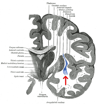
The claustrum is a thin sheet of neurons and supporting glial cells, that connects to the cerebral cortex and subcortical regions including the amygdala, hippocampus and thalamus of the brain. It is located between the insular cortex laterally and the putamen medially, encased by the extreme and external capsules respectively. Blood to the claustrum is supplied by the middle cerebral artery. It is considered to be the most densely connected structure in the brain, and thus hypothesized to allow for the integration of various cortical inputs such as vision, sound and touch, into one experience. Other hypotheses suggest that the claustrum plays a role in salience processing, to direct attention towards the most behaviorally relevant stimuli amongst the background noise. The claustrum is difficult to study given the limited number of individuals with claustral lesions and the poor resolution of neuroimaging.

<span class="mw-page-title-main">Insular cortex</span> Portion of the mammalian cerebral cortex

The insular cortex is a portion of the cerebral cortex folded deep within the lateral sulcus within each hemisphere of the mammalian brain.

<span class="mw-page-title-main">Orbitofrontal cortex</span> Region of the prefrontal cortex of the brain

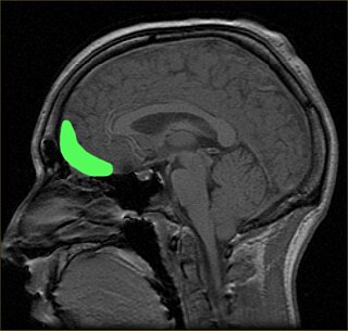
The orbitofrontal cortex (OFC) is a prefrontal cortex region in the frontal lobes of the brain which is involved in the cognitive process of decision-making. In non-human primates it consists of the association cortex areas Brodmann area 11, 12 and 13; in humans it consists of Brodmann area 10, 11 and 47.

Salience is the property by which some thing stands out. Salient events are an attentional mechanism by which organisms learn and survive; those organisms can focus their limited perceptual and cognitive resources on the pertinent subset of the sensory data available to them.

<span class="mw-page-title-main">Posterior cingulate cortex</span> Caudal part of the cingulate cortex of the brain

The posterior cingulate cortex (PCC) is the caudal part of the cingulate cortex, located posterior to the anterior cingulate cortex. This is the upper part of the "limbic lobe". The cingulate cortex is made up of an area around the midline of the brain. Surrounding areas include the retrosplenial cortex and the precuneus.

<span class="mw-page-title-main">Intraparietal sulcus</span> Sulcus on the lateral surface of the parietal lobe

The intraparietal sulcus (IPS) is located on the lateral surface of the parietal lobe, and consists of an oblique and a horizontal portion. The IPS contains a series of functionally distinct subregions that have been intensively investigated using both single cell neurophysiology in primates and human functional neuroimaging. Its principal functions are related to perceptual-motor coordination and visual attention, which allows for visually-guided pointing, grasping, and object manipulation that can produce a desired effect.

<span class="mw-page-title-main">Dorsal attention network</span> Large-scale brain network involved in voluntary orienting of attention

The dorsal attention network (DAN), also known anatomically as the dorsal frontoparietal network (D-FPN), is a large-scale brain network of the human brain that is primarily composed of the intraparietal sulcus (IPS) and frontal eye fields (FEF). It is named and most known for its role in voluntary orienting of visuospatial attention.

<span class="mw-page-title-main">Connectome</span> Comprehensive map of neural connections in the brain

A connectome is a comprehensive map of neural connections in the brain, and may be thought of as its "wiring diagram". An organism's nervous system is made up of neurons which communicate through synapses. A connectome is constructed by tracing the neuron in a nervous system and mapping where neurons are connected through synapses.

Connectomics is the production and study of connectomes: comprehensive maps of connections within an organism's nervous system. More generally, it can be thought of as the study of neuronal wiring diagrams with a focus on how structural connectivity, individual synapses, cellular morphology, and cellular ultrastructure contribute to the make up of a network. The nervous system is a network made of billions of connections and these connections are responsible for our thoughts, emotions, actions, memories, function and dysfunction. Therefore, the study of connectomics aims to advance our understanding of mental health and cognition by understanding how cells in the nervous system are connected and communicate. Because these structures are extremely complex, methods within this field use a high-throughput application of functional and structural neural imaging, most commonly magnetic resonance imaging (MRI), electron microscopy, and histological techniques in order to increase the speed, efficiency, and resolution of these nervous system maps. To date, tens of large scale datasets have been collected spanning the nervous system including the various areas of cortex, cerebellum, the retina, the peripheral nervous system and neuromuscular junctions.

<span class="mw-page-title-main">Default mode network</span> Large-scale brain network active when not focusing on an external task

In neuroscience, the default mode network (DMN), also known as the default network, default state network, or anatomically the medial frontoparietal network (M-FPN), is a large-scale brain network primarily composed of the dorsal medial prefrontal cortex, posterior cingulate cortex, precuneus and angular gyrus. It is best known for being active when a person is not focused on the outside world and the brain is at wakeful rest, such as during daydreaming and mind-wandering. It can also be active during detailed thoughts related to external task performance. Other times that the DMN is active include when the individual is thinking about others, thinking about themselves, remembering the past, and planning for the future.

<span class="mw-page-title-main">Resting state fMRI</span> Type of functional magnetic resonance imaging

Resting state fMRI is a method of functional magnetic resonance imaging (fMRI) that is used in brain mapping to evaluate regional interactions that occur in a resting or task-negative state, when an explicit task is not being performed. A number of resting-state brain networks have been identified, one of which is the default mode network. These brain networks are observed through changes in blood flow in the brain which creates what is referred to as a blood-oxygen-level dependent (BOLD) signal that can be measured using fMRI.

The biological basis of personality is a collection of brain systems and mechanisms that underlie human personality. Human neurobiology, especially as it relates to complex traits and behaviors, is not well understood, but research into the neuroanatomical and functional underpinnings of personality are an active field of research. Animal models of behavior, molecular biology, and brain imaging techniques have provided some insight into human personality, especially trait theories.

Dynamic functional connectivity (DFC) refers to the observed phenomenon that functional connectivity changes over a short time. Dynamic functional connectivity is a recent expansion on traditional functional connectivity analysis which typically assumes that functional networks are static in time. DFC is related to a variety of different neurological disorders, and has been suggested to be a more accurate representation of functional brain networks. The primary tool for analyzing DFC is fMRI, but DFC has also been observed with several other mediums. DFC is a recent development within the field of functional neuroimaging whose discovery was motivated by the observation of temporal variability in the rising field of steady state connectivity research.

<span class="mw-page-title-main">Salience network</span> Large-scale brain network involved in detecting and attending to relevant stimuli

The salience network (SN), also known anatomically as the midcingulo-insular network (M-CIN) or ventral attention network, is a large scale network of the human brain that is primarily composed of the anterior insula (AI) and dorsal anterior cingulate cortex (dACC). It is involved in detecting and filtering salient stimuli, as well as in recruiting relevant functional networks. Together with its interconnected brain networks, the SN contributes to a variety of complex functions, including communication, social behavior, and self-awareness through the integration of sensory, emotional, and cognitive information.

The sensorimotor network (SMN), also known as somatomotor network, is a large-scale brain network that primarily includes somatosensory and motor regions and extends to the supplementary motor areas (SMA). The auditory cortex may also be included. The SMN is activated during motor tasks, such as finger tapping, indicating that the network readies the brain when performing and coordinating motor tasks.

Social cognitive neuroscience is the scientific study of the biological processes underpinning social cognition. Specifically, it uses the tools of neuroscience to study "the mental mechanisms that create, frame, regulate, and respond to our experience of the social world". Social cognitive neuroscience uses the epistemological foundations of cognitive neuroscience, and is closely related to social neuroscience. Social cognitive neuroscience employs human neuroimaging, typically using functional magnetic resonance imaging (fMRI). Human brain stimulation techniques such as transcranial magnetic stimulation and transcranial direct-current stimulation are also used. In nonhuman animals, direct electrophysiological recordings and electrical stimulation of single cells and neuronal populations are utilized for investigating lower-level social cognitive processes.

Network neuroscience is an approach to understanding the structure and function of the human brain through an approach of network science, through the paradigm of graph theory. A network is a connection of many brain regions that interact with each other to give rise to a particular function. Network Neuroscience is a broad field that studies the brain in an integrative way by recording, analyzing, and mapping the brain in various ways. The field studies the brain at multiple scales of analysis to ultimately explain brain systems, behavior, and dysfunction of behavior in psychiatric and neurological diseases. Network neuroscience provides an important theoretical base for understanding neurobiological systems at multiple scales of analysis.

<span class="mw-page-title-main">Frontoparietal network</span> Large-scale brain network involved in sustained attention and complex cognition

The frontoparietal network (FPN), generally also known as the central executive network (CEN) or, more specifically, the lateral frontoparietal network (L-FPN), is a large-scale brain network primarily composed of the dorsolateral prefrontal cortex and posterior parietal cortex, around the intraparietal sulcus. It is involved in sustained attention, complex problem-solving and working memory.

Lucina Q. Uddin is an American cognitive neuroscientist who is a professor at the University of California, Los Angeles. Her research investigates the relationship between brain connectivity and cognition in typical and atypical development using network neuroscience approaches.

References

  1. 1 2 3 4 5 Riedl, Valentin; Utz, Lukas; Castrillón, Gabriel; Grimmer, Timo; Rauschecker, Josef P.; Ploner, Markus; Friston, Karl J.; Drzezga, Alexander; Sorg, Christian (January 12, 2016). "Metabolic connectivity mapping reveals effective connectivity in the resting human brain". PNAS. 113 (2): 428–433. Bibcode:2016PNAS..113..428R. doi: 10.1073/pnas.1513752113 . PMC   4720331 . PMID   26712010.
  2. Foster, Brett L.; Parvizi, Josef (2012-03-01). "Resting oscillations and cross-frequency coupling in the human posteromedial cortex". NeuroImage. 60 (1): 384–391. doi:10.1016/j.neuroimage.2011.12.019. ISSN   1053-8119. PMC   3596417 . PMID   22227048.
  3. Buckner, Randy L.; Andrews-Hanna, Jessica R.; Schacter, Daniel L. (2008). "The Brain's Default Network". Annals of the New York Academy of Sciences. 1124 (1): 1–38. Bibcode:2008NYASA1124....1B. doi:10.1196/annals.1440.011. ISSN   1749-6632. PMID   18400922. S2CID   3167595.
  4. Morris, Peter G.; Smith, Stephen M.; Barnes, Gareth R.; Stephenson, Mary C.; Hale, Joanne R.; Price, Darren; Luckhoo, Henry; Woolrich, Mark; Brookes, Matthew J. (2011-10-04). "Investigating the electrophysiological basis of resting state networks using magnetoencephalography". Proceedings of the National Academy of Sciences. 108 (40): 16783–16788. Bibcode:2011PNAS..10816783B. doi: 10.1073/pnas.1112685108 . ISSN   0027-8424. PMC   3189080 . PMID   21930901.
  5. 1 2 Petersen, Steven; Sporns, Olaf (October 2015). "Brain Networks and Cognitive Architectures". Neuron. 88 (1): 207–219. doi:10.1016/j.neuron.2015.09.027. PMC   4598639 . PMID   26447582.
  6. 1 2 3 4 5 6 Bressler, Steven L.; Menon, Vinod (June 2010). "Large scale brain networks in cognition: emerging methods and principles". Trends in Cognitive Sciences. 14 (6): 233–290. doi:10.1016/j.tics.2010.04.004. PMID   20493761. S2CID   5967761 . Retrieved 24 January 2016.
  7. Bressler, Steven L. (2008). "Neurocognitive networks". Scholarpedia. 3 (2): 1567. Bibcode:2008SchpJ...3.1567B. doi: 10.4249/scholarpedia.1567 .
  8. Uddin, Lucina (2022-10-10). "A Brain Network by Any Other Name". Journal of Cognitive Neuroscience. 2022 (10): 363–364. doi:10.1162/jocn_a_01925. PMID   36223250. S2CID   252844955.
  9. Uddin, LQ; Betzel, Richard F.; Cohen, Jessica R.; Damoiselastx, Jessica S.; De Brigard, Felipe; Eickhoff, Simon B.; Fornito, Alex; Gratton, Caterina; Gordon, Evan M.; Laird, Angela R.; Larson-Prior, Linda; McIntosh, A. Randal; Nickerson, Lisa D.; Pessoa, Luiz; Pinho, Ana Luísa; Poldrack, Russell A.; Razi, Adeel; Sadaghiani, Sepideh; Shine, James M.; Yendiki, Anastasia; Yeo, BTT; Spreng, RN (October 2023). "Controversies and progress on standardization of large-scale brain network nomenclature". Network Neuroscience. 7 (3): 864–903. doi:10.1162/netn_a_00323. PMC   10473266 . PMID   37781138.
  10. 1 2 3 4 5 6 7 8 9 10 Yeo, B. T. Thomas; Krienen, Fenna M.; Sepulcre, Jorge; Sabuncu, Mert R.; Lashkari, Danial; Hollinshead, Marisa; Roffman, Joshua L.; Smoller, Jordan W.; Zöllei, Lilla; Polimeni, Jonathan R.; Fischl, Bruce; Liu, Hesheng; Buckner, Randy L. (2011-09-01). "The organization of the human cerebral cortex estimated by intrinsic functional connectivity". Journal of Neurophysiology. 106 (3): 1125–1165. Bibcode:2011NatSD...2E0031H. doi:10.1152/jn.00338.2011. PMC   3174820 . PMID   21653723.
  11. Abou Elseoud, Ahmed; Littow, Harri; Remes, Jukka; Starck, Tuomo; Nikkinen, Juha; Nissilä, Juuso; Timonen, Markku; Tervonen, Osmo; Kiviniemi, Vesa (2011-06-03). "Group-ICA Model Order Highlights Patterns of Functional Brain Connectivity". Frontiers in Systems Neuroscience. 5: 37. doi: 10.3389/fnsys.2011.00037 . PMC   3109774 . PMID   21687724.
  12. 1 2 3 4 5 Bassett, Daniella; Bertolero, Max (July 2019). "How Matter Becomes Mind". Scientific American. 321 (1): 32. Retrieved 23 June 2019.
  13. Griffiths, Kristi R.; Braund, Taylor A.; Kohn, Michael R.; Clarke, Simon; Williams, Leanne M.; Korgaonkar, Mayuresh S. (2 March 2021). "Structural brain network topology underpinning ADHD and response to methylphenidate treatment". Translational Psychiatry. 11 (1): 150. doi:10.1038/s41398-021-01278-x. PMC   7925571 . PMID   33654073.
  14. Menon, Vinod (2011-09-09). "Large-scale brain networks and psychopathology: A unifying triple network model". Trends in Cognitive Sciences. 15 (10): 483–506. doi:10.1016/j.tics.2011.08.003. PMID   21908230. S2CID   26653572.
  15. 1 2 3 4 5 6 7 8 Heine, Lizette; Soddu, Andrea; Gomez, Francisco; Vanhaudenhuyse, Audrey; Tshibanda, Luaba; Thonnard, Marie; Charland-Verville, Vanessa; Kirsch, Murielle; Laureys, Steven; Demertzi, Athena (2012). "Resting state networks and consciousness. Alterations of multiple resting state network connectivity in physiological, pharmacological and pathological consciousness states". Frontiers in Psychology. 3: 295. doi: 10.3389/fpsyg.2012.00295 . PMC   3427917 . PMID   22969735.
  16. Eickhoff, SB; Yeo, BTT; Genon, S (November 2018). "Imaging-based parcellations of the human brain" (PDF). Nature Reviews. Neuroscience. 19 (11): 672–686. doi:10.1038/s41583-018-0071-7. PMID   30305712. S2CID   52954265.
  17. 1 2 3 4 5 Uddin, LQ; Yeo, BTT; Spreng, RN (November 2019). "Towards a Universal Taxonomy of Macro-scale Functional Human Brain Networks". Brain Topography. 32 (6): 926–942. doi:10.1007/s10548-019-00744-6. PMC   7325607 . PMID   31707621.
  18. Doucet, GE; Lee, WH; Frangou, S (2019-10-15). "Evaluation of the spatial variability in the major resting-state networks across human brain functional atlases". Human Brain Mapping. 40 (15): 4577–4587. doi:10.1002/hbm.24722. PMC   6771873 . PMID   31322303.
  19. Smith, SM; Fox, PT; Miller, KL; Glahn, DC; Fox, PM; Mackay, CE; Filippini, N; Watkins, KE; Toro, R; Laird, AR; Beckmann, CF (2009-08-04). "Correspondence of the brain's functional architecture during activation and rest". Proceedings of the National Academy of Sciences of the United States of America. 106 (31): 13040–5. Bibcode:2009PNAS..10613040S. doi: 10.1073/pnas.0905267106 . PMC   2722273 . PMID   19620724.
  20. Buckner, Randy L. (2012-08-15). "The serendipitous discovery of the brain's default network". NeuroImage. 62 (2): 1137–1145. doi:10.1016/j.neuroimage.2011.10.035. ISSN   1053-8119. PMID   22037421. S2CID   9880586.
  21. 1 2 3 4 5 6 7 Yuan, Rui; Di, Xin; Taylor, Paul A.; Gohel, Suril; Tsai, Yuan-Hsiung; Biswal, Bharat B. (30 April 2015). "Functional topography of the thalamocortical system in human". Brain Structure and Function. 221 (4): 1971–1984. doi:10.1007/s00429-015-1018-7. PMC   6363530 . PMID   25924563.
  22. 1 2 3 4 Bell, Peter T.; Shine, James M. (2015-11-09). "Estimating Large-Scale Network Convergence in the Human Functional Connectome". Brain Connectivity. 5 (9): 565–74. doi:10.1089/brain.2015.0348. PMID   26005099.
  23. 1 2 3 4 5 Shafiei, Golia; Zeighami, Yashar; Clark, Crystal A.; Coull, Jennifer T.; Nagano-Saito, Atsuko; Leyton, Marco; Dagher, Alain; Mišić, Bratislav (2018-10-01). "Dopamine Signaling Modulates the Stability and Integration of Intrinsic Brain Networks". Cerebral Cortex. 29 (1): 397–409. doi:10.1093/cercor/bhy264. PMC   6294404 . PMID   30357316.
  24. 1 2 3 4 Bailey, Stephen K.; Aboud, Katherine S.; Nguyen, Tin Q.; Cutting, Laurie E. (13 December 2018). "Applying a network framework to the neurobiology of reading and dyslexia". Journal of Neurodevelopmental Disorders. 10 (1): 37. doi: 10.1186/s11689-018-9251-z . PMC   6291929 . PMID   30541433.
  25. 1 2 3 4 5 6 7 Boerger, Timothy; Pahapill, Peter; Butts, Alissa; Arocho-Quinones, Elsa; Raghavan, Manoj; Krucoff, Max (2023-07-13). "Large-scale brain networks and intra-axial tumor surgery: a narrative review of functional mapping techniques, critical needs, and scientific opportunities". Frontiers in Human Neuroscience. 17. doi: 10.3389/fnhum.2023.1170419 . PMC   10372448 . PMID   37520929.
  26. Steimke, Rosa; Nomi, Jason S.; Calhoun, Vince D.; Stelzel, Christine; Paschke, Lena M.; Gaschler, Robert; Goschke, Thomas; Walter, Henrik; Uddin, Lucina Q. (2017-12-01). "Salience network dynamics underlying successful resistance of temptation". Social Cognitive and Affective Neuroscience. 12 (12): 1928–1939. doi: 10.1093/scan/nsx123 . ISSN   1749-5016. PMC   5716209 . PMID   29048582.
  27. 1 2 Menon, V. (2015-01-01), "Salience Network", in Toga, Arthur W. (ed.), Brain Mapping, Academic Press, pp. 597–611, doi:10.1016/B978-0-12-397025-1.00052-X, ISBN   978-0-12-397316-0 , retrieved 2019-12-08
  28. 1 2 3 4 5 Vossel, Simone; Geng, Joy J.; Fink, Gereon R. (2014). "Dorsal and Ventral Attention Systems: Distinct Neural Circuits but Collaborative Roles". The Neuroscientist. 20 (2): 150–159. doi:10.1177/1073858413494269. PMC   4107817 . PMID   23835449.
  29. Shulman, Gordon L.; McAvoy, Mark P.; Cowan, Melanie C.; Astafiev, Serguei V.; Tansy, Aaron P.; d'Avossa, Giovanni; Corbetta, Maurizio (2003-11-01). "Quantitative Analysis of Attention and Detection Signals During Visual Search". Journal of Neurophysiology. 90 (5): 3384–3397. doi:10.1152/jn.00343.2003. ISSN   0022-3077. PMID   12917383.
  30. 1 2 3 Hutton, John S.; Dudley, Jonathan; Horowitz-Kraus, Tzipi; DeWitt, Tom; Holland, Scott K. (1 September 2019). "Functional Connectivity of Attention, Visual, and Language Networks During Audio, Illustrated, and Animated Stories in Preschool-Age Children". Brain Connectivity. 9 (7): 580–592. doi:10.1089/brain.2019.0679. PMC   6775495 . PMID   31144523.
  31. Fox, Michael D.; Corbetta, Maurizio; Snyder, Abraham Z.; Vincent, Justin L.; Raichle, Marcus E. (2006-06-27). "Spontaneous neuronal activity distinguishes human dorsal and ventral attention systems". Proceedings of the National Academy of Sciences. 103 (26): 10046–10051. Bibcode:2006PNAS..10310046F. doi: 10.1073/pnas.0604187103 . ISSN   0027-8424. PMC   1480402 . PMID   16788060.
  32. Scolari, Miranda; Seidl-Rathkopf, Katharina N; Kastner, Sabine (2015-02-01). "Functions of the human frontoparietal attention network: Evidence from neuroimaging". Current Opinion in Behavioral Sciences. Cognitive control. 1: 32–39. doi:10.1016/j.cobeha.2014.08.003. ISSN   2352-1546. PMC   4936532 . PMID   27398396.
  33. Marek, Scott; Dosenbach, Nico U. F. (June 2018). "The frontoparietal network: function, electrophysiology, and importance of individual precision mapping". Dialogues in Clinical Neuroscience. 20 (2): 133–140. doi:10.31887/DCNS.2018.20.2/smarek. ISSN   1294-8322. PMC   6136121 . PMID   30250390.
  34. Yang, Yan-li; Deng, Hong-xia; Xing, Gui-yang; Xia, Xiao-luan; Li, Hai-fang (2015). "Brain functional network connectivity based on a visual task: visual information processing-related brain regions are significantly activated in the task state". Neural Regeneration Research. 10 (2): 298–307. doi: 10.4103/1673-5374.152386 . PMC   4392680 . PMID   25883631.