Related Research Articles

Eosin is the name of several fluorescent acidic compounds which bind to and form salts with basic, or eosinophilic, compounds like proteins containing amino acid residues such as arginine and lysine, and stains them dark red or pink as a result of the actions of bromine on eosin. In addition to staining proteins in the cytoplasm, it can be used to stain collagen and muscle fibers for examination under the microscope. Structures that stain readily with eosin are termed eosinophilic. In the field of histology, Eosin Y is the form of eosin used most often as a histologic stain.

Staining is a technique used to enhance contrast in samples, generally at the microscopic level. Stains and dyes are frequently used in histology, in cytology, and in the medical fields of histopathology, hematology, and cytopathology that focus on the study and diagnoses of diseases at the microscopic level. Stains may be used to define biological tissues, cell populations, or organelles within individual cells.
Methyl blue is a chemical compound with the molecular formula C37H27N3Na2O9S3. It is used as a stain in histology, and stains collagen blue in tissue sections. It can be used in some differential staining techniques such as Mallory's connective tissue stain and Gömöri trichrome stain, and can be used to mediate electron transfer in microbial fuel cells. Fungal cell walls are also stained by methyl blue.
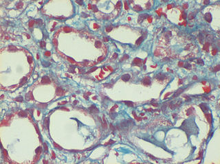
Trichrome staining is a histological staining method that uses two or more acid dyes in conjunction with a polyacid. Staining differentiates tissues by tinting them in contrasting colours. It increases the contrast of microscopic features in cells and tissues, which makes them easier to see when viewed through a microscope.

Fibrils are structural biological materials found in nearly all living organisms. Not to be confused with fibers or filaments, fibrils tend to have diameters ranging from 10–100 nanometers. Fibrils are not usually found alone but rather are parts of greater hierarchical structures commonly found in biological systems. Due to the prevalence of fibrils in biological systems, their study is of great importance in the fields of microbiology, biomechanics, and materials science.

Reticular fibers, reticular fibres or reticulin is a type of fiber in connective tissue composed of type III collagen secreted by reticular cells. They are mainly composed reticulin protein and form a network or mesh. Reticular fibers crosslink to form a fine meshwork (reticulin). This network acts as a supporting mesh in soft tissues such as liver, bone marrow, and the tissues and organs of the lymphatic system.

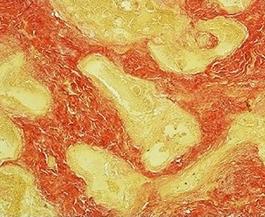
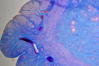
Masson's trichrome is a three-colour staining procedure used in histology. The recipes evolved from Claude L. Pierre Masson's (1880–1959) original formulation have different specific applications, but all are suited for distinguishing cells from surrounding connective tissue.
Aniline Blue WS, also called aniline blue, diphenylamine blue, China blue, or Soluble blue, is a mixture of methyl blue and water blue. It may also be either one of them. It is a soluble dye used as a biological dye, in fluorescence microscopy, appearing a yellow-green colour after excitation with violet light. It is a mixture of the trisulfonates of triphenyl rosaniline and of diphenyl rosaniline.

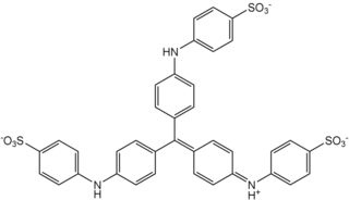
Water blue, also known as aniline blue, Acid blue 22, Soluble Blue 3M, Marine Blue V, or C.I. 42755, is a chemical compound used as a stain in histology. Water blue stains collagen blue in tissue sections. It is soluble in water and slightly soluble in ethanol.

Light green SF, also called C.I. 42095, light green SF yellowish, is a green triarylmethane dye.

Hematoxylin and eosin stain is one of the principal tissue stains used in histology. It is the most widely used stain in medical diagnosis and is often the gold standard. For example, when a pathologist looks at a biopsy of a suspected cancer, the histological section is likely to be stained with H&E.

Phosphotungstic acid (PTA) or tungstophosphoric acid (TPA), is a heteropoly acid with the chemical formula H3PW12O40]. It forms hydrates H3[PW12O40]·nH2O. It is normally isolated as the n = 24 hydrate but can be desiccated to the hexahydrate (n = 6). EPTA is the name of ethanolic phosphotungstic acid, its alcohol solution used in biology. It has the appearance of small, colorless-grayish or slightly yellow-green crystals, with melting point 89 °C (24 H2O hydrate). It is odorless and soluble in water (200 g/100 ml). It is not especially toxic, but is a mild acidic irritant. The compound is known by a variety of names and acronyms (see 'other names' section of infobox).
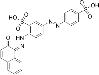
Van Gieson's stain is a mixture of picric acid and acid fuchsin. It is the simplest method of differential staining of collagen and other connective tissue. It was introduced to histology by American neuropsychiatrist and pathologist Ira Van Gieson.

Acid fuchsin or fuchsine acid, (also called Acid Violet 19 and C.I. 42685) is an acidic magenta dye with the chemical formula C20H17N3Na2O9S3. It is a sodium sulfonate derivative of fuchsine. Acid fuchsin has wide use in histology, and is one of the dyes used in Masson's trichrome stain. This method is commonly used to stain cytoplasm and nuclei of tissue sections in the histology laboratory in order to distinguish muscle from collagen. The muscle stains red with the acid fuchsin, and the collagen is stained green or blue with Light Green SF yellowish or methyl blue. It can also be used to identify growing bacteria.
Phosphomolybdic acid is the heteropolymetalate with the formula H3[Mo12PO40]·12H2O. It is a yellow solid, although even slightly impure samples have a greenish coloration. It is also known as dodeca molybdophosphoric acid or PMA, is a yellow-green chemical compound that is freely soluble in water and polar organic solvents such as ethanol. It is used as a stain in histology and in organic synthesis.
Biebrich scarlet is a molecule used in Lillie's trichrome.
Infantile digital fibromatosis (IDF), also termed inclusion body fibromatosis, Reye tumor, or Reye's tumor, usually occurs as a single, small, asymptomatic, nodule in the dermis on a finger or toe of infants and young children. IMF is a rare disorder with approximately 200 cases reported in the medical literature as of 2021. The World Health Organization in 2020 classified these nodules as a specific benign tumor type in the category of fibroblastic and myofibroblastic tumors. IDF was first described by the Australian pathologist, Douglas Reye, in 1965.
Gömöri trichrome stain is a histological stain used on muscle tissue.
Verhoeff's stain, also known as Verhoeff's elastic stain (VEG) or Verhoeff–Van Gieson stain (VVG), is a staining protocol used in histology, developed by American ophthalmic surgeon and pathologist Frederick Herman Verhoeff (1874–1968) in 1908. The formulation is used to demonstrate normal or pathologic elastic fibers.
Mallory's trichrome stain also called Mallory's Triple Stain is a stain utilized in histology to aid in revealing different macromolecules that make up the cell. It uses the three stains: aniline blue, acid fuchsin, and orange G. As a result, this staining technique can reveal collagen, ordinary cytoplasm, and red blood cells. It is used in examining the collagen of connective tissue.
References
- ↑ Lillie, R. D. (1940). "Further Experiments with the Masson Trichrome Modification of Mallory's Connective Tissue Stain". Stain Technology . 15 (1): 17–22. doi:10.3109/10520294009110327.
- ↑ Tchertkoff, Victor (1962-10-01). "Staining Procedures Used by the Biological Stain Commission, ed. 2. Revised by H. J. Conn, et al. Baltimore, Md., The Williams & Wilkins Co., 1960. $5.00". Clinical Chemistry. 8 (5): 548. doi: 10.1093/clinchem/8.5.548a . ISSN 0009-9147.
- ↑ LILLIE, WALTER I. (1940-11-01). "Prechiasmal Syndrome Produced by Chronic Local Arachnoiditis". Archives of Ophthalmology . 24 (5): 940. doi:10.1001/archopht.1940.00870050090006. ISSN 0093-0326.
- ↑ Cowie, Robert. "Masson's Trichrome Stain". Science Direct .