Pore-forming toxin

Last updated

a-hemolysin from S.aureus (PDB: 7AHL ) 7ahl.png
α-hemolysin from S.aureus ( PDB: 7AHL )

Pore-forming proteins (PFTs, also known as pore-forming toxins) are usually produced by bacteria, and include a number of protein exotoxins but may also be produced by other organisms such as apple snails that produce perivitellin-2 [1] [2] or earthworms, who produce lysenin. They are frequently cytotoxic (i.e., they kill cells), as they create unregulated pores in the membrane of targeted cells.

Contents

Types

PFTs can be divided into two categories, depending on the alpha-helical or beta-barrel architecture of their transmembrane channel [3] that can consist either of

Other categories:

According to TCDB, there are following families of pore-forming toxins:

Beta-pore-forming toxins

Leukocidin
Identifiers
SymbolLeukocidin
Pfam PF07968
InterPro IPR001340
TCDB 1.C.3
OPM superfamily 35
OPM protein 7ahl
Available protein structures:
Pfam   structures / ECOD  
PDB RCSB PDB; PDBe; PDBj
PDBsum structure summary

β-PFTs are so-named because of their structural characteristics: they are composed mostly of β-strand-based domains. They have divergent sequences, and are classified by Pfam into a number of families including Leukocidins, Etx-Mtx2, Toxin-10, and aegerolysin. X-ray crystallographic structures have revealed some commonalities: α-hemolysin [6] and Panton-Valentine leukocidin S [7] are structurally related. Similarly, aerolysin [8] and Clostridial Epsilon-toxin. [9] and Mtx2 are linked in the Etx/Mtx2 family. [10]

The ß-PFTs include a number of toxins of commercial interest for the control of pest insects. These toxins are potent but also highly specific to a limited range of target insects, making them safe biological control agents.

Insecticidal members of the Etx/Mtx2 family include Mtx2 [10] and Mtx3 [11] from Lysinibacillus sphaericus that can control mosquito vectors of human diseases and also Cry15, Cry23, Cry33, Cry38, Cry45, Cry51, Cry60, Cry64 and Cry74 from Bacillus thuringiensis [12] that control a range of insect pests that can cause great losses to agriculture.

Insecticidal toxins in the Toxin–10 family show an overall similarity to the aerolysin and Etx/Mtx2 toxin structures but differ in two notable features. While all of these toxins feature a head domain and a larger, extended beta-sheet tail domain, in the Toxin_10 family, the head is formed exclusively from the N-terminal region of the primary amino acid sequence whereas regions from throughout the protein sequence contribute to the head domain in Etx/Mtx2 toxins. In addition, the head domains of the Toxin_10 proteins show lectin-like features of carbohydrate binding domains. The only reported natural targets of Toxin_10 proteins are insects. With the exception of Cry36 [13] and Cry78, [12] the Toxin_10 toxins appear to act as two-part, binary toxins. The partner proteins in these combinations may belong to different structural groups, depending on the individual toxin: two Toxin_10 proteins (BinA and BinB) act together in the Bin mosquitocidal toxin of Lysinibacillus sphaericus; [14] the Toxin_10 Cry49 is co-dependent on the 3-domain toxin family member Cry48 for its activity against Culex mosquito larvae; [15] and the Bacillus thuringiensis Toxin_10 protein Cry35 interacts with the aegerolysin family Cry34 to kill Western Corn Rootworm. [16] This toxin pair has been included in insect resistant plants such as SmartStax corn.

Mode of action

Structural comparison of pore-form a-Hemolysin (pink/red) and soluble-form PVL (pale green/green). It is postulated that the green section in PVL 'flips out' to the 'red' conformation as seen in a-Haemolysin. (PDB: 7AHL, 1T5R ) Structural comparison of pore-form alpha-Haemolysin (pink-red) and soluble-form PVL (pale green-green) toxins - PDB 7AHL and 1T5R.png
Structural comparison of pore-form α-Hemolysin (pink/red) and soluble-form PVL (pale green/green). It is postulated that the green section in PVL 'flips out' to the 'red' conformation as seen in α-Haemolysin. ( PDB: 7AHL, 1T5R )

β-PFTs are dimorphic proteins that exist as soluble monomers and then assemble to form multimeric assemblies that constitute the pore. Figure 1 shows the pore-form of α-Hemolysin, the first crystal structure of a β-PFT in its pore-form. 7 α-Hemolysin monomers come together to create the mushroom-shaped pore. The 'cap' of the mushroom sits on the surface of the cell, and the 'stalk' of the mushroom penetrates the cell membrane, rendering it permeable (see later). The 'stalk' is composed of a 14-strand β-barrel, with two strands donated from each monomer.

A structure of the Vibrio cholerae cytolysin [17] in the pore form is also heptameric; however, Staphylococcus aureus gamma-hemolysin [18] reveals an octomeric pore, consequently with a 16-strand 'stalk'.

The Panton-Valentine leucocidin S structure [19] shows a highly related structure, but in its soluble monomeric state. This shows that the strands involved in forming the 'stalk' are in a very different conformation – shown in Fig 2.

Structural comparison of pore-form α-Hemolysin (pink/red) and soluble-form PVL (pale green/green). It is postulated that the green section in PVL 'flips out' to the 'red' conformation as seen in α-Haemolysin. (PDB: 7AHL, 1T5R) β-PFTs are dimorphic proteins that exist as soluble monomers and then assemble to form multimeric assemblies that constitute the pore. Figure 1 shows the pore-form of α-Hemolysin, the first crystal structure of a β-PFT in its pore-form. 7 α-Hemolysin monomers come together to create the mushroom-shaped pore. The 'cap' of the mushroom sits on the surface of the cell, and the 'stalk' of the mushroom penetrates the cell membrane, rendering it permeable (see later). The 'stalk' is composed of a 14-strand β-barrel, with two strands donated from each monomer. A structure of the Vibrio cholerae cytolysin PDB:3O44 [20] in the pore form is also heptameric; however, Staphylococcus aureus gamma-hemolysin (PDB:3B07) [21] reveals an octomeric pore, consequently with a 16-strand 'stalk'. The Panton-Valentine leucocidin S structure (PDB: 1T5R) [7] shows a highly related structure, but in its soluble monomeric state. This shows that the strands involved in forming the 'stalk' are in a very different conformation – shown in Fig 2. While the Bin toxin of Lysinibacillus sphaericus is able to form pores in artificial membranes [22] and mosquito cells in culture, [23] it also causes a series of other cellular changes including the uptake of toxin in recycling endosomes and the production of large, autophagic vesicles [24] and the ultimate cause of cell death may be apoptotic. [25] Similar effects on cell biology are also seen with other Toxin_10 activities [26] [27] but the roles of these events in toxicity remain to be established.

Assembly

The transition between soluble monomer and membrane-associated protomer to oligomer is not a trivial one: It is believed that β-PFTs, follow as similar assembly pathway as the CDCs (see Cholesterol-dependent cytolysins later), in that they must first assemble on the cell-surface (in a receptor-mediated fashion in some cases) in a pre-pore state. Following this, the large-scale conformational change occurs in which the membrane spanning section is formed and inserted into the membrane. The portion entering the membrane, referred to as the head, is usually apolar and hydrophobic, this produces an energetically favorable insertion of the pore-forming toxin. [3]

Specificity

Some β-PFTs such as clostridial ε-toxin and Clostridium perfringens enterotoxin (CPE) bind to the cell membrane via specific receptors – possibly certain claudins for CPE, [28] possibly GPI anchors or other sugars for ε-toxin – these receptors help raise the local concentration of the toxins, allowing oligomerisation and pore formation.

The BinB Toxin_10 component of the Lysinibacillus sphaericus Bin toxin specifically recognises a GPI anchored alpha glycosidase in the midgut of Culex [29] and Anopheles mosquitoes but not the related protein found in Aedes mosquitoes, [30] hence conferring specificity on the toxin.

The cyto-lethal effects of the pore

When the pore is formed, the tight regulation of what can and cannot enter/leave a cell is disrupted. Ions and small molecules, such as amino acids and nucleotides within the cell, flow out, and water from the surrounding tissue enters. The loss of important small molecules to the cell can disrupt protein synthesis and other crucial cellular reactions. The loss of ions, especially calcium, can cause cell signaling pathways to be spuriously activated or deactivated. The uncontrolled entry of water into a cell can cause the cell to swell up uncontrollably: this causes a process called blebbing, wherein large parts of the cell membrane are distorted and give way under the mounting internal pressure. In the end, this can cause the cell to burst. In particular, nuclear - free erythrocytes under the influence of alpha-staphylotoxin undergo hemolysis with the loss of a large protein hemoglobin.

Binary toxins

There are many different types of binary toxins. The term binary toxin simply implies a two part toxin where both components are necessary for toxic activity. Several β-PFTs form binary toxins.

As discussed above, the majority of the Toxin_10 family proteins act as part of binary toxins with partner proteins that may belong to the Toxin_10 or other structural families. The interplay of the individual components has not been well studied to date. Other beta sheet toxins of commercial importance are also binary. These include the Cry23/Cry37 toxin from Bacillus thuringiensis. [31] These toxins have some structural similarity to the Cry34/Cry35 binary toxin but neither component shows a match to established Pfam families and the features of the larger Cry23 protein have more in common with the Etx/Mtx2 family than the Toxin_10 family to which Cry35 belongs.

Enzymatic binary toxins

Some binary toxins are composed of an enzymatic component and a component that is involved in membrane interactions and entry of the enzymatic component into the cell. The membrane interacting component may have structural domains that are rich in beta sheets. Binary toxins, such as anthrax lethal and edema toxins (Main article: Anthrax toxin), C. perfringens iota toxin and C. difficile cyto-lethal toxins consist of two components (hence binary):

  • an enzymatic component – A
  • a membrane-altering component – B

In these enzymatic binary toxins, the B component facilitates the entry of the enzymatic 'payload' (A subunit) into the target cell, by forming homooligomeric pores, as shown above for βPFTs. The A component then enters the cytosol and inhibits normal cell functions by one of the following means:

ADP-ribosylation

ADP-ribosylation is a common enzymatic method used by different bacterial toxins from various species. Toxins such as C. perfringens iota toxin and C. botulinum C2 toxin, attach a ribosyl-ADP moiety to surface arginine residue 177 of G-actin. This prevents G-actin assembling to form F-actin, and, thus, the cytoskeleton breaks down, resulting in cell death. Insecticidal members of the ADP-ribosyltransferase family of toxins include the Mtx1 toxin of Lysinibacillus sphaericus [32] and the Vip1/Vip2 toxin of Bacillus thuringiensis and some members of the toxin complex (Tc) toxins from gram negative bacteria such as Photorhabdus and Xenorhabdus species. The beta sheet-rich regions of the Mtx1 protein are lectin-like sequences that may be involved in glycolipid interactions. [33]

Proteolysis of mitogen-activated protein kinase kinases (MAPKK)

The A component of anthrax toxin lethal toxin is zinc-metalloprotease, which shows specificity for a conserved family of mitogen-activated protein kinases. The loss of these proteins results in a breakdown of cell signaling, which, in turn, renders the cell insensitive to outside stimuli – therefore no immune response is triggered.

Increasing intracellular levels of cAMP

Anthrax toxin edema toxin triggers a calcium ion influx into the target cell. This subsequently elevates intracellular cAMP levels. This can profoundly alter any sort of immune response, by inhibiting leucocyte proliferation, phagocytosis, and proinflammatory cytokine release.

Cholesterol-dependent cytolysins

EM reconstruction of a Pneumolysin pre-pore Molecular model of the pre-pore form of a MACPF protein based upon the structure of pneunolysin.png
EM reconstruction of a Pneumolysin pre-pore
a) The structure of perfringolysin O and b) the structure of PluMACPF. In both proteins, the two small clusters of a-helices that unwind and pierce the membrane are in pink. (PDB: 1PFO, 2QP2 ) PFO and Plu-MACPF.png
a) The structure of perfringolysin O and b) the structure of PluMACPF. In both proteins, the two small clusters of α-helices that unwind and pierce the membrane are in pink. ( PDB: 1PFO, 2QP2 )

CDCs, such as pneumolysin, from S. pneumoniae , form pores as large as 260Å (26 nm), containing between 30 and 44 monomer units. [36] Electron microscopy studies of pneumolysin show that it assembles into large multimeric peripheral membrane complexes before undergoing a conformational change in which a group of α-helices in each monomer change into extended, amphipathic β-hairpins that span the membrane, in a manner reminiscent of α-haemolysin, albeit on a much larger scale (Fig 3). CDCs are homologous to the MACPF family of pore-forming toxins, and it is suggested that both families use a common mechanism (Fig 4). [35] Eukaryote MACPF proteins function in immune defence and are found in proteins such as perforin and complement C9 [37] though perivitellin-2 is a MACPF attached to a delivery lectin that has enterotoxic and neurotoxic properties toward mice. [1] [2] [38]

A family of highly conserved cholesterol-dependent cytolysins, closely related to perfringolysin from Clostridium perfringens are produced by bacteria from across the order Bacillales and include anthrolysin, alveolysin and sphaericolysin. [29] Sphaericolysin has been shown to exhibit toxicity to a limited range of insects injected with the purified protein. [39]

Biological function

Bacteria may invest much time and energy in making these toxins: CPE can account for up to 15% of the dry mass of C. perfringens at the time of sporulation. [ citation needed ] The purpose of toxins is thought to be one of the following:

See also

Related Research Articles

<span class="mw-page-title-main">Exotoxin</span> Toxin from bacteria that destroys or disrupts cells

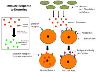
An exotoxin is a toxin secreted by bacteria. An exotoxin can cause damage to the host by destroying cells or disrupting normal cellular metabolism. They are highly potent and can cause major damage to the host. Exotoxins may be secreted, or, similar to endotoxins, may be released during lysis of the cell. Gram negative pathogens may secrete outer membrane vesicles containing lipopolysaccharide endotoxin and some virulence proteins in the bounding membrane along with some other toxins as intra-vesicular contents, thus adding a previously unforeseen dimension to the well-known eukaryote process of membrane vesicle trafficking, which is quite active at the host–pathogen interface.

<span class="mw-page-title-main">Enterotoxin</span> Toxin from a microorganism affecting the intestines

An enterotoxin is a protein exotoxin released by a microorganism that targets the intestines. They can be chromosomally or plasmid encoded. They are heat labile (>60⁰), of low molecular weight and water-soluble. Enterotoxins are frequently cytotoxic and kill cells by altering the apical membrane permeability of the mucosal (epithelial) cells of the intestinal wall. They are mostly pore-forming toxins, secreted by bacteria, that assemble to form pores in cell membranes. This causes the cells to die.

A latrotoxin is a high-molecular mass neurotoxin found in the venom of spiders of the genus Latrodectus as well as at least one species of another genus in the same family, Steatoda nobilis. Latrotoxins are the main active components of the venom and are responsible for the symptoms of latrodectism.

<span class="mw-page-title-main">Anthrax toxin</span> Tripartite protein complex secreted by virulent strains of Bacillus anthracis

Anthrax toxin is a three-protein exotoxin secreted by virulent strains of the bacterium, Bacillus anthracis—the causative agent of anthrax. The toxin was first discovered by Harry Smith in 1954. Anthrax toxin is composed of a cell-binding protein, known as protective antigen (PA), and two enzyme components, called edema factor (EF) and lethal factor (LF). These three protein components act together to impart their physiological effects. Assembled complexes containing the toxin components are endocytosed. In the endosome, the enzymatic components of the toxin translocate into the cytoplasm of a target cell. Once in the cytosol, the enzymatic components of the toxin disrupts various immune cell functions, namely cellular signaling and cell migration. The toxin may even induce cell lysis, as is observed for macrophage cells. Anthrax toxin allows the bacteria to evade the immune system, proliferate, and ultimately kill the host animal. Research on anthrax toxin also provides insight into the generation of macromolecular assemblies, and on protein translocation, pore formation, endocytosis, and other biochemical processes.

Tetanolysin is a toxin produced by Clostridium tetani bacteria. Its function is unknown, but it is believed to contribute to the pathogenesis of tetanus. The other C. tetani toxin, tetanospasmin, is more definitively linked to tetanus. It is sensitive to oxygen.

Cytolysin refers to the substance secreted by microorganisms, plants or animals that is specifically toxic to individual cells, in many cases causing their dissolution through lysis. Cytolysins that have a specific action for certain cells are named accordingly. For instance, the cytolysins responsible for the destruction of red blood cells, thereby liberating hemoglobins, are named hemolysins, and so on. Cytolysins may be involved in immunity as well as in venoms.

<span class="mw-page-title-main">Hemolysin</span> Molecule destroying the membrane of red blood cells

Hemolysins or haemolysins are lipids and proteins that cause lysis of red blood cells by disrupting the cell membrane. Although the lytic activity of some microbe-derived hemolysins on red blood cells may be of great importance for nutrient acquisition, many hemolysins produced by pathogens do not cause significant destruction of red blood cells during infection. However, hemolysins are often capable of lysing red blood cells in vitro.

Lysinibacillus sphaericus is a Gram-positive, mesophilic, rod-shaped bacterium commonly found on soil. It can form resistant endospores that are tolerant to high temperatures, chemicals and ultraviolet light and can remain viable for long periods of time. It is of particular interest to the World Health Organization due to the larvicide effect of some strains against two mosquito genera, more effective than Bacillus thuringiensis, frequently used as a biological pest control. L. sphaericus cells in a vegetative state are also effective against Aedes aegypti larvae, an important vector of yellow fever and dengue viruses.

The Membrane Attack Complex/Perforin (MACPF) superfamily, sometimes referred to as the MACPF/CDC superfamily, is named after a domain that is common to the membrane attack complex (MAC) proteins of the complement system and perforin (PF). Members of this protein family are pore-forming toxins (PFTs). In eukaryotes, MACPF proteins play a role in immunity and development.

<i>Staphylococcus aureus</i> alpha toxin

Alpha-toxin, also known as alpha-hemolysin (Hla), is the major cytotoxic agent released by bacterium Staphylococcus aureus and the first identified member of the pore forming beta-barrel toxin family. This toxin consists mostly of beta-sheets (68%) with only about 10% alpha-helices. The hly gene on the S. aureus chromosome encodes the 293 residue protein monomer, which forms heptameric units on the cellular membrane to form a complete beta-barrel pore. This structure allows the toxin to perform its major function, development of pores in the cellular membrane, eventually causing cell death.

<span class="mw-page-title-main">Delta endotoxins</span> Group of insecticidal toxins produced by the bacteria Bacillus thuringiensis

Delta endotoxins (δ-endotoxins) are a family of pore-forming toxins produced by Bacillus thuringiensis species of bacteria. They are useful for their insecticidal action and are the primary toxin produced by the genetically modified (GM) Bt maize/corn and other GM crops. During spore formation the bacteria produce crystals of such proteins that are also known as parasporal bodies, next to the endospores; as a result some members are known as a parasporin. The Cyt (cytolytic) toxin group is another group of delta-endotoxins formed in the cytoplasm. VIP toxins are formed at other stages of the life cycle.

Streptolysins are two hemolytic exotoxins from Streptococcus pyogenes. Types include streptolysin O, which is oxygen-labile, and streptolysin S, which is oxygen-stable.

<span class="mw-page-title-main">AB toxin</span>

The AB toxins are two-component protein complexes secreted by a number of pathogenic bacteria, though there is a pore-forming AB toxin found in the eggs of a snail. They can be classified as Type III toxins because they interfere with internal cell function. They are named AB toxins due to their components: the "A" component is usually the "active" portion, and the "B" component is usually the "binding" portion. The "A" subunit possesses enzyme activity, and is transferred to the host cell following a conformational change in the membrane-bound transport "B" subunit. These proteins consist of two independent polypeptides, which correspond to the A/B subunit moieties. The enzyme component (A) enters the cell through endosomes produced by the oligomeric binding/translocation protein (B), and prevents actin polymerisation through ADP-ribosylation of monomeric G-actin.

The RTX toxin superfamily is a group of cytolysins and cytotoxins produced by bacteria. There are over 1000 known members with a variety of functions. The RTX family is defined by two common features: characteristic repeats in the toxin protein sequences, and extracellular secretion by the type I secretion systems (T1SS). The name RTX refers to the glycine and aspartate-rich repeats located at the C-terminus of the toxin proteins, which facilitate export by a dedicated T1SS encoded within the rtx operon.

The thiol-activated Cholesterol-dependent Cytolysin(CDC) family is a member of the MACPF superfamily. Cholesterol dependent cytolysins are a family of β-barrel pore-forming exotoxins that are secreted by gram-positive bacteria. CDCs are secreted as water-soluble monomers of 50-70 kDa, that when bound to the target cell, form a circular homo-oligomeric complex containing as many as 40 monomers. Through multiple conformational changes, the β-barrel transmembrane structure is formed and inserted into the target cell membrane. The presence of cholesterol in the target membrane is required for pore formation, though the presence of cholesterol is not required by all CDCs for binding. For example, intermedilysin secreted by Streptococcus intermedius will bind only to target membranes containing a specific protein receptor, independent of the presence of cholesterol, but cholesterol is required by intermedilysin for pore formation. While the lipid environment of cholesterol in the membrane can affect toxin binding, the exact molecular mechanism that cholesterol regulates the cytolytic activity of the CDC is not fully understood.

<span class="mw-page-title-main">Cry6Aa</span>

Cry6Aa is a toxic crystal protein generated by the bacterial family Bacillus thuringiensis during sporulation. This protein is a member of the alpha pore forming toxins family, which gives it insecticidal qualities advantageous in agricultural pest control. Each Cry protein has some level of target specificity; Cry6Aa has specific toxic action against coleopteran insects and nematodes. The corresponding B. thuringiensis gene, cry6aa, is located on bacterial plasmids. Along with several other Cry protein genes, cry6aa can be genetically recombined in Bt corn and Bt cotton so the plants produce specific toxins. Insects are developing resistance to the most commonly inserted proteins like Cry1Ac. Since Cry6Aa proteins function differently than other Cry proteins, they are combined with other proteins to decrease the development of pest resistance. Recent studies suggest this protein functions better in combination with other virulence factors such as other Cry proteins and metalloproteinases.>

Cry34Ab1 is one member of a binary Bacillus thuringiensis (Bt) crystal protein set isolated from Bt strain PS149B1. The protein exists as a 14 kDa aegerolysin that, in presence of Cry35Ab1, exhibits insecticidal activity towards Western Corn Rootworm. The protein has been transformed into maize plants under the commercialized events 4114 (DP-ØØ4114-3) by Pioneer Hi-Bred and 59122 (DAS-59122-7) by Dow AgroSciences. These events have, in turn, been bred into multiple trait stacks in additional products.

Kausik Chattopadhyay is an Indian structural biologist, protein biologist, and a professor at the Department of Biological Sciences. He was the Dean of R&D at the Indian Institute of Science Education and Research, Mohali until May 2021. He is known for his studies on the Pore-forming protein toxins and T-cell costimulatory molecules. The Department of Biotechnology of the Government of India awarded him the National Bioscience Award for Career Development, one of the highest Indian science awards, for his contributions to biosciences, in 2014.

Lysenin is a pore-forming toxin (PFT) present in the coelomic fluid of the earthworm Eisenia fetida. Pore-forming toxins are a group of proteins that act as virulence factors of several pathogenic bacteria. Lysenin proteins are chiefly involved in the defense against cellular pathogens. Following the general mechanism of action of PFTs lysenin is segregated as a soluble monomer that binds specifically to a membrane receptor, sphingomyelin in the case of lysenin. After attaching to the membrane, the oligomerization begins, resulting in a nonamer on top of membrane, known as a prepore. After a conformational change, which could be triggered by a decrease of pH, the oligomer is inserted into the membrane in the so-called pore state.

<span class="mw-page-title-main">Cytotoxin K</span> This protein is one of the toxins excreted by bacillus cereus and causes abdominal symptoms

Cytotoxin-K (CytK) is a protein toxin produced by the gram-positive bacteria Bacillus cereus. It was first discovered in a certain Bacillus cereus strain which was isolated from a food poisoning epidemic that occurred in a French nursing home in 1998. There were six cases of bloody diarrhea, three of which were fatal. None of the known enterotoxins from B. cereus could be detected at this time. Later, this B. cereus strain and its relatives were classified as a brand-new species called Bacillus cytotoxicus, which is the thermo-tolerant member of the B. cereus genus. The cytotoxin-K gene is present in approximately 50% of Bacillus cereus isolates, and its expression is regulated by several factors, including temperature and nutrient availability.

References

  1. 1 2 Giglio ML, Ituarte S, Milesi V, Dreon MS, Brola TR, Caramelo J, et al. (August 2020). "Exaptation of two ancient immune proteins into a new dimeric pore-forming toxin in snails". Journal of Structural Biology. 211 (2): 107531. doi:10.1016/j.jsb.2020.107531. hdl: 11336/143650 . PMID   32446810. S2CID   218873723.
  2. 1 2 Giglio ML, Ituarte S, Ibañez AE, Dreon MS, Prieto E, Fernández PE, Heras H (13 March 2020). "Novel Role for Animal Innate Immune Molecules: Enterotoxic Activity of a Snail Egg MACPF-Toxin". Frontiers in Immunology. 11: 428. doi: 10.3389/fimmu.2020.00428 . PMC   7082926 . PMID   32231667.
  3. 1 2 Mueller M, Grauschopf U, Maier T, Glockshuber R, Ban N (June 2009). "The structure of a cytolytic alpha-helical toxin pore reveals its assembly mechanism". Nature. 459 (7247): 726–730. Bibcode:2009Natur.459..726M. doi:10.1038/nature08026. PMID   19421192. S2CID   205216795.
  4. Structure of Gastermin A channel in the lipid bilayer
  5. Cané, Lucía; Guzmán, Fanny; Balatti, Galo; Daza Millone, María Antonieta; Pucci Molineris, Melisa; Maté, Sabina; Martini, M. Florencia; Herlax, Vanesa (24 May 2023). "Biophysical Analysis to Assess the Interaction of CRAC and CARC Motif Peptides of Alpha Hemolysin of Escherichia coli with Membranes". Biochemistry. 62 (12): 1994–2011. doi:10.1021/acs.biochem.3c00164. ISSN   0006-2960. PMID   37224476.
  6. Song L, Hobaugh MR, Shustak C, Cheley S, Bayley H, Gouaux JE (December 1996). "Structure of staphylococcal alpha-hemolysin, a heptameric transmembrane pore". Science. 274 (5294): 1859–1866. Bibcode:1996Sci...274.1859S. doi:10.1126/science.274.5294.1859. PMID   8943190. S2CID   45663016.
  7. 1 2 Guillet V, Roblin P, Werner S, Coraiola M, Menestrina G, Monteil H, et al. (September 2004). "Crystal structure of leucotoxin S component: new insight into the Staphylococcal beta-barrel pore-forming toxins". The Journal of Biological Chemistry. 279 (39): 41028–41037. doi: 10.1074/jbc.M406904200 . PMID   15262988.
  8. Parker MW, Buckley JT, Postma JP, Tucker AD, Leonard K, Pattus F, Tsernoglou D (January 1994). "Structure of the Aeromonas toxin proaerolysin in its water-soluble and membrane-channel states". Nature. 367 (6460): 292–295. Bibcode:1994Natur.367..292P. doi:10.1038/367292a0. PMID   7510043. S2CID   4371932.
  9. Cole AR, Gibert M, Popoff M, Moss DS, Titball RW, Basak AK (August 2004). "Clostridium perfringens epsilon-toxin shows structural similarity to the pore-forming toxin aerolysin". Nature Structural & Molecular Biology. 11 (8): 797–8. doi:10.1038/nsmb804. PMID   15258571. S2CID   24508677.
  10. 1 2 Thanabalu T, Porter AG (April 1996). "A Bacillus sphaericus gene encoding a novel type of mosquitocidal toxin of 31.8 kDa". Gene. 170 (1): 85–89. doi:10.1016/0378-1119(95)00836-5. PMID   8621095.
  11. Liu JW, Porter AG, Wee BY, Thanabalu T (June 1996). "New gene from nine Bacillus sphaericus strains encoding highly conserved 35.8-kilodalton mosquitocidal toxins". Applied and Environmental Microbiology. 62 (6): 2174–2176. Bibcode:1996ApEnM..62.2174L. doi:10.1128/AEM.62.6.2174-2176.1996. PMC   167996 . PMID   8787415.
  12. 1 2 Berry C, Crickmore N (January 2017). "Structural classification of insecticidal proteins - Towards an in silico characterisation of novel toxins" (PDF). Journal of Invertebrate Pathology. 142: 16–22. doi:10.1016/j.jip.2016.07.015. PMID   27480403.
  13. US 7078592,Rupar MJ, Donovan WP, Chu CR, Pease E, Tan Y, Slaney AC, Malvar TM, Baum JA,"Nucleic acids encoding coleopteran-toxic polypeptides and insect-resistant transgenic plants comprising them.",issued 2007, assigned to Monsanto Technology LLC (St. Louis, MO)
  14. Oei C, Hindley J, Berry C (November 1990). "An analysis of the genes encoding the 51.4- and 41.9-kDa toxins of Bacillus sphaericus 2297 by deletion mutagenesis: the construction of fusion proteins". FEMS Microbiology Letters. 60 (3): 265–273. doi: 10.1016/0378-1097(90)90315-h . PMID   2083839.
  15. Jones GW, Nielsen-Leroux C, Yang Y, Yuan Z, Dumas VF, Monnerat RG, Berry C (December 2007). "A new Cry toxin with a unique two-component dependency from Bacillus sphaericus". FASEB Journal. 21 (14): 4112–4120. doi: 10.1096/fj.07-8913com . PMID   17646596. S2CID   15049423.
  16. Ellis RT, Stockhoff BA, Stamp L, Schnepf HE, Schwab GE, Knuth M, et al. (March 2002). "Novel Bacillus thuringiensis binary insecticidal crystal proteins active on western corn rootworm, Diabrotica virgifera virgifera LeConte". Applied and Environmental Microbiology. 68 (3): 1137–1145. Bibcode:2002ApEnM..68.1137E. doi:10.1128/AEM.68.3.1137-1145.2002. PMC   123759 . PMID   11872461.
  17. PDB 3o44 De S, Olson R (May 2011). "Crystal structure of the Vibrio cholerae cytolysin heptamer reveals common features among disparate pore-forming toxins". Proceedings of the National Academy of Sciences of the United States of America. 108 (18): 7385–7390. Bibcode:2011PNAS..108.7385D. doi: 10.1073/pnas.1017442108 . PMC   3088620 . PMID   21502531.
  18. PDB 3b07 Yamashita K, Kawai Y, Tanaka Y, Hirano N, Kaneko J, Tomita N, et al. (October 2011). "Crystal structure of the octameric pore of staphylococcal γ-hemolysin reveals the β-barrel pore formation mechanism by two components". Proceedings of the National Academy of Sciences of the United States of America. 108 (42): 17314–17319. Bibcode:2011PNAS..10817314Y. doi: 10.1073/pnas.1110402108 . PMC   3198349 . PMID   21969538.
  19. PDB 1T5R Guillet V, Roblin P, Werner S, Coraiola M, Menestrina G, Monteil H, et al. (September 2004). "Crystal structure of leucotoxin S component: new insight into the Staphylococcal beta-barrel pore-forming toxins". The Journal of Biological Chemistry. 279 (39): 41028–41037. doi: 10.1074/jbc.M406904200 . PMID   15262988.
  20. De S, Olson R (May 2011). "Crystal structure of the Vibrio cholerae cytolysin heptamer reveals common features among disparate pore-forming toxins". Proceedings of the National Academy of Sciences of the United States of America. 108 (18): 7385–7390. Bibcode:2011PNAS..108.7385D. doi: 10.1073/pnas.1017442108 . PMC   3088620 . PMID   21502531.
  21. Yamashita K, Kawai Y, Tanaka Y, Hirano N, Kaneko J, Tomita N, et al. (October 2011). "Crystal structure of the octameric pore of staphylococcal γ-hemolysin reveals the β-barrel pore formation mechanism by two components". Proceedings of the National Academy of Sciences of the United States of America. 108 (42): 17314–17319. Bibcode:2011PNAS..10817314Y. doi: 10.1073/pnas.1110402108 . PMC   3198349 . PMID   21969538.
  22. Schwartz JL, Potvin L, Coux F, Charles JF, Berry C, Humphreys MJ, et al. (November 2001). "Permeabilization of model lipid membranes by Bacillus sphaericus mosquitocidal binary toxin and its individual components". The Journal of Membrane Biology. 184 (2): 171–183. doi:10.1007/s00232-001-0086-1. PMID   11719853. S2CID   22113520.
  23. Cokmus C, Davidson EW, Cooper K (May 1997). "Electrophysiological effects of Bacillus sphaericus binary toxin on cultured mosquito cells". Journal of Invertebrate Pathology. 69 (3): 197–204. doi:10.1006/jipa.1997.4660. PMID   9170345.
  24. Opota O, Gauthier NC, Doye A, Berry C, Gounon P, Lemichez E, Pauron D (February 2011). "Bacillus sphaericus binary toxin elicits host cell autophagy as a response to intoxication". PLOS ONE. 6 (2): e14682. Bibcode:2011PLoSO...614682O. doi: 10.1371/journal.pone.0014682 . PMC   3038859 . PMID   21339824.
  25. Tangsongcharoen C, Chomanee N, Promdonkoy B, Boonserm P (June 2015). "Lysinibacillus sphaericus binary toxin induces apoptosis in susceptible Culex quinquefasciatus larvae". Journal of Invertebrate Pathology. 128: 57–63. doi:10.1016/j.jip.2015.04.008. PMID   25958262.
  26. de Melo JV, Jones GW, Berry C, Vasconcelos RH, de Oliveira CM, Furtado AF, et al. (July 2009). "Cytopathological effects of Bacillus sphaericus Cry48Aa/Cry49Aa toxin on binary toxin-susceptible and -resistant Culex quinquefasciatus larvae". Applied and Environmental Microbiology. 75 (14): 4782–4789. Bibcode:2009ApEnM..75.4782D. doi:10.1128/AEM.00811-09. PMC   2708442 . PMID   19502449.
  27. Narva KE, Wang NX, Herman R (January 2017). "Safety considerations derived from Cry34Ab1/Cry35Ab1 structure and function". Journal of Invertebrate Pathology. 142: 27–33. doi:10.1016/j.jip.2016.07.019. PMID   27480405.
  28. Fujita K, Katahira J, Horiguchi Y, Sonoda N, Furuse M, Tsukita S (July 2000). "Clostridium perfringens enterotoxin binds to the second extracellular loop of claudin-3, a tight junction integral membrane protein". FEBS Letters. 476 (3): 258–261. doi:10.1016/S0014-5793(00)01744-0. hdl: 2433/150516 . PMID   10913624. S2CID   10333473.
  29. 1 2 Silva-Filha MH, Nielsen-LeRoux C, Charles JF (August 1999). "Identification of the receptor for Bacillus sphaericus crystal toxin in the brush border membrane of the mosquito Culex pipiens (Diptera: Culicidae)". Insect Biochemistry and Molecular Biology. 29 (8): 711–721. doi:10.1016/S0965-1748(99)00047-8. PMID   10451923.
  30. Ferreira LM, Romão TP, de-Melo-Neto OP, Silva-Filha MH (August 2010). "The orthologue to the Cpm1/Cqm1 receptor in Aedes aegypti is expressed as a midgut GPI-anchored α-glucosidase, which does not bind to the insecticidal binary toxin". Insect Biochemistry and Molecular Biology. 40 (8): 604–610. doi:10.1016/j.ibmb.2010.05.007. PMID   20685335.
  31. Donovan WP, Donovan JC, Slaney AC (2000). "Bacillus thuringiensis cryET33 and cryET34 compositions and uses therefor". Monsanto Company (Patent).
  32. Thanabalu T. Cloning and characterisation of a gene encoding a 100 kDa toxin from Bacillus sphaericus SSII-1 and expression of insecticidal toxins in Caulobacter crescentus (Ph.D. thesis). Institute of Molecular and Cell Biology, National University of Singapore.
  33. Treiber N, Reinert DJ, Carpusca I, Aktories K, Schulz GE (August 2008). "Structure and mode of action of a mosquitocidal holotoxin". Journal of Molecular Biology. 381 (1): 150–159. doi:10.1016/j.jmb.2008.05.067. PMID   18586267.
  34. Rossjohn J, Feil SC, McKinstry WJ, Tweten RK, Parker MW (May 1997). "Structure of a cholesterol-binding, thiol-activated cytolysin and a model of its membrane form". Cell. 89 (5): 685–692. doi: 10.1016/S0092-8674(00)80251-2 . PMID   9182756. S2CID   8345038.
  35. 1 2 Rosado CJ, Buckle AM, Law RH, Butcher RE, Kan WT, Bird CH, et al. (September 2007). "A common fold mediates vertebrate defense and bacterial attack". Science. 317 (5844): 1548–1551. Bibcode:2007Sci...317.1548R. doi: 10.1126/science.1144706 . PMID   17717151. S2CID   20372720.
  36. Tilley SJ, Orlova EV, Gilbert RJ, Andrew PW, Saibil HR (April 2005). "Structural basis of pore formation by the bacterial toxin pneumolysin". Cell. 121 (2): 247–256. doi: 10.1016/j.cell.2005.02.033 . PMID   15851031. S2CID   1613454.
  37. Tschopp J, Masson D, Stanley KK (1986). "Structural/functional similarity between proteins involved in complement- and cytotoxic T-lymphocyte-mediated cytolysis". Nature. 322 (6082): 831–834. Bibcode:1986Natur.322..831T. doi:10.1038/322831a0. PMID   2427956. S2CID   4330219.
  38. Dreon MS, Frassa MV, Ceolín M, Ituarte S, Qiu JW, Sun J, et al. (30 May 2013). van der Goot FG (ed.). "Novel animal defenses against predation: a snail egg neurotoxin combining lectin and pore-forming chains that resembles plant defense and bacteria attack toxins". PLOS ONE. 8 (5): e63782. Bibcode:2013PLoSO...863782D. doi: 10.1371/journal.pone.0063782 . PMC   3667788 . PMID   23737950.
  39. Nishiwaki H, Nakashima K, Ishida C, Kawamura T, Matsuda K (May 2007). "Cloning, functional characterization, and mode of action of a novel insecticidal pore-forming toxin, sphaericolysin, produced by Bacillus sphaericus". Applied and Environmental Microbiology. 73 (10): 3404–3411. Bibcode:2007ApEnM..73.3404N. doi:10.1128/AEM.00021-07. PMC   1907092 . PMID   17400778.
  40. 1 2 Alberts B, Johnson A, Lewis J, Raff M, Roberts K, Walter P (March 2002). Molecular Biology of the Cell (hardcover; weight 7.6 pounds) (4th ed.). Routledge. ISBN   978-0-8153-3218-3.

Further reading