Cholera toxin

Last updated
Cholera toxin mechanism CholeraToxin.png
Cholera toxin mechanism

Cholera toxin (also known as choleragen and sometimes abbreviated to CTX, Ctx or CT) is an AB5 multimeric protein complex secreted by the bacterium Vibrio cholerae . [1] [2] CTX is responsible for the massive, watery diarrhea characteristic of cholera infection. [3] It is a member of the heat-labile enterotoxin family.

Contents

History

Robert Koch, a German physician and microbiologist, was the first person to postulate the existence of cholera toxin. In 1886, Koch proposed that Vibrio cholerae secreted a substance which caused the symptoms of Cholera. [4] Koch's postulation was proven correct by Indian microbiologist Sambhu Nath De, whom in 1951 studied and documented the effects of injecting rabbits with heat-killed cholerae bacteria. [5] De concluded from this experiment that an endotoxin liberated upon disintegration of the bacteria was the cause of the symptoms of cholera. [5] In 1959, De conducted another experiment, this time using a bactaria-free culture-filtrate from V. Cholera injected into the small intestines of rabbits. [6] The resulting build up of fluid in the intestines conclusively proved the existence of a toxin. [7]

Structure

Cholera toxin B pentamer, Vibrio cholerae 1chq.jpg
Cholera toxin B pentamer, Vibrio cholerae

The complete toxin is a hexamer made up of a single copy of the A subunit (part A, enzymatic, P01555 ), and five copies of the B subunit (part B, receptor binding, P01556 ), denoted as AB5. Subunit B binds while subunit A activates the G protein which activates adenylate cyclase. The three-dimensional structure of the toxin was determined using X-ray crystallography by Zhang et al. in 1995. [8]

The five B subunits—each weighing 11 kDa, form a five-membered ring. The A subunit which is 28 kDa, has two important segments. The A1 portion of the chain (CTA1) is a globular enzyme payload that ADP-ribosylates G proteins, while the A2 chain (CTA2) forms an extended alpha helix which sits snugly in the central pore of the B subunit ring. [9]

This structure is similar in shape, mechanism, and sequence to the heat-labile enterotoxin secreted by some strains of the Escherichia coli bacterium.

Pathogenesis

Cholera toxin acts by the following mechanism: First, the B subunit ring of the cholera toxin binds to GM1 gangliosides on the surface of target cells. If a cell lacks GM1, the toxin most likely binds to other types of glycans, such as Lewis Y and Lewis X, attached to proteins instead of lipids. [10] [11] [12] Once bound, the entire toxin complex is endocytosed by the cell and the reduction of a disulfide bridge releases the cholera toxin A1 (CTA1) chain. The endosome is moved to the Golgi apparatus, where the A1 protein is recognized by the endoplasmic reticulum (ER) chaperone, protein disulfide isomerase. The A1 chain is then unfolded and delivered to the membrane, where Ero1 triggers the release of the A1 protein by oxidation of protein disulfide isomerase complex. [13] As the A1 protein moves from the ER into the cytoplasm by the Sec61 channel, it refolds and avoids deactivation as a result of ubiquitination.

CTA1 is then free to bind with a human partner protein called ARF6 (ADP-ribosylation factor 6); binding to Arf6 drives a change in the shape of CTA1 which exposes its active site and enables its catalytic activity. [14] The CTA1 fragment catalyses ADP-ribosylation of the Gs alpha subunit (Gαs) proteins using NAD. The ADP-ribosylation causes the Gαs subunit to lose its catalytic activity of GTP hydrolysis into GDP + Pi, thus maintaining Gαs in its activated state. Increased Gαs activation leads to increased adenylate cyclase activity, which increases the intracellular concentration of 3',5'-cyclic AMP (cAMP) to more than 100-fold over normal and over-activates cytosolic PKA. These active PKA then phosphorylate the cystic fibrosis transmembrane conductance regulator (CFTR) chloride channel proteins, which leads to ATP-mediated efflux of chloride ions and leads to secretion of H2O, Na+, K+, and HCO3 into the intestinal lumen. In addition, the entry of Na+ and consequently the entry of water into enterocytes are diminished. The combined effects result in rapid fluid loss from the intestine, up to 2 liters per hour, leading to severe dehydration and other factors associated with cholera, including a rice-water stool. [15]

The pertussis toxin (also an AB5 protein) produced by Bordetella pertussis acts in a similar manner with the exception that it ADP-ribosylates the i subunit, rendering it unable to inhibit cAMP production. [16]

Origin

Comparison between RS1, CTXph and f1 filamentous phages. RS1, CTXph, and f1.gif
Comparison between RS1, CTXφ and f1 filamentous phages.

The gene encoding the cholera toxin was introduced into V. cholerae by horizontal gene transfer. Virulent strains of V. cholerae (the O1 and O139 Serogroups) hold a genes from a virus known as the CTXφ bacteriophage. [17] The integrated CTXφ gene contains many of the genes of RS1, a filamentous "satellite" phage, including elements for replication (RstA), integration (RstB), and regulation of gene expression (RstR), as well as genes coding for proteins needed for phage packaging and secretion (Psh, Cep, OrfU, Ace and Zot), which are very similar to the genes of Ff filamentous coliphages. [17] These genes (and others) enable the replication and later secretion of the CTXφ bacteriophage, as well as coding for CTX, enabling the horizontal gene transfer of CTXφ to other susceptible celles. [17]

Applications

Because the B subunit appears to be relatively non-toxic, researchers have found a number of applications for it in cell and molecular biology. It is routinely used as a neuronal tracer. [18]

Treatment of cultured rodent neural stem cells with cholera toxin induces changes in the localization of the transcription factor Hes3 and increases their numbers. [19]

GM1 gangliosides are found in lipid rafts on the cell surface. B subunit complexes labelled with fluorescent tags or subsequently targeted with antibodies can be used to identify rafts.

Vaccine

There are currently two vaccines for cholera: Dukoral and Shanchol. Both vaccines use whole killed V. cholerae cells however, Dukoral also contains recombinant cholera toxin β (rCTB). Some studies suggest that the inclusion of rCTB may improve vaccine efficacy in young children (2-10) and increase the duration of protection. This is countered by the costs of protecting and storing rCTB against degradation. [20]

Vaccine adjuvant

Another application of the CTB subunit may be as a vaccine adjuvant to other vaccines. It has been shown that coupling CTB and antigens improves the response of the vaccine. Currently the adjuvant potential of CTB has been shown in large animal models so further research is needed. This may allow for CTB to be used as an adjuvant for vaccinating against many kinds of diseases. This may include bacterial and viral infections, allergy and diabetes. Of note, as CTB has shown to induce mucosal humoral immune responses, vaccines against mucosal viruses such as HIV are a potential target. [20]

Membrane biology

Lipid rafts

Since cholera toxin has been shown to preferentially bind to GM1 gangliosides, this characteristic can be utilized for membrane studies. Lipid rafts are difficult to study as they vary in size and lifetime, as well being part of an extremely dynamic component of cells. Using cholera toxin β as a marker, we can get a better understanding of the properties and functions of lipid rafts. [21]

Endocytosis

Endocytosis is broadly divided into clathrin-dependent and clathrin-independent process, and the cholera toxin utilizes both pathways. Cholera toxin has been shown to enter cells via endocytosis in multiple pathways. These pathways include caveolae, clathrin-coated pits, clathrin-independent carriers (CLICs), and GPI-Enriched Endocytic Compartments (GEECs) pathway, ARF6-mediated endocytosis and Fast Endophilin-Mediated Endocytosis (FEME). How cholera toxin triggers these endocytosis pathways is not fully understood, but the fact that cholera toxin triggers these pathways shows the use of the toxin as an important marker to investigate these mechanisms. [21]

Retrograde trafficking

One of the most important aspects of cholera toxin is the retrograde traffic mechanism that transports the toxin from the cell membrane back to the trans-Golgi network and the endoplasmic reticulum. Since both cholera toxin and GM1 species can be tagged with a fluorescent tags, the mechanism of retrograde traffic can be monitored. This opens up the potential to monitor the mechanism in real time. This may open up new discoveries on how intracellular transport works and how protein and lipid sorting work in the endocytotic pathway. [21]

See also

Related Research Articles

<span class="mw-page-title-main">Cyclic adenosine monophosphate</span> Cellular second messenger

Cyclic adenosine monophosphate is a second messenger, or cellular signal occurring within cells, that is important in many biological processes. cAMP is a derivative of adenosine triphosphate (ATP) and used for intracellular signal transduction in many different organisms, conveying the cAMP-dependent pathway.

<span class="mw-page-title-main">Cholera</span> Bacterial infection of the small intestine

Cholera is an infection of the small intestine by some strains of the bacterium Vibrio cholerae. Symptoms may range from none, to mild, to severe. The classic symptom is large amounts of watery diarrhea lasting a few days. Vomiting and muscle cramps may also occur. Diarrhea can be so severe that it leads within hours to severe dehydration and electrolyte imbalance. This may result in sunken eyes, cold skin, decreased skin elasticity, and wrinkling of the hands and feet. Dehydration can cause the skin to turn bluish. Symptoms start two hours to five days after exposure.

<i>Vibrio cholerae</i> Species of bacterium

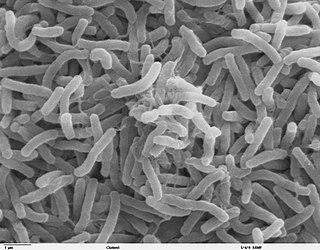
Vibrio cholerae is a species of Gram-negative, facultative anaerobe and comma-shaped bacteria. The bacteria naturally live in brackish or saltwater where they attach themselves easily to the chitin-containing shells of crabs, shrimp, and other shellfish. Some strains of V. cholerae are pathogenic to humans and cause a deadly disease called cholera, which can be derived from the consumption of undercooked or raw marine life species or drinking contaminated water.

<span class="mw-page-title-main">Exotoxin</span> Toxin from bacteria that destroys or disrupts cells

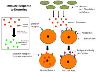
An exotoxin is a toxin secreted by bacteria. An exotoxin can cause damage to the host by destroying cells or disrupting normal cellular metabolism. They are highly potent and can cause major damage to the host. Exotoxins may be secreted, or, similar to endotoxins, may be released during lysis of the cell. Gram negative pathogens may secrete outer membrane vesicles containing lipopolysaccharide endotoxin and some virulence proteins in the bounding membrane along with some other toxins as intra-vesicular contents, thus adding a previously unforeseen dimension to the well-known eukaryote process of membrane vesicle trafficking, which is quite active at the host–pathogen interface.

<span class="mw-page-title-main">Enterotoxin</span> Toxin from a microorganism affecting the intestines

An enterotoxin is a protein exotoxin released by a microorganism that targets the intestines. They can be chromosomally or plasmid encoded. They are heat labile (>60⁰), of low molecular weight and water-soluble. Enterotoxins are frequently cytotoxic and kill cells by altering the apical membrane permeability of the mucosal (epithelial) cells of the intestinal wall. They are mostly pore-forming toxins, secreted by bacteria, that assemble to form pores in cell membranes. This causes the cells to die.

<span class="mw-page-title-main">ARF6</span> Protein-coding gene in the species Homo sapiens

ADP-ribosylation factor 6 (ARF6) is a member of the ADP ribosylation factor family of GTP-binding proteins. ARF6 has a variety of cellular functions that are frequently involved in trafficking of biological membranes and transmembrane protein cargo. ARF6 has specifically been implicated in endocytosis of plasma membrane proteins and also, to a lesser extent, plasma membrane protein recycling.

<span class="mw-page-title-main">ADP ribosylation factor</span> Group of proteins

ADP ribosylation factors (ARFs) are members of the ARF family of GTP-binding proteins of the Ras superfamily. ARF family proteins are ubiquitous in eukaryotic cells, and six highly conserved members of the family have been identified in mammalian cells. Although ARFs are soluble, they generally associate with membranes because of N-terminus myristoylation. They function as regulators of vesicular traffic and actin remodelling.

<span class="mw-page-title-main">GM1</span> Biochemical compound important in the brain and intestines

GM1 (monosialotetrahexosylganglioside) the "prototype" ganglioside, is a member of the ganglio series of gangliosides which contain one sialic acid residue. GM1 has important physiological properties and impacts neuronal plasticity and repair mechanisms, and the release of neurotrophins in the brain. Besides its function in the physiology of the brain, GM1 acts as the site of binding for both cholera toxin and E. coli heat-labile enterotoxin.

<span class="mw-page-title-main">Pore-forming toxin</span> Protein-produced toxins that create pores in cell membrane

Pore-forming proteins are usually produced by bacteria, and include a number of protein exotoxins but may also be produced by other organisms such as apple snails that produce perivitellin-2 or earthworms, who produce lysenin. They are frequently cytotoxic, as they create unregulated pores in the membrane of targeted cells.

The AB5 toxins are six-component protein complexes secreted by certain pathogenic bacteria known to cause human diseases such as cholera, dysentery, and hemolytic–uremic syndrome. One component is known as the A subunit, and the remaining five components are B subunits. All of these toxins share a similar structure and mechanism for entering targeted host cells. The B subunit is responsible for binding to receptors to open up a pathway for the A subunit to enter the cell. The A subunit is then able to use its catalytic machinery to take over the host cell's regular functions.

<span class="mw-page-title-main">ADP-ribosylation</span> Addition of one or more ADP-ribose moieties to a protein.

ADP-ribosylation is the addition of one or more ADP-ribose moieties to a protein. It is a reversible post-translational modification that is involved in many cellular processes, including cell signaling, DNA repair, gene regulation and apoptosis. Improper ADP-ribosylation has been implicated in some forms of cancer. It is also the basis for the toxicity of bacterial compounds such as cholera toxin, diphtheria toxin, and others.

<span class="mw-page-title-main">ARF1</span> Protein-coding gene in the species Homo sapiens

ADP-ribosylation factor 1 is a protein that in humans is encoded by the ARF1 gene.

<span class="mw-page-title-main">Sambhu Nath De</span>

Sambhunath De ; was an Indian medical scientist and researcher, who discovered the cholera toxin, the animal model of cholera, and successfully demonstrated the method of transmission of cholera pathogen Vibrio cholerae.

Microbial toxins are toxins produced by micro-organisms, including bacteria, fungi, protozoa, dinoflagellates, and viruses. Many microbial toxins promote infection and disease by directly damaging host tissues and by disabling the immune system. Endotoxins most commonly refer to the lipopolysaccharide (LPS) or lipooligosaccharide (LOS) that are in the outer plasma membrane of Gram-negative bacteria. The botulinum toxin, which is primarily produced by Clostridium botulinum and less frequently by other Clostridium species, is the most toxic substance known in the world. However, microbial toxins also have important uses in medical science and research. Currently, new methods of detecting bacterial toxins are being developed to better isolate and understand these toxins. Potential applications of toxin research include combating microbial virulence, the development of novel anticancer drugs and other medicines, and the use of toxins as tools in neurobiology and cellular biology.

<span class="mw-page-title-main">Heat-labile enterotoxin family</span> Family of toxic protein complexes

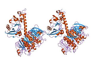
In molecular biology, the heat-labile enterotoxin family includes Escherichia coli heat-labile enterotoxin and cholera toxin (Ctx) secreted by Vibrio cholerae.

The CTXφ bacteriophage is a filamentous bacteriophage. It is a positive-strand DNA virus with single-stranded DNA (ssDNA).

Vibrio campbellii is a Gram-negative, curved rod-shaped, marine bacterium closely related to its sister species, Vibrio harveyi. It is an emerging pathogen in aquatic organisms.

John Mekalanos is a microbiologist who is primarily known for leading one of the first teams that reported the discovery of the type VI secretion system as well as his work on the pathogenicity of the bacterial species Vibrio cholerae, its toxin, and its secretion systems. Since 1998, he has been a member of the National Academy of Sciences.

Jan Roland Holmgren is a Swedish physician, microbiologist, immunologist, and vaccinologist, known for his research on cholera and mucosal immunology, specifically, for his leadership in developing "the world's first effective oral cholera vaccine".

Clathrin-independent endocytosis refers to the cellular process by which cells internalize extracellular molecules and particles through mechanisms that do not rely on the protein clathrin, playing a crucial role in diverse physiological processes such as nutrient uptake, membrane turnover, and cellular signaling.

References

  1. Ryan KJ; Ray CG, eds. (2004). Sherris Medical Microbiology (4th ed.). McGraw Hill. p. 375. ISBN   978-0-8385-8529-0.
  2. Faruque, Shah M; Nair, G. Balakrish, eds. (July 2008). Vibrio cholerae: Genomics and Molecular Biology. Caister Academic Press. ISBN   978-1-904455-33-2.
  3. Aizpurua-Olaizola, Oier; Sastre Torano, Javier; Pukin, Aliaksei; Fu, Ou; Boons, Geert Jan; de Jong, Gerhardus J.; Pieters, Roland J. (2018). "Affinity capillary electrophoresis for the assessment of binding affinity of carbohydrate-based cholera toxin inhibitors". Electrophoresis. 39 (2): 344–347. doi:10.1002/elps.201700207. ISSN   1522-2683. PMID   28905402. S2CID   33657660.
  4. Bharati, Kaushik; Ganguly, Nirmal K. (February 2011). "Cholera toxin: A paradigm of a multifunctional protein". The Indian Journal of Medical Research. 133 (2): 179–187. ISSN   0971-5916. PMC   3089049 . PMID   21415492.
  5. 1 2 De, S. N.; Sarkar, J. K.; Tribedi, B. P. (1951). "An experimental study of the action of cholera toxin". The Journal of Pathology and Bacteriology. 63 (4): 707–717. doi:10.1002/path.1700630417. PMID   14898376.
  6. De, S. N. (1959-05-30). "Enterotoxicity of bacteria-free culture-filtrate of Vibrio cholerae". Nature. 183 (4674): 1533–1534. doi:10.1038/1831533a0. ISSN   0028-0836. PMID   13666809.
  7. Bharati, Kaushik; Ganguly, Nirmal K. (February 2011). "Cholera toxin: A paradigm of a multifunctional protein". The Indian Journal of Medical Research. 133 (2): 179–187. ISSN   0971-5916. PMC   3089049 . PMID   21415492.
  8. Zhang R, Scott D, Westbrook M, Nance S, Spangler B, Shipley G, Westbrook E (1995). "The three-dimensional crystal structure of cholera toxin". J Mol Biol. 251 (4): 563–73. doi: 10.1006/jmbi.1995.0456 . PMID   7658473.
  9. De Haan L, Hirst TR (2004). "Cholera toxin: a paradigm for multi-functional engagement of cellular mechanisms (Review)". Mol. Membr. Biol. 21 (2): 77–92. doi: 10.1080/09687680410001663267 . PMID   15204437. S2CID   22270979.
  10. Amberlyn M Wands; Akiko Fujita (October 2015). "Fucosylation and protein glycosylation create functional receptors for Cholera toxin". eLife . 4: e09545. doi: 10.7554/eLife.09545 . PMC   4686427 . PMID   26512888.
  11. Cervin J, Wands AM, Casselbrant A, Wu H, Krishnamurthy S, Cvjetkovic A, et al. (2018). "GM1 ganglioside-independent intoxication by Cholera toxin". PLOS Pathogens . 14 (2): e1006862. doi: 10.1371/journal.ppat.1006862 . PMC   5825173 . PMID   29432456.
  12. Wands AM, Cervin J, Huang H, Zhang Y, Youn G, Brautigam CA, Matson Dzebo M, Björklund P, Wallenius V, Bright DK, Bennett CS, Wittung-Stafshede P, Sampson NS, Yrlid U, Kohler JJ (February 7, 2018). "Fucosylated Molecules Competitively Interfere with Cholera Toxin Binding to Host Cells". ACS Infectious Diseases . 4 (5): 758–770. doi:10.1021/acsinfecdis.7b00085. PMC   5948155 . PMID   29411974.
  13. Tsai, Billy; Rapoport, Tom A. (2002). "Unfolded cholera toxin is transferred to the ER membrane and released from protein disulfide isomerase upon oxidation by Ero1". Journal of Cell Biology. 159 (2): 207–216. doi:10.1083/jcb.200207120. PMC   2173060 . PMID   12403808. S2CID   3111310.
  14. O'Neal C, Jobling M, Holmes R, Hol W (2005). "Structural basis for the activation of cholera toxin by human ARF6-GTP". Science. 309 (5737): 1093–6. Bibcode:2005Sci...309.1093O. doi:10.1126/science.1113398. PMID   16099990. S2CID   8669389.
  15. Joaquín Sánchez; Jan Holmgren (February 2011). "Cholera toxin – A foe & a friend" (PDF). Indian Journal of Medical Research. 133: 158. Archived from the original (PDF) on 2013-02-03. Retrieved 2013-06-09.
  16. Boron, W. F., & Boulpaep, E. L. (2009). Medical physiology: a cellular and molecular approach (2nd ed.). Philadelphia, Pennsylvania: Saunders/Elsevier.
  17. 1 2 3 4 Davis, Brigid M; Waldor, Matthew K (2003-02-01). "Filamentous phages linked to virulence of Vibrio cholerae". Current Opinion in Microbiology. 6 (1): 35–42. doi:10.1016/S1369-5274(02)00005-X. ISSN   1369-5274.
  18. Pierre-Hervé Luppi. "The Discovery of Cholera-Toxin as a Powerful Neuroanatomical Tool" . Retrieved 2011-03-23.
  19. Androutsellis-Theotokis A, Walbridge S, Park DM, Lonser RR, McKay RD (2010). "Cholera toxin regulates a signaling pathway critical for the expansion of neural stem cell cultures from the fetal and adult rodent brains". PLOS ONE. 5 (5): e10841. Bibcode:2010PLoSO...510841A. doi: 10.1371/journal.pone.0010841 . PMC   2877108 . PMID   20520777.
  20. 1 2 Baldauf, Keegan; Royal, Joshua; Hamorsky, Krystal; Matoba, Nobuyuki (2015). "Cholera Toxin B: One Subunit with Many Pharmaceutical Applications". Toxins. 7 (3): 974–996. doi: 10.3390/toxins7030974 . PMC   4379537 . PMID   25802972.
  21. 1 2 3 Kenworthy, Anne K.; Schmieder, Stefanie S.; Raghunathan, Krishnan; Tiwari, Ajit; Wang, Ting; Kelly, Christopher V.; Lencer, Wayne I. (2021). "Cholera Toxin as a Probe for Membrane Biology". Toxins. 13 (8): 543. doi: 10.3390/toxins13080543 . PMC   8402489 . PMID   34437414.