Spermatocele

Last updated
Spermatocele
Spermatocele.JPG
Ultrasound of a testicle (grey) and a spermatocele (black).
Pronunciation
Specialty Urology

Spermatocele is a fluid-filled cyst that develops in the epididymis. [3] The fluid is usually a clear or milky white color and may contain sperm. [4] Spermatoceles are typically filled with spermatozoa [5] and they can vary in size from several millimeters to many centimeters. Small spermatoceles are relatively common, occurring in an estimated 30 percent of males. [6] They are generally not painful. However, some people may experience discomfort such as a dull pain in the scrotum from larger spermatoceles. [7] They are not cancerous, nor do they cause an increased risk of testicular cancer. Additionally, unlike varicoceles, they do not reduce fertility. [7]

Contents

History

"Spermatocele" is originally derived from the Greek term spermatos (sperm) and kele (cavity or mass). [8] Oftentimes, "epididymal cyst" has been used interchangeably with "spermatocele." However, it is important to note their differences. Epididymal cysts may appear anywhere along or within the epididymis and do not contain sperm, whereas spermatoceles may contain sperm. Epididymal cysts have been shown to occur more frequently in children before reaching puberty. [9]

Epidemiology

Spermatoceles usually affect men who are middle-aged and can, although rarely, affect children during puberty. [10] The incidence rate is around 5-20% for children. [11] It is estimated that approximately 30 percent of men have been diagnosed with small spermatoceles while less have larger spermatoceles. The incidence of spermatoceles increases as men age. [12] Before puberty, children from the male sex may develop a similar benign mass called epididymis cyst. Although both epididymis cyst and spermatocele may be referred as the same, the epididymis cyst does not contain sperm and it can occur anywhere within the epididymis. It can be differentiated through an ultrasound imagining. Epididymis cysts larger than 10mm in diameter are recommended for surgery but if there is no problem then surgery is discouraged as it can affect fertility in the future. [9]

Chronic infectious epididymitis

Chronic infectious epididymitis is rare. [13] Some signs and symptoms include localized tenderness and swelling in the epididymis, which are different from any tenderness/abnormality present in the testis, these are usually not found in lower urinary tract. Chronic infectious epididymitis may be diagnosed in healthy adolescents as well as men. Some factors that predispose individuals to chronic infectious epididymitis include sexual activity, heavy physical exertion, and bicycle or motorcycle riding. [14] Those diagnosed with chronic or recurrent epididymitis should receive a CT scan with contrast and a prostate ultrasonography to rule out structural abnormality of the urinary tract. If suspected to have chronic infectious epididymitis, one should consider getting a urinalysis, urine culture, and urine nucleic acid amplification tests for presence of Neisseria gonorrhoeae and Chlamydia trachomatis. [15] Management of chronic infectious epididymitis is similar to management of acute infectious epididymitis, rarely does treatment extend to surgical management. [16]

Chronic noninfectious epididymitis

Chronic noninfectious epididymitis – Trauma, autoimmune disease, or vasculitis can cause chronic noninfectious epididymitis, but no clear cause or origin of the disease is found in most cases. Noninfectious epididymitis that happens spontaneously might be caused by the reflux of urine through the ejaculatory ducts and vas deferens into the epididymis, producing inflammation that leads to swelling and ductal obstruction. [15] Men with history of vasectomy are also predisposed to chronic nonifectious epididymitis. Typical inciting factors include prolonged periods of sitting (long plane or car travel, sedentary desk jobs) or vigorous exercise (heavy lifting). Acute infectious epididymitis are often associated with more tenderness and swelling, whereas chronic noninfectious epididymitis tend to have less tenderness and swelling upon examination. Thorough past medical history and physical examination can help with determining the diagnosis. It is often that individuals with chronic noninfectious epididymitis will present with a history of a lack of symptom improvement while on antibiotic therapy. Management of chronic noninfectious epididymitis includes scrotal elevation, nonsteroidal anti-inflammatory drugs (NSAIDs) such as ibuprofen (unless unable to take for medical reasons), and it is recommended that individuals avoid physical activities that may cause said symptoms. Those with sedentary jobs or often experience prolonged periods of time sitting should practice physical mobility more frequently. [16]

Risk Factors

It is not well known what may be causing the growth of a spermatocele. It has been observed that pregnant women who were prescribed diethylstilbestrol (DES) to prevent pregnancy complications, such as miscarriage, and gave birth to a son most likely increased the risk of the son to develop a spermatocele in the future. However, doctors stopped prescribing this medication in 1971 since it increased the risk of women developing a rare vaginal cancer. [17]

Causes

Spermatoceles can originate as diverticulum from the tubules found in the head of the epididymis. Sperm accumulation gradually causes the diverticulum to increase in size, causing a spermatocele. While there are many tubules connecting the epididymis to the testis, a blockage in one of the tubules may lead to formation of a cyst. [18] In many instances they appear to occur spontaneously without any preceding instances of injury. [6]

Scarring of any part of the epididymis due to trauma or inflammation can cause it to become obstructed and in turn form a spermatocele. [4]

Diagnosis

Micrograph of a spermatocele. The characteristic sperm are present (black dots - left of image). H&E stain. Spermatocele - very high mag.jpg
Micrograph of a spermatocele. The characteristic sperm are present (black dots - left of image). H&E stain.
Dilated rete testis containing spermatozoa within cyst lumen. H&E stain 20x Spermato20x.jpg
Dilated rete testis containing spermatozoa within cyst lumen. H&E stain 20x

Spermatoceles can be discovered as incidental scrotal masses found on physical examination by a physician or by self-inspection of the scrotum and testicles. [7] The various types of diagnosis for spermatocele types include hydrocele, varicocele, hernia, simple epididymal cyst, and neoplasm. [8]

The primary care physician may diagnose and manage benign causes of scrotal masses such as hydrocele, varicocele and spermatocele. However, if a "must not miss" diagnosis related to testicular masses such as testicular torsion, epididymitis, acute orchitis, strangulated hernia and testicular cancer is suspected, the family physician must refer to a urologist. [19] Finding a painless, cystic mass at the head of the epididymis that is clearly separate from the testicle can indicate a spermatocele. Shining a light through the mass, a process known as transillumination, can also help differentiate between a fluid-filled cyst and a tumor, which would not allow as much light to pass. [20] If uncertainty exists, ultrasonography of the scrotum can confirm the presence of a spermatocele. [7] The location and history of any scrotal masses are crucial in determining whether or not the mass is benign or malignant. [21] Lab tests such as a complete blood count (CBC test) or urine test can also be conducted to check for any possible infection or signs of inflammation. [22]

Spermatoceles come in varying sizes and shapes. Some spermatoceles are very small and can only be detected through an ultrasound. More commonly seen are spermatoceles that are a pea-sized lump. They tend to form above or behind a testicle and have a shape and size that looks like a pea. Larger growths have been reported to look similar to a third testicle and can be very discomforting. [22] For those who are affected with large spermatoceles, some have reported feeling pain, heaviness, and fullness in the affected testicle. [20]

When to see a doctor

If a person is experiencing pain and/or swelling in the scrotum or if a person notices a physical difference such as a mass during a testicular self-exam then they must seek medical attention quickly to help rule out any other causes and begin treatment. [20]

Special Populations

Spermatoceles in Children

Epididymal cysts may arise in children due to numerous different reasons. They may even originate from a disturbance in endocrine factors during the development of the embryo. [9] The incidence of these cysts are possibly linked to boys who are exposed to diethylstilbestrol, an estrogen medication, while in the fetus. [9]

Although the evidence and information for incidence of spermatoceles in children is lacking, there are general management guidelines for caregivers who may encounter spermatoceles in their children. [9] Epididymal cysts are typically benign in nature. However, caregivers should take note of any discomfort and symptoms in children, including but not limited to, erythema, scrotal edema or swelling. [9] Epididymal cysts appear in approximately 70% of boys who present as asymptomatic. [9] The diagnosis of epididymal cysts in children can be discovered upon physical examination and eventually confirmed via ultrasound. [9]

Treatment

Small cysts as well as asymptomatic larger cysts are better left alone and carefully observed. However, treatment can be considered if the cysts are causing discomfort/pain, enlarging in size, or per patient request. There are a few different treatment options, ranging in levels of invasiveness. [7]

Medications

Certain drugs such as oral analgesics or anti-inflammatory medications can be taken by mouth to decrease spermatocele-related symptoms such as pain and/or swelling. At the moment, there is no medication that can inhibit the production of a spermatocele or cure it. Common medications recommended by doctors are acetaminophen (Tylenol) that can treat pain. To treat the inflammation and pain, non-steroidal anti-inflammatory drugs (NSAIDs) are recommended such as ibuprofen (Motrin, Advil), naproxen (Aleve), and others. [20] Antibiotics may also be prescribed in cases where risk of infection and discomfort is indicated. [8]

Medical Procedures

Aspiration and sclerotherapy are treatments that remove fluid from the spermatocele and seal the spermatocele sac closed from further fluid build-up, respectively. [7] Due to a higher risk of epididymis damage, fertility problems, and further recurrences, these procedures are not recommended and not commonly used.

Surgical Treatments

Surgical treatments must be thoroughly discussed with people prior to any final decisions. Different types of surgical procedures may be chosen based on the extent of risk. [23]

A short procedure called a spermatocelectomy can be performed for spermatoceles that cause irritating symptoms. This standard procedure can be performed in an outpatient setting with the use of local or general anesthesia. This procedure typically consists of removing the spermatocele and a portion of the epididymis. [7] The risk of epididymal injury is 17.12%. This incident may happen when the spermatocele is being dissected from the head of the epididymis. [6] Spermatocelectomy can cause complications such as hematoma, wound infection, scrotal abscess as well as recurrence. [8] After surgery, the doctor may recommend to apply ice packs for several days to help reduce swelling. Oral pain medications can also be taken for a few days to reduce discomfort. [7] In addition, the patient returns 2–6 weeks after to inspect any progress/complications. [8] If received a vasectomy, then two semen samples must be collected and analyzed after 6–12 weeks post-operation and several ejaculations. The centrifuged semen samples are used to look for the absence of viable sperm in the semen. [24]

Risk of Complications

After surgical removal, it is possible that the pain will persist and recurrence can occur. Fertility may be compromised with these surgical procedures, so people may consider postponing surgery until after having children. [20] If the pain is intolerable and the person would like to get the spermatocele removed immediately, the person should talk to his doctor about the possibility of freezing or donating sperm in case infertility occurs. [25]

Male Reproductive System

Image of the male reproductive system Human male reproductive system en.svg
Image of the male reproductive system

Spermatoceles are important to not be ignored as it can affect the male reproductive system. The testes are organs inside the scrotum that create sperm as well as sex hormones and testosterone. The sperm in the testes move to the epididymis which is a long, coiled tube behind the testes. The primary function is to store and mature sperm so it can fertilize the egg. However, if the epididymis becomes injured, then there is a chance that the sperm will not mature and a man will not be able to reproduce with a woman. Therefore, serious thought must be considered when undergoing spermatocelectomy. [26]

Prevention and Screening

There is no way to prevent a spermatocele from forming, but there are routines that can be established to help identify any changes in a person's scrotum such as masses, abnormalities, or discomfort. Performing a monthly testicular self-exam can improve the person's chances of identifying spermatoceles or any abnormalities quickly.

It is best to perform a testicular self-exam after a warm shower to help the scrotum relax. To properly exam the scrotum, look for any swelling on the skin and examine each testicle by rolling the testicle between the thumbs and the fingers. A normal testicle is oval-shaped and will usually feel smooth and firm. It is also not uncommon for the testicles to be different sizes. [4]

Upon physical examination, if a 'lump' is found during the testicular exam, further screening may be performed via ultrasound to eliminate testicular cancer. [18]

See also

Related Research Articles

<span class="mw-page-title-main">Testicular torsion</span> Medical condition

Testicular torsion occurs when the spermatic cord twists, cutting off the blood supply to the testicle. The most common symptom in children is sudden, severe testicular pain. The testicle may be higher than usual in the scrotum and vomiting may occur. In newborns, pain is often absent and instead the scrotum may become discolored or the testicle may disappear from its usual place.

<span class="mw-page-title-main">Hydrocele testis</span> Medical condition

A hydrocele testis is an accumulation of clear fluid within the cavum vaginale, the potential space between the layers of the tunica vaginalis of the testicle. It is the most common form of hydrocele and is often referred to simply as a "hydrocele". A primary hydrocele testis causes a painless enlargement in the scrotum on the affected side and is thought to be due to the defective absorption of fluid secreted between the two layers of the tunica vaginalis. A secondary hydrocele is secondary to either inflammation or a neoplasm in the testis.

<span class="mw-page-title-main">Epididymitis</span> Medical condition

Epididymitis is a medical condition characterized by inflammation of the epididymis, a curved structure at the back of the testicle. Onset of pain is typically over a day or two. The pain may improve with raising the testicle. Other symptoms may include swelling of the testicle, burning with urination, or frequent urination. Inflammation of the testicle is commonly also present.

<span class="mw-page-title-main">Epididymal hypertension</span> Condition that arises during male sexual arousal when seminal fluid is not ejaculated

Epididymal hypertension (EH), informally referred to as blue balls for males or blue vulva for females, is a harmless but uncomfortable sensation in the genital regions during a prolonged state of sexual arousal. It usually resolves within hours unless relieved through an orgasm.

<span class="mw-page-title-main">Testicular self-examination</span>

Testicular self-examination (TSE) is a procedure where a man examines his own testicles and scrotum for possible lumps or swelling. It is usually undertaken at home while standing in front of a mirror and after having a warm bath or shower. Monthly self-examination of the testicles starting at puberty may be an effective way of detecting testicular cancer at an early, potentially treatable stage, which can lead to a 5-year survival rate of 98%. In men aged 15 to 40, testicular cancer is the most common cancer, and the annual rate of increase over the last 10 years in cases of testicular cancer has been shown to be approximately 1% each year. Testicular cancer typically presents with a painless testicular swelling or lump or any change in shape or texture of the testicles.

Orchitis is inflammation of the testicles. It can also involve swelling, pains and frequent infection, particularly of the epididymis, as in epididymitis. The term is from the Ancient Greek ὄρχις meaning "testicle"; same root as orchid.

<span class="mw-page-title-main">Testicular atrophy</span> Reduction in the size and function of the testicles

Testicular atrophy is a medical condition in which one or both testicles diminish in size and may be accompanied by reduced testicular function. Testicular atrophy is not related to the temporary shrinkage of the surrounding scrotum, which might occur in response to cold temperature.

<span class="mw-page-title-main">Polyorchidism</span> Medical condition

Polyorchidism is the incidence of more than two testicles. It is a very rare congenital disorder, with fewer than 200 cases reported in medical literature and six cases in veterinary literature.

<span class="mw-page-title-main">Hydrocele</span> Accumulation of fluid in a body cavity

A hydrocele is an accumulation of serous fluid in a body cavity. A hydrocele testis, the most common form of hydrocele, is the accumulation of fluids around a testicle. It is often caused by fluid collecting within a layer wrapped around the testicle, called the tunica vaginalis, which is derived from peritoneum. Provided there is no hernia present, it goes away without treatment in the first year. Although hydroceles usually develop in males, rare instances have been described in females in the canal of Nuck.

<span class="mw-page-title-main">Varicocele</span> Medical condition

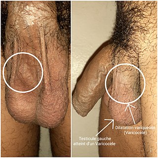
A varicocele is, in a male person, an abnormal enlargement of the pampiniform venous plexus in the scrotum; in a female person, it is an abnormal painful swelling to the embryologically identical pampiniform venous plexus; it is more commonly called pelvic compression syndrome. In the male varicocele, this plexus of veins drains blood from the testicles back to the heart. The vessels originate in the abdomen and course down through the inguinal canal as part of the spermatic cord on their way to the testis. Varicoceles occur in around 15% to 20% of all men. The incidence of varicocele increase with age.

<span class="mw-page-title-main">Hematocele</span> Medical condition

A hematocele is a collections of blood in a body cavity or potential space. The term most commonly refers to the collection of blood in the tunica vaginalis around the testes, known as a scrotal hematocele. Hematoceles can also occur in the abdominal cavity and other body cavities. Hematoceles are rare, making them harder to diagnose and treat. They are very common especially as slowly growing masses in the scrotum usually in men older than 50 years.

<span class="mw-page-title-main">Testicular pain</span> Medical condition

Testicular pain, also known as scrotal pain, occurs when part or all of either one or both testicles hurt. Pain in the scrotum is also often included. Testicular pain may be of sudden onset or of long duration.

<span class="mw-page-title-main">Epididymal cyst</span> Medical condition

Epididymal cyst is a harmless sac in the testicles filled with fluid. The most frequent clinical presentation occurs when a routine physical examination yields an unexpected finding, which is then confirmed by scrotal ultrasonography. Although the exact cause of epididymal cysts is unknown, it is likely a congenital anomaly associated with hormonal imbalances during the embryonic stage of development.

Post-vasectomy pain syndrome (PVPS) is a chronic and sometimes debilitating genital pain condition that may develop immediately or several years after vasectomy. Because this condition is a syndrome, there is no single treatment method, therefore efforts focus on mitigating/relieving the individual patient's specific pain. When pain in the epididymides is the primary symptom, post-vasectomy pain syndrome is often described as congestive epididymitis.

<span class="mw-page-title-main">Sperm granuloma</span> Lump of extravasated sperm found in some vasectomized men

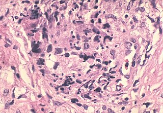
A sperm granuloma is a lump of leaked sperm that appears along the vasa deferentia or epididymides in vasectomized individuals. While the majority of sperm granulomas are present along the vas deferens, the rest of them form at the epididymis. Sperm granulomas range in size, from one millimeter to one centimeter. They consist of a central mass of degenerating sperm surrounded by tissue containing blood vessels and immune system cells. Sperm granulomas may also have a yellow, white, or cream colored center when cut open. While some sperm granulomas can be painful, most of them are painless and asymptomatic. Sperm granulomas can appear as a result of surgery, trauma, or an infection. They can appear as early as four days after surgery and fully formed ones can appear as late as 208 days later.

<span class="mw-page-title-main">Scrotum</span> Sac of skin that protects the testicles

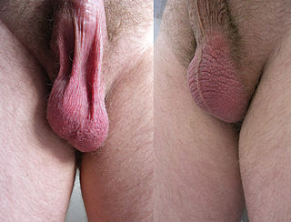
In most terrestrial mammals, the scrotum or scrotal sac is a part of the external male genitalia located at the base of the penis. It consists of a sac of skin containing the external spermatic fascia, testicles, epididymides, and vasa deferentia. The scrotum will usually tighten during penile erection and when exposed to cold temperatures.

Vasectomy reversal is a term used for surgical procedures that reconnect the male reproductive tract after interruption by a vasectomy. Two procedures are possible at the time of vasectomy reversal: vasovasostomy and vasoepididymostomy. Although vasectomy is considered a permanent form of contraception, advances in microsurgery have improved the success of vasectomy reversal procedures. The procedures remain technically demanding and may not restore the pre-vasectomy condition.

<span class="mw-page-title-main">Scrotal ultrasound</span> Medical ultrasound examination of the scrotum.

Scrotalultrasound is a medical ultrasound examination of the scrotum. It is used in the evaluation of testicular pain, and can help identify solid masses.

Male genital examination is a physical examination of the genital in males to detect ailments and to assess sexual development, and is normally a component of an annual physical examination. The examination includes checking the penis, scrotum, and urethral meatus. A comprehensive assessment of the male genitals assesses the pubic hair based on Sexual Maturity Rating and the size of the testicles and penis. The exam can also be conducted to verify a person's age and biological sex. The genitourinary system can also be assessed as part of the male genital examination. During a genital examination, the doctor can detect any of the following: structural abnormalities, urethral opening abnormalities, problems related to not being circumcised, lumps, tumors, redness, excoriation, edema, lesions, swelling, cancer, hair-related issues, and many others. In some instances where a physical examination of the male genitals is not sufficient to diagnose an individual, then an internal genital examination using imaging or ultrasounds will be needed for further evaluation.

References

  1. Entry "spermatocele" in Merriam-Webster Online Dictionary .
  2. OED 2nd edition, 1989.
  3. "Spermatocele: Practice Essentials, History of the Procedure, Problem". 18 May 2022. Retrieved 26 April 2023.
  4. 1 2 3 "Spermatocele - Symptoms and causes". Mayo Clinic. Retrieved 2020-07-28.
  5. Rioja, Jorge; Sánchez-Margallo, Francisco M.; Usón, Jesús; Rioja, Luis A. (2011). "Adult hydrocele and spermatocele". BJU International. 107 (11): 1852–1864. doi: 10.1111/j.1464-410X.2011.10353.x . ISSN   1464-410X. PMID   21592287. S2CID   27569964.
  6. 1 2 3 "Spermatocele: Symptoms, Causes, Treatments". Cleveland Clinic. Retrieved 2020-07-28.
  7. 1 2 3 4 5 6 7 8 "Spermatoceles: Symptoms, Diagnosis & Treatment - Urology Care Foundation". www.urologyhealth.org. Retrieved 2020-07-28.
  8. 1 2 3 4 5 "Spermatocele: Practice Essentials, History of the Procedure, Problem". 2021.{{cite journal}}: Cite journal requires |journal= (help)
  9. 1 2 3 4 5 6 7 8 Boscarelli, Alessandro; Bellini, Tommaso (2021). "Epididymal cyst in children". European Journal of Pediatrics. 180 (9): 2723–2729. doi:10.1007/s00431-021-04080-5. ISSN   1432-1076. PMID   33851241. S2CID   233225219.
  10. "Epididymal Cysts. Be clear about Epididymal Cysts. Patient". patient.info. Retrieved 2021-07-29.
  11. Bleve, Cosimo; Conighi, Maria Luisa; Bucci, Valeria; Costa, Lorenzo; Chiarenza, Salvatore Fabio (2018). "Torsion of huge epididymal cyst in a 16-year-old boy: case report and review of the literature". La Pediatria Medica e Chirurgica: Medical and Surgical Pediatrics. 40 (1). doi: 10.4081/pmc.2018.162 . ISSN   2420-7748. PMID   29871476. S2CID   46942959.
  12. "UroPartners - Spermatoceles". uropartners.com. Retrieved 2021-07-29.
  13. Rupp, Timothy J.; Leslie, Stephen W. (2021), "Epididymitis", StatPearls, Treasure Island (FL): StatPearls Publishing, PMID   28613565 , retrieved 2021-08-03
  14. "Epididymitis". Cancer Therapy Advisor. 2019. Retrieved 2021-08-03.
  15. 1 2 Trojian, Thomas; Lishnak, Timothy S.; Heiman, Diana L. (2009). "Epididymitis and Orchitis: An Overview". American Family Physician. 79 (7): 583–587. ISSN   0002-838X. PMID   19378875.
  16. 1 2 Holcomb, George (1988). "Epididymitis in infants and boys: Underlying urogenital anomalies and efficacy of imaging modalities". Journal of Pediatric Surgery. 23 (7): 684. doi:10.1016/s0022-3468(88)80691-2. ISSN   0022-3468.
  17. "Spermatocele". www.mayoclinic.org. Retrieved 2021-08-01.
  18. 1 2 "Spermatocele". The Urology Group of Virginia. Retrieved 2021-08-02.
  19. Junnila, Jennifer; Lassen, Patrick (1998). "Testicular Masses". American Family Physician. 57 (4): 685–692. ISSN   0002-838X. PMID   9490992.
  20. 1 2 3 4 5 "Spermatocele - Diagnosis and treatment - Mayo Clinic". www.mayoclinic.org. Retrieved 2021-08-01.
  21. Lee, Jerry C.; Bhatt, Shweta; Dogra, Vikram S. (2008). "Imaging of the Epididymis". Ultrasound Quarterly. 24 (1): 03–16. doi:10.1097/RUQ.0b013e318168f116. ISSN   0894-8771. PMID   18362528. S2CID   37488407.
  22. 1 2 "Spermatocele: Symptoms, Causes, Treatments". Cleveland Clinic. Retrieved 2021-08-01.
  23. Stuckmann, G (2006). "Sonographie des Skrotums". Praxis. 95 (18): 709–720. doi:10.1024/0369-8394.95.18.709. ISSN   1661-8157. PMID   16722401.
  24. Kliesch, S. (2014). "[Hydrocele, spermatocele, and vasectomy: management of complications]". Der Urologe. Ausg. A. 53 (5): 671–675. doi:10.1007/s00120-014-3486-4. ISSN   1433-0563. PMID   24806800.
  25. "Spermatocele Disease Reference Guide". Drugs.com. Retrieved 2021-08-01.
  26. "Male Reproductive System: Structure & Function". Cleveland Clinic. Retrieved 2021-08-01.