Trehalase

Last updated
trehalase (brush-border membrane glycoprotein)
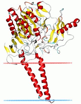
Trehalase.jpg
Trehalase [1]
Identifiers
SymbolTREH
NCBI gene 11181
HGNC 12266
OMIM 275360
RefSeq NM_007180
UniProt O43280
Other data
EC number 3.2.1.28
Locus Chr. 11 q23.3
Search for
Structures Swiss-model
Domains InterPro

The enzyme Trehalase is a glycoside hydrolase, produced by cells in the brush border of the small intestine, which catalyzes the conversion of trehalose to glucose. [2] [3] [4] [5] It is found in most animals.

Contents

The non-reducing disaccharide trehalose (α-D-glucopyranosyl-1,1-α-D-glucopyranoside) is one of the most important storage carbohydrates, and is produced by almost all forms of life except mammals. The disaccharide is hydrolyzed into two molecules of glucose by the enzyme trehalase. There are two types of trehalases found in Saccharomyces cerevisiae , viz. neutral trehalase (NT) and acid trehalase (AT) classified according to their pH optima [4]. NT has an optimum pH of 7.0, while that of AT is 4.5.

Recently it has been reported that more than 90% of total AT activity in S. cerevisiae is extracellular and cleaves extracellular trehalose into glucose in the periplasmic space.

Trehalose hydrolysis

One molecule of trehalose is hydrolyzed to two molecules of glucose by the enzyme trehalase. Enzymatic hydrolysis of trehalose was first observed in Aspergillus niger by Bourquelot in 1893. Fischer reported this reaction in S. cerevisiae in 1895. Since then the trehalose hydrolyzing enzyme, trehalase (α, α-trehalose-1-C-glucohydrolase, EC 3.2.1.28) has been reported from many other organisms including plants and animals. [6] Though trehalose is not known to be produced by mammals, trehalase enzyme is found to be present in the kidney brush border membrane and the intestinal villi membranes. In the intestine the function of this enzyme is to hydrolyze ingested trehalose. Individuals with a defect in their intestinal trehalase have diarrhea when they eat foods with high trehalose content, such as mushrooms. [6] Trehalose hydrolysis by trehalase enzyme is an important physiological process for various organisms, such as fungal spore germination, insect flight, and the resumption of growth in resting cells. [6]

Bacterial trehalase

Trehalose has been reported to be present as a storage carbohydrate in Pseudomonas, Bacillus, Rhizobium and in several actinomycetes and may be partially responsible for their resistance properties. Most of the trehalase enzymes isolated from bacteria have as optimum pH of 6.5–7.5. The trehalase enzyme of Mycobacterium smegmatis is a membrane bound protein. Periplasmic trehalase of Escherichia coli K12 is induced by growth at high osmolarity. The hydrolysis of trehalose into glucose takes place in the periplasm, and the glucose is then transported into the bacterial cell. Another cytoplasmic trehalase has also been reported from E. coli. The gene, which encodes this cytoplasmic trehalase, exhibits high homology to the periplasmic trehalase.

Trehalase in plants

In plant kingdom, though trehalose has been reported from several pteridophytes including Selaginella lepidophylla and Botrychium lunaria; the sugar is rare in vascular plants and reported only in ripening fruits of several members of Apiaceae and in the leaves of the desiccation-tolerant angiosperm Myrothamnus flabellifolius. But, the enzyme trehalase is ubiquitous in plants. This is puzzling that trehalase is present in higher plants, though its substrate is absent. No clear role has been demonstrated for trehalase activity in plants. However, trehalose inhibits the synthesis of its precursor, trehalose 6-phosphate, a plant metabolism regulator; [7] hence, its removal may be required. [8] It has been suggested that trehalases could play a role in defense mechanisms or the enzyme could play a role in the degradation of trehalose derived from plant-associated microorganisms.

Fungal trehalase

Two distinct trehalases have been reported from S. cerevisiae. One is regulated by cAMP-dependent phosphorylation and localized to the cytosol. The second trehalase activity is found in the vacuoles. The pH optimum of cytosolic trehalse was determined to be approximately 7.0 and is thus referred to as 'neutral trehalase' (NT); whereas the vacuolar trehalase is most active at pH 4.5 and consequently termed 'acid trehalase' (AT). These enzymes are encoded by two different genes – NTH1 and ATH1 respectively.

Neutral trehalase

The cytosolic trehalase enzyme, NT, has been purified and characterized extensively from S. cerevisiae. In non-denaturing gels this enzyme protein exhibited a molecular mass of 160 kDa, while in sodium dodecyl sulfate-polyacrylamide gel electrophoresis (SDS-PAGE) it showed a mass of 80 kDa. This hydrolase enzyme is specific for trehalose. The Km of NT has been reported to be 5.7mM. The gene responsible for trehalase activity in S. cerevisiae is NTH1. This gene is with an open reading frame of 2079 base pairs (bp), encoding a protein of 693 amino acids, corresponding to a molecular mass of 79569 Da.

NT activity is regulated by protein phosphorylation-dephosphorylation. Phosphorylation with cAMP-dependent protein kinase activates NT. Dephosphorylation of the purified phosphorylated enzyme by alkaline phosphatase caused an almost complete inactivation of the enzyme activity; but a recovery of the enzyme activity could be observed by rephosphorylation while incubating with ATP and protein kinase. The activity of NT in crude extracts is enhanced by polycations, while the activity of purified phosphorylated NT is inhibited by them. The activation of crude extracts was found to be due to the removal of polyphosphates, both of which inhibit NT activity.

Acid Trehalase

The molecular weight of AT was found to be 218 kDa by gel filtration chromatography. AT is a glycoprotein. It has 86% carbohydrate content. It has been reported that the maturation of AT is a stepwise process beginning with a carbohydrate-free 41 kDa protein; this form is then core-glycosylated in the endoplasmic reticulum to form a 76 kDa glycol-protein. In Golgi bodies, the protein is further glycosylated yielding a 180 kDa form, which ultimately attains a maturity in vacuoles, where its molecular weight becomes around 220 kDa. The 41 kDa carbohydrate free protein moiety of the enzyme was obtained by Endoglycosidase H treatment of purified AT, resulted after sodium dodecyl sulfate gel electrophoresis27. AT exhibited an apparent Km for trehalose of about 4.7 mM at pH 4.5. The gene responsible for AT activity in S. cerevisiae is ATH1.

Ath1p, i.e. AT, has been reported to be necessary for S. cerevisiae to utilize extracellular trehalose as carbon source16. ATH1 deletion mutant of the yeast could not grow in the medium with trehalose as the carbon source.

Researchers have suggested that AT moves from its site of synthesis to the periplasmic space, where it binds exogenous trehalose to internalize it and hydrolyze it in the vacuoles. Recently it has been shown that more than 90% of AT activity in S. cerevisiae is extracellular and the hydrolysis of trehalose into glucose takes place at the periplasmic space. Previously, a highly glycosylated protein, gp37, which is the product of YGP1 gene, was reported to be co-purified with AT activity. Invertase activity was also reported to be co-purified with AT activity. The physical association of AT with these two proteins was thought to suffice for the AT to be secreted by invertase and gp37 secretion pathways in absence of any known secretion signal for Ath1p.

In a Candida utils strain, one regulatory a one non-regulatory trehalase were also reported. These two enzymes were reported to be distinguishable by their molecular weight, behavior in ion-exchange chromatography and kinetic properties. The regulatory trehalase appeared to be a cytoplasmic enzyme and the nonregulatory enzyme was mostly detected in vacuoles. But, in a more recent report, a C. utils strain was demonstrated to lack any detectable AT activity but contain only NT activity. AT activity was not detectable in this strain, though the strain was shown to utilize extracellular trehalose as carbon source.

Related Research Articles

<span class="mw-page-title-main">Endomembrane system</span> Membranes in the cytoplasm of a eukaryotic cell

The endomembrane system is composed of the different membranes (endomembranes) that are suspended in the cytoplasm within a eukaryotic cell. These membranes divide the cell into functional and structural compartments, or organelles. In eukaryotes the organelles of the endomembrane system include: the nuclear membrane, the endoplasmic reticulum, the Golgi apparatus, lysosomes, vesicles, endosomes, and plasma (cell) membrane among others. The system is defined more accurately as the set of membranes that forms a single functional and developmental unit, either being connected directly, or exchanging material through vesicle transport. Importantly, the endomembrane system does not include the membranes of plastids or mitochondria, but might have evolved partially from the actions of the latter.

Hydrolysis is any chemical reaction in which a molecule of water breaks one or more chemical bonds. The term is used broadly for substitution, elimination, and solvation reactions in which water is the nucleophile.

<span class="mw-page-title-main">Lysosome</span> Cell organelle

A lysosome is a membrane-bound organelle found in many animal cells. They are spherical vesicles that contain hydrolytic enzymes that can break down many kinds of biomolecules. A lysosome has a specific composition, of both its membrane proteins, and its lumenal proteins. The lumen's pH (~4.5–5.0) is optimal for the enzymes involved in hydrolysis, analogous to the activity of the stomach. Besides degradation of polymers, the lysosome is involved in various cell processes, including secretion, plasma membrane repair, apoptosis, cell signaling, and energy metabolism.

<span class="mw-page-title-main">Cellulase</span> Class of enzymes

Cellulase is any of several enzymes produced chiefly by fungi, bacteria, and protozoans that catalyze cellulolysis, the decomposition of cellulose and of some related polysaccharides:

<span class="mw-page-title-main">Maltase</span> Enzyme

Maltase is one type of alpha-glucosidase enzymes located in the brush border of the small intestine. This enzyme catalyzes the hydrolysis of disaccharide maltose into two simple sugars of glucose. Maltase is found in plants, bacteria, yeast, humans, and other vertebrates. It is thought to be synthesized by cells of the mucous membrane lining the intestinal wall.

Deoxyribonuclease refers to a group of glycoprotein endonucleases which are enzymes that catalyze the hydrolytic cleavage of phosphodiester linkages in the DNA backbone, thus degrading DNA. The role of the DNase enzyme in cells includes breaking down extracellular DNA (ecDNA) excreted by apoptosis, necrosis, and neutrophil extracellular traps (NET) of cells to help reduce inflammatory responses that otherwise are elicited. A wide variety of deoxyribonucleases are known and fall into one of two families, which differ in their substrate specificities, chemical mechanisms, and biological functions. Laboratory applications of DNase include purifying proteins when extracted from prokaryotic organisms. Additionally, DNase has been applied as a treatment for diseases that are caused by ecDNA in the blood plasma. Assays of DNase are emerging in the research field as well.

<span class="mw-page-title-main">Trehalose</span> Chemical compound

Trehalose is a sugar consisting of two molecules of glucose. It is also known as mycose or tremalose. Some bacteria, fungi, plants and invertebrate animals synthesize it as a source of energy, and to survive freezing and lack of water.

<span class="mw-page-title-main">Cathepsin</span> Family of proteases

Cathepsins are proteases found in all animals as well as other organisms. There are approximately a dozen members of this family, which are distinguished by their structure, catalytic mechanism, and which proteins they cleave. Most of the members become activated at the low pH found in lysosomes. Thus, the activity of this family lies almost entirely within those organelles. There are, however, exceptions such as cathepsin K, which works extracellularly after secretion by osteoclasts in bone resorption. Cathepsins have a vital role in mammalian cellular turnover.

<span class="mw-page-title-main">Glycogen debranching enzyme</span> Mammalian protein found in Homo sapiens

A debranching enzyme is a molecule that helps facilitate the breakdown of glycogen, which serves as a store of glucose in the body, through glucosyltransferase and glucosidase activity. Together with phosphorylases, debranching enzymes mobilize glucose reserves from glycogen deposits in the muscles and liver. This constitutes a major source of energy reserves in most organisms. Glycogen breakdown is highly regulated in the body, especially in the liver, by various hormones including insulin and glucagon, to maintain a homeostatic balance of blood-glucose levels. When glycogen breakdown is compromised by mutations in the glycogen debranching enzyme, metabolic diseases such as Glycogen storage disease type III can result.

<span class="mw-page-title-main">Phytase</span> Class of enzymes

A phytase is any type of phosphatase enzyme that catalyzes the hydrolysis of phytic acid – an indigestible, organic form of phosphorus that is found in many plant tissues, especially in grains and oil seeds – and releases a usable form of inorganic phosphorus. While phytases have been found to occur in animals, plants, fungi and bacteria, phytases have been most commonly detected and characterized from fungi.

Phospholipase D (EC 3.1.4.4, lipophosphodiesterase II, lecithinase D, choline phosphatase, PLD; systematic name phosphatidylcholine phosphatidohydrolase) is an enzyme of the phospholipase superfamily that catalyses the following reaction
<span class="mw-page-title-main">Sulfatase</span> Class of enzymes

Sulfatases EC 3.1.6.- are enzymes of the esterase class that catalyze the hydrolysis of sulfate esters. These may be found on a range of substrates, including steroids, carbohydrates and proteins. Sulfate esters may be formed from various alcohols and amines. In the latter case the resultant N-sulfates can also be termed sulfamates.

<span class="mw-page-title-main">Hormone-sensitive lipase</span> Enzyme

Hormone-sensitive lipase (EC 3.1.1.79, HSL), also previously known as cholesteryl ester hydrolase (CEH), sometimes referred to as triacylglycerol lipase, is an enzyme that, in humans, is encoded by the LIPE gene, and catalyzes the following reaction:

<span class="mw-page-title-main">Myrosinase</span>

Myrosinase is a family of enzymes involved in plant defense against herbivores, specifically the mustard oil bomb. The three-dimensional structure has been elucidated and is available in the PDB.

<span class="mw-page-title-main">Sucrase-isomaltase</span>

Oligo-1,6-glucosidase is a glucosidase enzyme located on the brush border of the small intestine, which catalyses the following reaction:
<span class="mw-page-title-main">UTP—glucose-1-phosphate uridylyltransferase</span> Class of enzymes

UTP—glucose-1-phosphate uridylyltransferase also known as glucose-1-phosphate uridylyltransferase is an enzyme involved in carbohydrate metabolism. It synthesizes UDP-glucose from glucose-1-phosphate and UTP; i.e.,

An α,α-trehalase (EC 3.2.1.28) is an enzyme with systematic name α,α-trehalose glucohydrolase. This enzyme catalyzes the chemical reaction

In molecular biology, glycoside hydrolase family 37 is a family of glycoside hydrolases.

<span class="mw-page-title-main">Zingibain</span>

Zingibain, zingipain, or ginger protease is a cysteine protease enzyme found in ginger rhizomes. It catalyses the preferential cleavage of peptides with a proline residue at the P2 position. It has two distinct forms, ginger protease I (GP-I) and ginger protease II (GP-II).

<span class="mw-page-title-main">Trehalosamine</span>

Trehalosamines are amino sugars in which a hydroxyl group of trehalose is replaced with an amino group. While 2-, 3-, and 4-trehalosamine derived from actinomycetes have been reported as natural compounds, 6-trehalosamine has been reported as a synthetic compound. They have weak antimicrobial activity and could be considered as a class of aminoglycoside antibiotics. The properties and functions of 4-trehalosamine have been well investigated as follows.

References

  1. Gibson RP, Gloster TM, Roberts S, Warren RA, Storch de Gracia I, García A, et al. (2007). "Molecular basis for trehalase inhibition revealed by the structure of trehalase in complex with potent inhibitors". Angewandte Chemie. 46 (22): 4115–9. doi:10.1002/anie.200604825. PMID   17455176.
  2. Myrbäck K, Örtenblad B (1937). "Trehalose und Hefe. II. Trehalasewirkung von Hefepräparaten". Biochem. Z. 291: 61–69.
  3. Kalf GF, Rieder SV (February 1958). "The purification and properties of trehalase". The Journal of Biological Chemistry. 230 (2): 691–8. doi: 10.1016/S0021-9258(18)70491-2 . PMID   13525386.
  4. Hehre EJ, Sawai T, Brewer CF, Nakano M, Kanda T (June 1982). "Trehalase: stereocomplementary hydrolytic and glucosyl transfer reactions with alpha- and beta-D-glucosyl fluoride". Biochemistry. 21 (13): 3090–7. doi:10.1021/bi00256a009. PMID   7104311.
  5. Mori H, Lee JH, Okuyama M, Nishimoto M, Ohguchi M, Kim D, et al. (November 2009). "Catalytic reaction mechanism based on alpha-secondary deuterium isotope effects in hydrolysis of trehalose by European honeybee trehalase". Bioscience, Biotechnology, and Biochemistry. 73 (11): 2466–73. doi: 10.1271/bbb.90447 . PMID   19897915. S2CID   22774772.
  6. 1 2 3 Elbein AD, Pan YT, Pastuszak I, Carroll D (April 2003). "New insights on trehalose: a multifunctional molecule". Glycobiology. 13 (4): 17R–27R. doi: 10.1093/glycob/cwg047 . PMID   12626396.
  7. Paul MJ, Primavesi LF, Jhurreea D, Zhang Y (2008). "Trehalose metabolism and signaling". Annual Review of Plant Biology. 59: 417–41. doi:10.1146/annurev.arplant.59.032607.092945. PMID   18257709.
  8. Loewus FA, Tanner W, eds. (December 2012). Plant carbohydrates I: Intracellular carbohydrates. Berlin: Springer Science & Business Media. p. 274. doi:10.1007/978-3-642-68275-9_7. ISBN   978-3-642-68277-3.

See also