


DNA repair protein XRCC1, also known as X-ray repair cross-complementing protein 1, is a protein that in humans is encoded by the XRCC1 gene. XRCC1 is involved in DNA repair, where it complexes with DNA ligase III.
DNA repair protein XRCC1, also known as X-ray repair cross-complementing protein 1, is a protein that in humans is encoded by the XRCC1 gene. XRCC1 is involved in DNA repair, where it complexes with DNA ligase III.
| XRCC1_N | |||||||||
|---|---|---|---|---|---|---|---|---|---|
 nmr solution structure of the single-strand break repair protein xrcc1-n-terminal domain | |||||||||
| Identifiers | |||||||||
| Symbol | XRCC1_N | ||||||||
| Pfam | PF01834 | ||||||||
| Pfam clan | CL0202 | ||||||||
| InterPro | IPR002706 | ||||||||
| SCOP2 | 1xnt / SCOPe / SUPFAM | ||||||||
| |||||||||
XRCC1 is involved in the efficient repair of DNA single-strand breaks formed by exposure to ionizing radiation and alkylating agents. This protein interacts with DNA ligase III, polymerase beta and poly (ADP-ribose) polymerase to participate in the base excision repair pathway. It may play a role in DNA processing during meiogenesis, i.e. during the induction of meiosis and recombination in germ cells. A rare microsatellite polymorphism in this gene is associated with cancer in patients of varying radiosensitivity. [5]
The XRCC1 protein does not have enzymatic activity, but acts as a scaffolding protein that interacts with multiple repair enzymes. The scaffolding allows these repair enzymes to then carry out their enzymatic steps in repairing DNA. XRCC1 is involved in single-strand break repair, base excision repair and nucleotide excision repair. [6]
As reviewed by London, [6] XRCC1 protein has three globular domains connected by two linker segments of ~150 and 120 residues. The XRCC1 N-terminal domain binds to DNA polymerase beta, the C-terminal BRCT domain interacts with DNA ligase III alpha and the central domain contains a poly(ADP-ribose) binding motif. This central domain allows recruitment of XRCC1 to polymeric ADP-ribose that forms on PARP1 after PARP1 binds to single strand breaks. The first linker contains a nuclear localization sequence and also has a region that interacts with DNA repair protein REV1, and REV1 recruits translesion polymerases. The second linker interacts with polynucleotide kinase phosphatase ( PNKP) (that processes DNA broken ends during base excision repair), aprataxin (active in single-strand DNA repair and non-homologous end joining) and a third protein designated aprataxin- and PNKP-like factor.
XRCC1 has an essential role in microhomology-mediated end joining (MMEJ) repair of double strand breaks. MMEJ is a highly error-prone DNA repair pathway that results in deletion mutations. XRCC1 is one of 6 proteins required for this pathway. [7]
XRCC1 is over-expressed in non-small-cell lung carcinoma (NSCLC), [8] and at an even higher level in metastatic lymph nodes of NSCLC. [9]
Deficiency in XRCC1, due to being heterozygous for a mutated XRCC1 gene coding for a truncated XRCC1 protein, suppresses tumor growth in mice. [10] Under three experimental conditions for inducing three types of cancer (colon cancer, melanoma or breast cancer), mice heterozygous for this XRCC1 mutation had substantially lower tumor volume or number than wild type mice undergoing the same carcinogenic treatments.
Cancers are very often deficient in expression of one or more DNA repair genes, but over-expression of a DNA repair gene is less usual in cancer. For instance, at least 36 DNA repair proteins, when mutationally defective in germ line cells, cause increased risk of cancer (hereditary cancer syndromes).[ citation needed ] (Also see DNA repair-deficiency disorder.) Similarly, at least 12 DNA repair genes have frequently been found to be epigenetically repressed in one or more cancers.[ citation needed ] (See also Epigenetically reduced DNA repair and cancer.) Ordinarily, deficient expression of a DNA repair enzyme results in increased un-repaired DNA damages which, through replication errors (translesion synthesis), lead to mutations and cancer. However, XRCC1 mediated MMEJ repair is directly mutagenic, so in this case, over-expression, rather than under-expression, apparently leads to cancer. Reduction of mutagenic XRCC1 mediated MMEJ repair leads to reduced progression of cancer.
In aged human adipose-derived stem cells, base excision repair (BER), but not DNA double-strand break repair, is impaired. The XRCC1 protein, but not other BER factors, showed an age-associated decline. [11] Overexpression of XRCC1 reversed the age-associated decline of BER function.
Oxidative stress is increased in the brain during ischemic stroke leading to an increased burden on stress resistance mechanisms, including those for repairing oxidatively damaged DNA. Consequently any loss of a repair system that would ordinarily restore damaged DNA may impede survival and normal function of brain neurons. Ghosh et al. [12] reported that partial loss of XRCC1 function causes increased DNA damage in the brain and reduced recovery from ischemic stroke. This finding indicates that XRCC1-mediated base excision repair is important for speedy recovery from stroke.
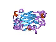
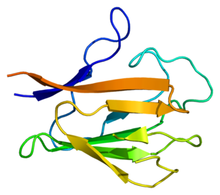
The NMR solution structure of the Xrcc1 N-terminal domain (Xrcc1 NTD) shows that the structural core is a beta-sandwich with beta-strands connected by loops, three helices and two short two-stranded beta-sheets at each connection side. The Xrcc1 NTD specifically binds single-strand break DNA (gapped and nicked) and a gapped DNA-beta-Pol complex. [13]
XRCC1 has been shown to interact with:

DNA ligase is a type of enzyme that facilitates the joining of DNA strands together by catalyzing the formation of a phosphodiester bond. It plays a role in repairing single-strand breaks in duplex DNA in living organisms, but some forms may specifically repair double-strand breaks. Single-strand breaks are repaired by DNA ligase using the complementary strand of the double helix as a template, with DNA ligase creating the final phosphodiester bond to fully repair the DNA.

DNA repair is a collection of processes by which a cell identifies and corrects damage to the DNA molecules that encodes its genome. In human cells, both normal metabolic activities and environmental factors such as radiation can cause DNA damage, resulting in tens of thousands of individual molecular lesions per cell per day. Many of these lesions cause structural damage to the DNA molecule and can alter or eliminate the cell's ability to transcribe the gene that the affected DNA encodes. Other lesions induce potentially harmful mutations in the cell's genome, which affect the survival of its daughter cells after it undergoes mitosis. As a consequence, the DNA repair process is constantly active as it responds to damage in the DNA structure. When normal repair processes fail, and when cellular apoptosis does not occur, irreparable DNA damage may occur. This can eventually lead to malignant tumors, or cancer as per the two-hit hypothesis.
RecQ helicase is a family of helicase enzymes initially found in Escherichia coli that has been shown to be important in genome maintenance. They function through catalyzing the reaction ATP + H2O → ADP + P and thus driving the unwinding of paired DNA and translocating in the 3' to 5' direction. These enzymes can also drive the reaction NTP + H2O → NDP + P to drive the unwinding of either DNA or RNA.

Nucleotide excision repair is a DNA repair mechanism. DNA damage occurs constantly because of chemicals, radiation and other mutagens. Three excision repair pathways exist to repair single stranded DNA damage: Nucleotide excision repair (NER), base excision repair (BER), and DNA mismatch repair (MMR). While the BER pathway can recognize specific non-bulky lesions in DNA, it can correct only damaged bases that are removed by specific glycosylases. Similarly, the MMR pathway only targets mismatched Watson-Crick base pairs.

Poly (ADP-ribose) polymerase (PARP) is a family of proteins involved in a number of cellular processes such as DNA repair, genomic stability, and programmed cell death.
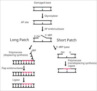
Base excision repair (BER) is a cellular mechanism, studied in the fields of biochemistry and genetics, that repairs damaged DNA throughout the cell cycle. It is responsible primarily for removing small, non-helix-distorting base lesions from the genome. The related nucleotide excision repair pathway repairs bulky helix-distorting lesions. BER is important for removing damaged bases that could otherwise cause mutations by mispairing or lead to breaks in DNA during replication. BER is initiated by DNA glycosylases, which recognize and remove specific damaged or inappropriate bases, forming AP sites. These are then cleaved by an AP endonuclease. The resulting single-strand break can then be processed by either short-patch or long-patch BER.

MUTYH is a human gene that encodes a DNA glycosylase, MUTYH glycosylase. It is involved in oxidative DNA damage repair and is part of the base excision repair pathway. The enzyme excises adenine bases from the DNA backbone at sites where adenine is inappropriately paired with guanine, cytosine, or 8-oxo-7,8-dihydroguanine, a common form of oxidative DNA damage.

DNA repair protein XRCC4 also known as X-ray repair cross-complementing protein 4 or XRCC4 is a protein that in humans is encoded by the XRCC4 gene. In addition to humans, the XRCC4 protein is also expressed in many other metazoans, fungi and in plants. The X-ray repair cross-complementing protein 4 is one of several core proteins involved in the non-homologous end joining (NHEJ) pathway to repair DNA double strand breaks (DSBs).

Histone-modifying enzymes are enzymes involved in the modification of histone substrates after protein translation and affect cellular processes including gene expression. To safely store the eukaryotic genome, DNA is wrapped around four core histone proteins, which then join to form nucleosomes. These nucleosomes further fold together into highly condensed chromatin, which renders the organism's genetic material far less accessible to the factors required for gene transcription, DNA replication, recombination and repair. Subsequently, eukaryotic organisms have developed intricate mechanisms to overcome this repressive barrier imposed by the chromatin through histone modification, a type of post-translational modification which typically involves covalently attaching certain groups to histone residues. Once added to the histone, these groups elicit either a loose and open histone conformation, euchromatin, or a tight and closed histone conformation, heterochromatin. Euchromatin marks active transcription and gene expression, as the light packing of histones in this way allows entry for proteins involved in the transcription process. As such, the tightly packed heterochromatin marks the absence of current gene expression.
ADP-ribosylation is the addition of one or more ADP-ribose moieties to a protein. It is a reversible post-translational modification that is involved in many cellular processes, including cell signaling, DNA repair, gene regulation and apoptosis. Improper ADP-ribosylation has been implicated in some forms of cancer. It is also the basis for the toxicity of bacterial compounds such as cholera toxin, diphtheria toxin, and others.

Poly [ADP-ribose] polymerase 1 (PARP-1) also known as NAD+ ADP-ribosyltransferase 1 or poly[ADP-ribose] synthase 1 is an enzyme that in humans is encoded by the PARP1 gene. It is the most abundant of the PARP family of enzymes, accounting for 90% of the NAD+ used by the family. PARP1 is mostly present in cell nucleus, but cytosolic fraction of this protein was also reported.

DNA ligase 1 is an enzyme that in humans is encoded by the LIG1 gene. DNA ligase I is the only known eukaryotic DNA ligase involved in both DNA replication and repair, making it the most studied of the ligases.

DNA polymerase beta, also known as POLB, is an enzyme present in eukaryotes. In humans, it is encoded by the POLB gene.

DNA excision repair protein ERCC-6 is a protein that in humans is encoded by the ERCC6 gene. The ERCC6 gene is located on the long arm of chromosome 10 at position 11.23.

Aprataxin is a protein that in humans is encoded by the APTX gene.

The gene polymerase delta 1 (POLD1) encodes the large, POLD1/p125, catalytic subunit of the DNA polymerase delta (Polδ) complex. The Polδ enzyme is responsible for synthesizing the lagging strand of DNA, and has also been implicated in some activities at the leading strand. The POLD1/p125 subunit encodes both DNA polymerizing and exonuclease domains, which provide the protein an important second function in proofreading to ensure replication accuracy during DNA synthesis, and in a number of types of replication-linked DNA repair following DNA damage.
Bifunctional polynucleotide phosphatase/kinase is an enzyme that in humans is encoded by the PNKP gene. A detailed structural study of the crystallized mouse protein examined both the 5´-polynucleotide kinase and 3’-polynucleotide phosphatase activities. Additional features of the peptide sequence include a forkhead association (FHA) domain, ATP binding site and nuclear and mitochondrial localization sequences.

Poly [ADP-ribose] polymerase 2 is an enzyme that in humans is encoded by the PARP2 gene. It is one of the PARP family of enzymes.
Microhomology-mediated end joining (MMEJ), also known as alternative nonhomologous end-joining (Alt-NHEJ) is one of the pathways for repairing double-strand breaks in DNA. As reviewed by McVey and Lee, the foremost distinguishing property of MMEJ is the use of microhomologous sequences during the alignment of broken ends before joining, thereby resulting in deletions flanking the original break. MMEJ is frequently associated with chromosome abnormalities such as deletions, translocations, inversions and other complex rearrangements.

DNA ligase 3 is an enzyme that, in humans, is encoded by the LIG3 gene. The human LIG3 gene encodes ATP-dependent DNA ligases that seal interruptions in the phosphodiester backbone of duplex DNA.
PDB gallery | |
|---|---|
|