Microhomology-mediated end joining

Last updated

Microhomology-mediated end joining (MMEJ), also known as alternative nonhomologous end-joining (Alt-NHEJ) is one of the pathways for repairing double-strand breaks in DNA. As reviewed by McVey and Lee, [1] the foremost distinguishing property of MMEJ is the use of microhomologous sequences during the alignment of broken ends before joining, thereby resulting in deletions flanking the original break. MMEJ is frequently associated with chromosome abnormalities such as deletions, translocations, inversions and other complex rearrangements.

Contents

There are multiple pathways for repairing double strand breaks, mainly non-homologous end joining (NHEJ), homologous recombination (HR), and MMEJ. NHEJ directly joins both ends of the double strand break and is relatively accurate, although small (usually less than a few nucleotides) insertions or deletions sometimes occur. HR is highly accurate and uses the sister chromatid as a template for accurate repair of the DSB. MMEJ is distinguished from these other repair mechanisms by its use of microhomologous sequences to align the broken strands. This results in frequent deletions and occasionally insertions which are much larger than those produced by NHEJ [ citation needed ]. MMEJ is completely independent from classical NHEJ and does not rely on NHEJ core factors such as Ku protein, DNA-PK, or Ligase IV. [2]

In MMEJ, repair of the DSB is initiated by end resection by the MRE nuclease, leaving single stranded overhangs. [3] These single stranded overhangs anneal at microhomologies, which are short regions of complementarity, often 5–25 base pairs, between the two strands. A specialized form of MMEJ, called polymerase theta-mediated end-joining (TMEJ), is able to repair breaks using ≥1 bp of homology. [4] [5] The helicase domain of DNA polymerase theta possesses ATP-dependent single-strand annealing activity and may promote annealing of microhomologies. [6] Following annealing, any overhanging bases (flaps) are removed by nucleases such as Fen1 and gaps are filled in by DNA polymerase theta. [7] This gap-filling ability of polymerase theta helps to stabilize the annealing of ends with minimal complementarity. Besides microhomology footprints, polymerase theta's mutational signature also consists of (infrequent) templated inserts, which are thought to be the result of aborted template-dependent extension, followed by re-annealing at secondary homologous sequences. [5]

Cell cycle regulation

MMEJ repair is low in G0/G1 phase but is increased during S-phase and G2 phase of the cell cycle. [3] In contrast, NHEJ operates throughout the cell cycle, and homologous recombination (HR) operates only in late S and G2.

Double strand break repair pathway choice

The choice of which pathway is used for double strand break repair is complex. In most cases, MMEJ accounts for a minor proportion (10%) of double strand break repair, most likely in cases where the double strand break is resected but a sister chromatid is not available for homologous recombination. [3] Cells which are deficient in either classical NHEJ or HR typically display increased MMEJ. Human homologous recombination factors suppress mutagenic MMEJ following double-strand break resection. [8]

Genes required

A biochemical assay system shows that at least 6 genes are required for microhomology-mediated end joining: FEN1, Ligase III, MRE11, NBS1, PARP1 and XRCC1. [9] All six of these genes are up-regulated in one or more cancers. In humans, DNA polymerase theta, encoded by the POLQ gene, plays a central role in microhomology-mediated end joining. [7] Polymerase theta utilizes its helicase domain to displace replication protein A (RPA) from DNA ends and promote microhomology annealing. [6] Polymerase theta also uses its polymerase activity to conduct fill-in synthesis, which helps to stabilize paired ends.

Helicase Q, which is conserved in humans, is necessary for polymerase theta-independent MMEJ, as shown using mutational footprint analyses in Caenorrhabditis elegans. [10]

In cancer

Approximately half of all ovarian cancers are deficient in homologous recombination (HR). These HR-deficient tumors upregulate polymerase theta (POLQ), resulting in an increase in MMEJ. [11] These tumors are hyper-reliant upon MMEJ, so that knockdown of polymerase theta results in substantial lethality. In most cell types, MMEJ makes a minor contribution to double strand break repair. The hyper-reliance of HR-deficient tumors upon MMEJ may represent a possible drug target for cancer treatment.

MMEJ always involves insertions or deletions, so that it is a mutagenic pathway. [12] Cells with increased MMEJ may have higher genomic instability and a predisposition towards cancer development, although this has not been demonstrated directly.

In a crustacean

Penaeus monodon is a marine crustacean widely consumed for its nutritional value. Repair of double-strand breaks in this organism can occur by HRR, but NHEJ is undetectable. [13] While HRR appears to be the major double-strand break repair pathway, MMEJ was also found to play a significant role in repair of DNA double-strand breaks. [13]

Related Research Articles

<span class="mw-page-title-main">DNA repair</span> Cellular mechanism

DNA repair is a collection of processes by which a cell identifies and corrects damage to the DNA molecules that encode its genome. In human cells, both normal metabolic activities and environmental factors such as radiation can cause DNA damage, resulting in tens of thousands of individual molecular lesions per cell per day. Many of these lesions cause structural damage to the DNA molecule and can alter or eliminate the cell's ability to transcribe the gene that the affected DNA encodes. Other lesions induce potentially harmful mutations in the cell's genome, which affect the survival of its daughter cells after it undergoes mitosis. As a consequence, the DNA repair process is constantly active as it responds to damage in the DNA structure. When normal repair processes fail, and when cellular apoptosis does not occur, irreparable DNA damage may occur, including double-strand breaks and DNA crosslinkages. This can eventually lead to malignant tumors, or cancer as per the two hit hypothesis.

RecQ helicase is a family of helicase enzymes initially found in Escherichia coli that has been shown to be important in genome maintenance. They function through catalyzing the reaction ATP + H2O → ADP + P and thus driving the unwinding of paired DNA and translocating in the 3' to 5' direction. These enzymes can also drive the reaction NTP + H2O → NDP + P to drive the unwinding of either DNA or RNA.

<span class="mw-page-title-main">Non-homologous end joining</span> Pathway that repairs double-strand breaks in DNA

Non-homologous end joining (NHEJ) is a pathway that repairs double-strand breaks in DNA. NHEJ is referred to as "non-homologous" because the break ends are directly ligated without the need for a homologous template, in contrast to homology directed repair(HDR), which requires a homologous sequence to guide repair. NHEJ is active in both non-dividing and proliferating cells, while HDR is not readily accessible in non-dividing cells. The term "non-homologous end joining" was coined in 1996 by Moore and Haber.

<i>Mycobacterium smegmatis</i> Species of bacterium

Mycobacterium smegmatis is an acid-fast bacterial species in the phylum Actinomycetota and the genus Mycobacterium. It is 3.0 to 5.0 µm long with a bacillus shape and can be stained by Ziehl–Neelsen method and the auramine-rhodamine fluorescent method. It was first reported in November 1884 by Lustgarten, who found a bacillus with the staining appearance of tubercle bacilli in syphilitic chancres. Subsequent to this, Alvarez and Tavel found organisms similar to that described by Lustgarten also in normal genital secretions (smegma). This organism was later named M. smegmatis.

<span class="mw-page-title-main">Homologous recombination</span> Genetic recombination between identical or highly similar strands of genetic material

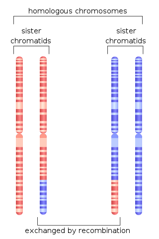
Homologous recombination is a type of genetic recombination in which genetic information is exchanged between two similar or identical molecules of double-stranded or single-stranded nucleic acids.

<span class="mw-page-title-main">Werner syndrome helicase</span>

Werner syndrome ATP-dependent helicase, also known as DNA helicase, RecQ-like type 3, is an enzyme that in humans is encoded by the WRN gene. WRN is a member of the RecQ Helicase family. Helicase enzymes generally unwind and separate double-stranded DNA. These activities are necessary before DNA can be copied in preparation for cell division. Helicase enzymes are also critical for making a blueprint of a gene for protein production, a process called transcription. Further evidence suggests that Werner protein plays a critical role in repairing DNA. Overall, this protein helps maintain the structure and integrity of a person's DNA.

<span class="mw-page-title-main">Ku (protein)</span>

Ku is a dimeric protein complex that binds to DNA double-strand break ends and is required for the non-homologous end joining (NHEJ) pathway of DNA repair. Ku is evolutionarily conserved from bacteria to humans. The ancestral bacterial Ku is a homodimer. Eukaryotic Ku is a heterodimer of two polypeptides, Ku70 (XRCC6) and Ku80 (XRCC5), so named because the molecular weight of the human Ku proteins is around 70 kDa and 80 kDa. The two Ku subunits form a basket-shaped structure that threads onto the DNA end. Once bound, Ku can slide down the DNA strand, allowing more Ku molecules to thread onto the end. In higher eukaryotes, Ku forms a complex with the DNA-dependent protein kinase catalytic subunit (DNA-PKcs) to form the full DNA-dependent protein kinase, DNA-PK. Ku is thought to function as a molecular scaffold to which other proteins involved in NHEJ can bind, orienting the double-strand break for ligation.

<span class="mw-page-title-main">Mutant</span> Phenotypically-different organism resulting from a mutation

In biology, and especially in genetics, a mutant is an organism or a new genetic character arising or resulting from an instance of mutation, which is generally an alteration of the DNA sequence of the genome or chromosome of an organism. It is a characteristic that would not be observed naturally in a specimen. The term mutant is also applied to a virus with an alteration in its nucleotide sequence whose genome is in the nuclear genome. The natural occurrence of genetic mutations is integral to the process of evolution. The study of mutants is an integral part of biology; by understanding the effect that a mutation in a gene has, it is possible to establish the normal function of that gene.

<span class="mw-page-title-main">DNA repair protein XRCC4</span> Protein-coding gene in the species Homo sapiens

DNA repair protein XRCC4 also known as X-ray repair cross-complementing protein 4 or XRCC4 is a protein that in humans is encoded by the XRCC4 gene. In addition to humans, the XRCC4 protein is also expressed in many other metazoans, fungi and in plants. The X-ray repair cross-complementing protein 4 is one of several core proteins involved in the non-homologous end joining (NHEJ) pathway to repair DNA double strand breaks (DSBs).

<span class="mw-page-title-main">PARP1</span> Mammalian protein found in Homo sapiens

Poly [ADP-ribose] polymerase 1 (PARP-1) also known as NAD+ ADP-ribosyltransferase 1 or poly[ADP-ribose] synthase 1 is an enzyme that in humans is encoded by the PARP1 gene. It is the most abundant of the PARP family of enzymes, accounting for 90% of the NAD+ used by the family. PARP1 is mostly present in cell nucleus, but cytosolic fraction of this protein was also reported.

<span class="mw-page-title-main">Flap structure-specific endonuclease 1</span>

Flap endonuclease 1 is an enzyme that in humans is encoded by the FEN1 gene.

<span class="mw-page-title-main">DNA polymerase lambda</span> Protein-coding gene in the species Homo sapiens

DNA polymerase lambda, also known as Pol λ, is an enzyme found in all eukaryotes. In humans, it is encoded by the POLL gene.

<span class="mw-page-title-main">ERCC4</span> Protein-coding gene in the species Homo sapiens

ERCC4 is a protein designated as DNA repair endonuclease XPF that in humans is encoded by the ERCC4 gene. Together with ERCC1, ERCC4 forms the ERCC1-XPF enzyme complex that participates in DNA repair and DNA recombination.

<span class="mw-page-title-main">POLQ</span>

DNA polymerase theta is an enzyme that in humans is encoded by the POLQ gene. This polymerase plays a key role in one of the three major double strand break repair pathways: theta-mediated end joining (TMEJ). Most double-strand breaks are repaired by non-homologous end joining (NHEJ) or homology directed repair (HDR). However, in some contexts, NHEJ and HR are insufficient and TMEJ is the only solution to repair the break. TMEJ is often described as alternative NHEJ, but differs in that it lacks a requirement for the Ku heterodimer, and it can only act on resected DNA ends. Following annealing of short regions on the DNA overhangs, DNA polymerase theta catalyzes template-dependent DNA synthesis across the broken ends, stabilizing the paired structure.

<span class="mw-page-title-main">DNA polymerase mu</span> Protein-coding gene

DNA polymerase mu is a polymerase enzyme found in eukaryotes. In humans, this protein is encoded by the POLM gene.

<span class="mw-page-title-main">Homology directed repair</span>

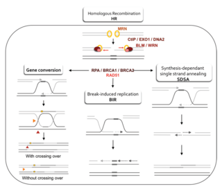
Homology-directed repair (HDR) is a mechanism in cells to repair double-strand DNA lesions. The most common form of HDR is homologous recombination. The HDR mechanism can only be used by the cell when there is a homologous piece of DNA present in the nucleus, mostly in G2 and S phase of the cell cycle. Other examples of homology-directed repair include single-strand annealing and breakage-induced replication. When the homologous DNA is absent, another process called non-homologous end joining (NHEJ) takes place instead.

<span class="mw-page-title-main">LIG3</span> Protein-coding gene in the species Homo sapiens

DNA ligase 3 is an enzyme that, in humans, is encoded by the LIG3 gene. The human LIG3 gene encodes ATP-dependent DNA ligases that seal interruptions in the phosphodiester backbone of duplex DNA.

<span class="mw-page-title-main">Synthesis-dependent strand annealing</span>

Synthesis-dependent strand annealing (SDSA) is a major mechanism of homology-directed repair of DNA double-strand breaks (DSBs). Although many of the features of SDSA were first suggested in 1976, the double-Holliday junction model proposed in 1983 was favored by many researchers. In 1994, studies of double-strand gap repair in Drosophila were found to be incompatible with the double-Holliday junction model, leading researchers to propose a model they called synthesis-dependent strand annealing. Subsequent studies of meiotic recombination in S. cerevisiae found that non-crossover products appear earlier than double-Holliday junctions or crossover products, challenging the previous notion that both crossover and non-crossover products are produced by double-Holliday junctions and leading the authors to propose that non-crossover products are generated through SDSA.

<span class="mw-page-title-main">DNA end resection</span> Biochemical process

DNA end resection, also called 5′–3′ degradation, is a biochemical process where the blunt end of a section of double-stranded DNA (dsDNA) is modified by cutting away some nucleotides from the 5' end to produce a 3' single-stranded sequence. The presence of a section of single-stranded DNA (ssDNA) allows the broken end of the DNA to line up accurately with a matching sequence, so that it can be accurately repaired.

<span class="mw-page-title-main">Double-strand break repair model</span>

A double-strand break repair model refers to the various models of pathways that cells undertake to repair double strand-breaks (DSB). DSB repair is an important cellular process, as the accumulation of unrepaired DSB could lead to chromosomal rearrangements, tumorigenesis or even cell death. In human cells, there are two main DSB repair mechanisms: Homologous recombination (HR) and non-homologous end joining (NHEJ). HR relies on undamaged template DNA as reference to repair the DSB, resulting in the restoration of the original sequence. NHEJ modifies and ligates the damaged ends regardless of homology. In terms of DSB repair pathway choice, most mammalian cells appear to favor NHEJ rather than HR. This is because the employment of HR may lead to gene deletion or amplification in cells which contains repetitive sequences. In terms of repair models in the cell cycle, HR is only possible during the S and G2 phases, while NHEJ can occur throughout whole process. These repair pathways are all regulated by the overarching DNA damage response mechanism. Besides HR and NHEJ, there are also other repair models which exists in cells. Some are categorized under HR, such as synthesis-dependent strain annealing, break-induced replication, and single-strand annealing; while others are an entirely alternate repair model, namely, the pathway microhomology-mediated end joining (MMEJ).

References

  1. McVey M, Lee SE (November 2008). "MMEJ repair of double-strand breaks (director's cut): deleted sequences and alternative endings". Trends in Genetics. 24 (11): 529–538. doi:10.1016/j.tig.2008.08.007. PMC   5303623 . PMID   18809224.
  2. Simsek D, Jasin M (April 2010). "Alternative end-joining is suppressed by the canonical NHEJ component Xrcc4-ligase IV during chromosomal translocation formation". Nature Structural & Molecular Biology. 17 (4): 410–416. doi:10.1038/nsmb.1773. PMC   3893185 . PMID   20208544.
  3. 1 2 3 Truong LN, Li Y, Shi LZ, Hwang PY, He J, Wang H, et al. (May 2013). "Microhomology-mediated End Joining and Homologous Recombination share the initial end resection step to repair DNA double-strand breaks in mammalian cells". Proceedings of the National Academy of Sciences of the United States of America. 110 (19): 7720–7725. Bibcode:2013PNAS..110.7720T. doi: 10.1073/pnas.1213431110 . PMC   3651503 . PMID   23610439.
  4. Roerink SF, van Schendel R, Tijsterman M (June 2014). "Polymerase theta-mediated end joining of replication-associated DNA breaks in C. elegans". Genome Research. 24 (6): 954–962. doi:10.1101/gr.170431.113. PMC   4032859 . PMID   24614976.
  5. 1 2 Schimmel J, van Schendel R, den Dunnen JT, Tijsterman M (September 2019). "Templated Insertions: A Smoking Gun for Polymerase Theta-Mediated End Joining". Trends in Genetics. 35 (9): 632–644. doi:10.1016/j.tig.2019.06.001. PMID   31296341. S2CID   195892718.
  6. 1 2 Mateos-Gomez PA, Kent T, Deng SK, McDevitt S, Kashkina E, Hoang TM, et al. (December 2017). "The helicase domain of Polθ counteracts RPA to promote alt-NHEJ". Nature Structural & Molecular Biology. 24 (12): 1116–1123. doi:10.1038/nsmb.3494. PMC   6047744 . PMID   29058711.
  7. 1 2 Sfeir A, Symington LS (November 2015). "Microhomology-Mediated End Joining: A Back-up Survival Mechanism or Dedicated Pathway?". Trends in Biochemical Sciences. 40 (11): 701–714. doi:10.1016/j.tibs.2015.08.006. PMC   4638128 . PMID   26439531.
  8. Ahrabi S, Sarkar S, Pfister SX, Pirovano G, Higgins GS, Porter AC, Humphrey TC (July 2016). "A role for human homologous recombination factors in suppressing microhomology-mediated end joining". Nucleic Acids Research. 44 (12): 5743–5757. doi:10.1093/nar/gkw326. PMC   4937322 . PMID   27131361.
  9. Sharma S, Javadekar SM, Pandey M, Srivastava M, Kumari R, Raghavan SC (March 2015). "Homology and enzymatic requirements of microhomology-dependent alternative end joining". Cell Death & Disease. 6 (3): e1697. doi:10.1038/cddis.2015.58. PMC   4385936 . PMID   25789972.
  10. Kamp JA, Lemmens BB, Romeijn RJ, Changoer SC, van Schendel R, Tijsterman M (December 2021). "Helicase Q promotes homology-driven DNA double-strand break repair and prevents tandem duplications". Nature Communications. 12 (1): 7126. Bibcode:2021NatCo..12.7126K. doi:10.1038/s41467-021-27408-z. PMC   8654963 . PMID   34880204.
  11. Ceccaldi R, Liu JC, Amunugama R, Hajdu I, Primack B, Petalcorin MI, et al. (February 2015). "Homologous-recombination-deficient tumours are dependent on Polθ-mediated repair". Nature. 518 (7538): 258–262. Bibcode:2015Natur.518..258C. doi:10.1038/nature14184. PMC   4415602 . PMID   25642963.
  12. Liang L, Deng L, Chen Y, Li GC, Shao C, Tischfield JA (September 2005). "Modulation of DNA end joining by nuclear proteins". The Journal of Biological Chemistry. 280 (36): 31442–31449. doi: 10.1074/jbc.M503776200 . PMID   16012167.
  13. 1 2 Srivastava S, Dahal S, Naidu SJ, Anand D, Gopalakrishnan V, Kooloth Valappil R, Raghavan SC (April 2017). "DNA double-strand break repair in Penaeus monodon is predominantly dependent on homologous recombination". DNA Research. 24 (2): 117–128. doi:10.1093/dnares/dsw059. PMC   5397610 . PMID   28431013.

General references