Z-DNA

Last updated
The Z-DNA structure. Proteopedia Z-DNA Z-DNA orbit animated small.gif
The Z-DNA structure. Proteopedia Z-DNA

Z-DNA is one of the many possible double helical structures of DNA. It is a left-handed double helical structure in which the helix winds to the left in a zigzag pattern, instead of to the right, like the more common B-DNA form. Z-DNA is thought to be one of three biologically active double-helical structures along with A-DNA and B-DNA.

Contents

History

Left-handed DNA was first proposed by Robert Wells and colleagues, as the structure of a repeating polymer of inosinecytosine. [1] They observed a "reverse" circular dichroism spectrum for such DNAs, and interpreted this incorrectly to mean that the strands wrapped around one another in a left-handed fashion. The relationship between Z-DNA and the more familiar B-DNA was indicated by the work of Pohl and Jovin, [2] who showed that the ultraviolet circular dichroism of poly(dG-dC) was nearly inverted in 4 M sodium chloride solution and that the structure of poly d(I–C)·poly d(I–C) was in fact a right-handed D-DNA conformation. The suspicion that this was the result of a conversion from B-DNA to Z-DNA was confirmed by examining the Raman spectra of these solutions and the Z-DNA crystals. [3] Subsequently, a crystal structure of "Z-DNA" was published which turned out to be the first single-crystal X-ray structure of a DNA fragment (a self-complementary DNA hexamer d(CG)3). It was resolved as a left-handed double helix with two antiparallel chains that were held together by Watson–Crick base pairs (see X-ray crystallography). It was solved by Andrew H. J. Wang, Alexander Rich, and coworkers in 1979 at MIT. [4] The crystallisation of a B- to Z-DNA junction in 2005 [5] provided a better understanding of the potential role Z-DNA plays in cells. Whenever a segment of Z-DNA forms, there must be B–Z junctions at its two ends, interfacing it to the B-form of DNA found in the rest of the genome.

In 2007, the RNA version of Z-DNA, Z-RNA, was described as a transformed version of an A-RNA double helix into a left-handed helix. [6] The transition from A-RNA to Z-RNA, however, was already described in 1984. [7]

Structure

B-Z junction bound to a Z-DNA binding domain. Note the two highlighted extruded bases. From PDB: 2ACJ . B-, Z-DNA junction 2ACJ.png
B–Z junction bound to a Z-DNA binding domain. Note the two highlighted extruded bases. From PDB: 2ACJ .

Z-DNA is quite different from the right-handed forms. In fact, Z-DNA is often compared against B-DNA in order to illustrate the major differences. The Z-DNA helix is left-handed and has a structure that repeats every other base pair. The major and minor grooves, unlike A- and B-DNA, show little difference in width. Formation of this structure is generally unfavourable, although certain conditions can promote it; such as alternating purinepyrimidine sequence (especially poly(dGC)2), negative DNA supercoiling or high salt and some cations (all at physiological temperature, 37 °C, and pH 7.3–7.4). Z-DNA can form a junction with B-DNA (called a "B-to-Z junction box") in a structure which involves the extrusion of a base pair. [8] The Z-DNA conformation has been difficult to study because it does not exist as a stable feature of the double helix. Instead, it is a transient structure that is occasionally induced by biological activity and then quickly disappears. [9]

Predicting Z-DNA structure

It is possible to predict the likelihood of a DNA sequence forming a Z-DNA structure. An algorithm for predicting the propensity of DNA to flip from the B-form to the Z-form, ZHunt, was written by P. Shing Ho in 1984 at MIT. [10] This algorithm was later developed by Tracy Camp, P. Christoph Champ, Sandor Maurice, and Jeffrey M. Vargason for genome-wide mapping of Z-DNA (with Ho as the principal investigator). [11]

Pathway of formation of Z-DNA from B-DNA

Since the discovery and crystallization of Z-DNA in 1979, the configuration has left scientists puzzled about the pathway and mechanism from the B-DNA configuration to the Z-DNA configuration. [12] The conformational change from B-DNA to the Z-DNA structure was unknown at the atomic level, but in 2010, computer simulations conducted by Lee et al. were able to computationally determine that the step-wise propagation of a B-to-Z transition would provide a lower energy barrier than the previously hypothesized concerted mechanism. [13] Since this was computationally proven, the pathway would still need to be tested experimentally in the lab for further confirmation and validity, in which Lee et al. specifically states in their journal article, "The current [computational] result could be tested by Single-molecule FRET (smFRET) experiments in the future." [13] In 2018, the pathway from B-DNA to Z-DNA was experimentally proven using smFRET assays. [14] This was performed by measuring the intensity values between the donor and acceptor fluorescent dyes, also known as Fluorophores, in relation to each other as they exchange electrons, while tagged onto a DNA molecule. [15] [16] The distances between the fluorophores could be used to quantitatively calculate the changes in proximity of the dyes and conformational changes in the DNA. A Z-DNA high affinity binding protein, hZαADAR1, [17] was used at varying concentrations to induce the transformation from B-DNA to Z-DNA. [14] The smFRET assays revealed a B* transition state, which formed as the binding of hZαADAR1 accumulated on the B-DNA structure and stabilized it. [14] This step occurs to avoid high junction energy, in which the B-DNA structure is allowed to undergo a conformational change to the Z-DNA structure without a major, disruptive change in energy. This result coincides with the computational results of Lee et al. proving the mechanism to be step-wise and its purpose being that it provides a lower energy barrier for the conformational change from the B-DNA to Z-DNA configuration. [13] Contrary to the previous notion, the binding proteins do not actually stabilize the Z-DNA conformation after it is formed, but instead they actually promote the formation of the Z-DNA directly from the B* conformation, which is formed by the B-DNA structure being bound by high affinity proteins. [14]

Biological significance

A biological role for Z-DNA in the regulation of type I interferon responses has been confirmed in studies of three well-characterized rare Mendelian Diseases: Dyschromatosis Symmetrica Hereditaria (OMIM: 127400), Aicardi-Goutières syndrome (OMIM: 615010) and Bilateral Striatal Necrosis/Dystonia. Families with haploid ADAR transcriptome enabled mapping of Zα variants directly to disease, showing that genetic information is encoded in DNA by both shape and sequence. [18] A role in regulating type I interferon responses in cancer is also supported by findings that 40% of a panel of tumors were dependent on the ADAR enzyme for survival. [19]

In previous studies, Z-DNA was linked to both Alzheimer's disease and systemic lupus erythematosus. To showcase this, a study was conducted on the DNA found in the hippocampus of brains that were normal, moderately affected with Alzheimer's disease, and severely affected with Alzheimer's disease. Through the use of circular dichroism, this study showed the presence of Z-DNA in the DNA of those severely affected. [20] In this study it was also found that major portions of the moderately affected DNA was in the B-Z intermediate conformation. This is significant because from these findings it was concluded that the transition from B-DNA to Z-DNA is dependent on the progression of Alzheimer's disease. [20] Additionally, Z-DNA is associated with systemic lupus erythematosus (SLE) through the presence of naturally occurring antibodies. Significant amounts of anti Z-DNA antibodies were found in SLE patients and were not present in other rheumatic diseases. [21] There are two types of these antibodies. Through radioimmunoassay, it was found that one interacts with the bases exposed on the surface of Z-DNA and denatured DNA, while the other exclusively interacts with the zig-zag backbone of only Z-DNA. Similar to that found in Alzheimer's disease, the antibodies vary depending on the stage of the disease, with maximal antibodies in the most active stages of SLE.

Z-DNA in transcription

Z-DNA is commonly believed to provide torsional strain relief during transcription, and it is associated with negative supercoiling. [5] [22] However, while supercoiling is associated with both DNA transcription and replication, Z-DNA formation is primarily linked to the rate of transcription. [23]

A study of human chromosome 22 showed a correlation between Z-DNA forming regions and promoter regions for nuclear factor I. This suggests that transcription in some human genes may be regulated by Z-DNA formation and nuclear factor I activation. [11]

Z-DNA sequences upstream of promoter regions have been shown to stimulate transcription. The greatest increase in activity is observed when the Z-DNA sequence is placed three helical turns after the promoter sequence. Furthermore, using micrococcal nuclease-crosslinking technique, [24] Z-DNA is unlikely to form nucleosomes, which are often located before and/or after a Z-DNA forming sequence. Because of this property, Z-DNA is hypothesized to code for the boundary in nucleosome positioning. Since the placement of nucleosomes influences the binding of transcription factors, Z-DNA is thought to regulate the rate of transcription. [24]

Developed behind the pathway of RNA polymerase through negative supercoiling, Z-DNA formed via active transcription has been shown to increase genetic instability, creating a propensity towards mutagenesis near promoters. [25] A study on Escherichia coli found that gene deletions spontaneously occur in plasmid regions containing Z-DNA-forming sequences. [26] In mammalian cells, the presence of such sequences was found to produce large genomic fragment deletions due to chromosomal double-strand breaks. Both of these genetic modifications have been linked to the gene translocations found in cancers such as leukemia and lymphoma, since breakage regions in tumor cells have been plotted around Z-DNA-forming sequences. [25] However, the smaller deletions in bacterial plasmids have been associated with replication slippage, while the larger deletions associated with mammalian cells are caused by non-homologous end-joining repair, which is known to be prone to error. [25] [26]

The toxic effect of ethidium bromide (EtBr) on trypanosomas is caused by shift of their kinetoplastid DNA to Z-form. The shift is caused by intercalation of EtBr and subsequent loosening of DNA structure that leads to unwinding of DNA, shift to Z-form and inhibition of DNA replication. [27]

Discovery of the Zα domain

The first domain to bind Z-DNA with high affinity was discovered in ADAR1 using an approach developed by Alan Herbert. [28] [29] Crystallographic and NMR studies confirmed the biochemical findings that this domain bound Z-DNA in a non-sequence-specific manner. [30] [31] [32] Related domains were identified in a number of other proteins through sequence homology. [29] The identification of the Zα domain provided a tool for other crystallographic studies that lead to the characterization of Z-RNA and the B–Z junction. Biological studies suggested that the Z-DNA binding domain of ADAR1 may localize this enzyme that modifies the sequence of the newly formed RNA to sites of active transcription. [33] [34] A role for Zα, Z-DNA and Z-RNA in defense of the genome against the invasion of Alu retro-elements in humans has evolved into a mechanism for the regulation of innate immune responses to dsRNA. Mutations in Zα are causal for human interferonopathies such as the Mendelian Aicardi-Goutières Syndrome. [35] [18] Additionally, Zα domains are demonstrated to localize at the stress granules because of their innate ability in binding nucleic acid. Furthermore, different Zα domains bind to the Z conformation of nucleic acid differently providing important avenues for specific targeting in drug discovery.

Consequences of Z-DNA binding to vaccinia E3L protein

As Z-DNA has been researched more thoroughly, it has been discovered that the structure of Z-DNA can bind to Z-DNA binding proteins through london dispersion and hydrogen bonding. [36] One example of a Z-DNA binding protein is the vaccinia E3L protein, which is a product of the E3L gene and mimics a mammalian protein that binds Z-DNA. [37] [38] Not only does the E3L protein have affinity to Z-DNA, it has also been found to play a role in the level of severity of virulence in mice caused by vaccinia virus, a type of poxvirus. Two critical components to the E3L protein that determine virulence are the N-terminus and the C-terminus. The N-terminus is made of up a sequence similar to that of the Zα domain, also called Adenosine deaminase z-alpha domain, while the C-terminus is composed of a double stranded RNA binding motif. [37] Through research done by Kim, Y. et al. at the Massachusetts Institute of Technology, it was shown that replacing the N-terminus of the E3L protein with a Zα domain sequence, containing 14 Z-DNA binding residues similar to E3L, had little to no effect on pathogenicity of the virus in mice. [37] In Contrast, Kim, Y. et al. also found that deleting all 83 residues of the E3L N-terminus resulted in decreased virulence. This supports their claim that the N-terminus containing the Z-DNA binding residues is necessary for virulence. [37] Overall, these findings show that the similar Z-DNA binding residues within the N-terminus of the E3L protein and the Zα domain are the most important structural factors determining virulence caused by the vaccinia virus, while amino acid residues not involved in Z-DNA binding have little to no effect. A future implication of these findings includes reducing Z-DNA binding of E3L in vaccines containing the vaccinia virus so negative reactions to the virus can be minimized in humans. [37]

Furthermore, Alexander Rich and Jin-Ah Kwon found that E3L acts as a transactivator for human IL-6, NF-AT, and p53 genes. Their results show that HeLa cells containing E3L had increased expression of human IL-6, NF-AT, and p53 genes and point mutations or deletions of certain Z-DNA binding amino acid residues decreased that expression. [36] Specifically, mutations in Tyr 48 and Pro 63 were found to reduce transactivation of the previously mentioned genes, as a result of loss of hydrogen bonding and london dispersion forces between E3L and the Z-DNA. [36] Overall, these results show that decreasing the bonds and interactions between Z-DNA and Z-DNA binding proteins decreases both virulence and gene expression, hence showing the importance of having bonds between Z-DNA and the E3L binding protein.

Comparison geometries of some DNA forms

Side view of A-, B-, and Z-DNA. A-DNA, B-DNA and Z-DNA.png
Side view of A-, B-, and Z-DNA.
The helix axis of A-, B-, and Z-DNA. B&Z&A DNA formula.svg
The helix axis of A-, B-, and Z-DNA.
Geometry attributes of A-, B, and Z-DNA [39] [40] [41]
A-formB-formZ-form
Helix senseright-handedright-handedleft-handed
Repeating unit1 bp1 bp2 bp
Rotation/bp32.7°34.3°30°
bp/turn111012
Inclination of bp to axis+19°−1.2°−9°
Rise/bp along axis2.3 Å (0.23 nm)3.32 Å (0.332 nm)3.8 Å (0.38 nm)
Pitch/turn of helix28.2 Å (2.82 nm)33.2 Å (3.32 nm)45.6 Å (4.56 nm)
Mean propeller twist+18°+16°
Glycosyl angle antiantiC: anti,
G: syn
Sugar pucker C3′-endoC2′-endoC: C2′-endo,
G: C3′-endo
Diameter23 Å (2.3 nm)20 Å (2.0 nm)18 Å (1.8 nm)

See also

Related Research Articles

Polyadenylation is the addition of a poly(A) tail to an RNA transcript, typically a messenger RNA (mRNA). The poly(A) tail consists of multiple adenosine monophosphates; in other words, it is a stretch of RNA that has only adenine bases. In eukaryotes, polyadenylation is part of the process that produces mature mRNA for translation. In many bacteria, the poly(A) tail promotes degradation of the mRNA. It, therefore, forms part of the larger process of gene expression.

<span class="mw-page-title-main">Tryptophan repressor</span> Transcription factor

Tryptophan repressor is a transcription factor involved in controlling amino acid metabolism. It has been best studied in Escherichia coli, where it is a dimeric protein that regulates transcription of the 5 genes in the tryptophan operon. When the amino acid tryptophan is plentiful in the cell, it binds to the protein, which causes a conformational change in the protein. The repressor complex then binds to its operator sequence in the genes it regulates, shutting off the genes.

<span class="mw-page-title-main">Nucleic acid double helix</span> Structure formed by double-stranded molecules

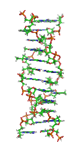
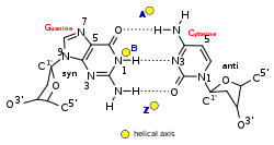
In molecular biology, the term double helix refers to the structure formed by double-stranded molecules of nucleic acids such as DNA. The double helical structure of a nucleic acid complex arises as a consequence of its secondary structure, and is a fundamental component in determining its tertiary structure.The structure was discovered by Rosalind Franklin and her student Raymond Gosling, but the term "double helix" entered popular culture with the publication in 1968 of The Double Helix: A Personal Account of the Discovery of the Structure of DNA by James Watson.

<i>trp</i> operon Operon that codes for the components for production of tryptophan

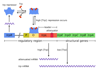
The trp operon is a group of genes that are transcribed together, encoding the enzymes that produce the amino acid tryptophan in bacteria. The trp operon was first characterized in Escherichia coli, and it has since been discovered in many other bacteria. The operon is regulated so that, when tryptophan is present in the environment, the genes for tryptophan synthesis are repressed.

Alexander Rich was an American biologist and biophysicist. He was the William Thompson Sedgwick Professor of Biophysics at MIT and Harvard Medical School. Rich earned an A.B. and an M.D. from Harvard University. He was a post-doc of Linus Pauling. During this time he was a member of the RNA Tie Club, a social and discussion group which attacked the question of how DNA encodes proteins. He had over 600 publications to his name.

<i>Fok</i>I

The restriction endonuclease Fok1, naturally found in Flavobacterium okeanokoites, is a bacterial type IIS restriction endonuclease consisting of an N-terminal DNA-binding domain and a non sequence-specific DNA cleavage domain at the C-terminal. Once the protein is bound to duplex DNA via its DNA-binding domain at the 5'-GGATG-3' recognition site, the DNA cleavage domain is activated and cleaves the DNA at two locations, regardless of the nucleotide sequence at the cut site. The DNA is cut 9 nucleotides downstream of the motif on the forward strand, and 13 nucleotides downstream of the motif on the reverse strand, producing two sticky ends with 4-bp overhangs.

<span class="mw-page-title-main">N-Myc</span> Protein-coding gene in the species Homo sapiens

N-myc proto-oncogene protein also known as N-Myc or basic helix-loop-helix protein 37 (bHLHe37), is a protein that in humans is encoded by the MYCN gene.

cAMP receptor protein Regulatory protein in bacteria

cAMP receptor protein is a regulatory protein in bacteria. CRP protein binds cAMP, which causes a conformational change that allows CRP to bind tightly to a specific DNA site in the promoters of the genes it controls. CRP then activates transcription through direct protein–protein interactions with RNA polymerase.

<span class="mw-page-title-main">Eukaryotic transcription</span> Transcription is heterocatalytic function of DNA

Eukaryotic transcription is the elaborate process that eukaryotic cells use to copy genetic information stored in DNA into units of transportable complementary RNA replica. Gene transcription occurs in both eukaryotic and prokaryotic cells. Unlike prokaryotic RNA polymerase that initiates the transcription of all different types of RNA, RNA polymerase in eukaryotes comes in three variations, each translating a different type of gene. A eukaryotic cell has a nucleus that separates the processes of transcription and translation. Eukaryotic transcription occurs within the nucleus where DNA is packaged into nucleosomes and higher order chromatin structures. The complexity of the eukaryotic genome necessitates a great variety and complexity of gene expression control.

<span class="mw-page-title-main">PCBP1</span> Protein-coding gene in the species Homo sapiens

Poly(rC)-binding protein 1 is a protein that in humans is encoded by the PCBP1 gene.

<span class="mw-page-title-main">ADAR</span> Mammalian protein found in Homo sapiens

The double-stranded RNA-specific adenosine deaminase enzyme family are encoded by the ADAR family genes. ADAR stands for adenosine deaminase acting on RNA. This article focuses on the ADAR proteins; This article details the evolutionary history, structure, function, mechanisms and importance of all proteins within this family.

<span class="mw-page-title-main">G3BP1</span> Protein-coding gene in the species Homo sapiens

Ras GTPase-activating protein-binding protein 1 is an enzyme that in humans is encoded by the G3BP1 gene.

<i>CSDA</i> (gene) Protein-coding gene in humans

DNA-binding protein A is a protein that in humans is encoded by the CSDA gene.

<span class="mw-page-title-main">PCBP3</span> Gene of the species Homo sapiens

Poly(rC)-binding protein 3 is a protein that in humans is encoded by the PCBP3 gene.

<span class="mw-page-title-main">NFX1</span> Protein-coding gene in the species Homo sapiens

Transcriptional repressor NF-X1 is a protein that in humans is encoded by the NFX1 gene.

<span class="mw-page-title-main">ZBP1</span> Protein-coding gene in the species Homo sapiens

Z-DNA-binding protein 1, also known as DNA-dependent activator of IFN-regulatory factors (DAI) and DLM-1, is a protein that in humans is encoded by the ZBP1 gene.

<span class="mw-page-title-main">B recognition element</span>

The B recognition element (BRE) is a DNA sequence found in the promoter region of most genes in eukaryotes and Archaea. The BRE is a cis-regulatory element that is found immediately near TATA box, and consists of 7 nucleotides. There are two sets of BREs: one (BREu) found immediately upstream of the TATA box, with the consensus SSRCGCC; the other (BREd) found around 7 nucleotides downstream, with the consensus RTDKKKK.

Zinc finger transcription factors or ZF-TFs, are transcription factors composed of a zinc finger-binding domain and any of a variety of transcription-factor effector-domains that exert their modulatory effect in the vicinity of any sequence to which the protein domain binds.

<span class="mw-page-title-main">Adenosine deaminase z-alpha domain</span>

In molecular biology, the protein domain Adenosine deaminase z-alpha domain refers to an evolutionary conserved protein domain. This family consists of the N-terminus and thus the z-alpha domain of double-stranded RNA-specific adenosine deaminase (ADAR), an RNA-editing enzyme. The z-alpha domain is a Z-DNA binding domain, and binding of this region to B-DNA has been shown to be disfavoured by steric hindrance.

Kevin Struhl is an American molecular biologist and the David Wesley Gaiser Professor of Biological Chemistry and Molecular Pharmacology at Harvard Medical School. Struhl is primarily known for his work on transcriptional regulatory mechanisms in yeast using molecular, genetic, biochemical, and genomic approaches. More recently, he has used related approaches to study transcriptional regulatory circuits involved in cellular transformation and the formation of cancer stem cells.

References

  1. Mitsui, Y.; Langridge, R.; Shortle, B. E.; Cantor, C. R.; Grant, R. C.; Kodama, M.; Wells, R. D. (1970). "Physical and enzymatic studies on poly d(I–C)·poly d(I–C), an unusual double-helical DNA". Nature. 228 (5277): 1166–1169. Bibcode:1970Natur.228.1166M. doi:10.1038/2281166a0. PMID   4321098. S2CID   4248932.
  2. Pohl, F. M.; Jovin, T. M. (1972). "Salt-induced co-operative conformational change of a synthetic DNA: equilibrium and kinetic studies with poly(dG-dC)". Journal of Molecular Biology. 67 (3): 375–396. doi:10.1016/0022-2836(72)90457-3. PMID   5045303.
  3. Thamann, T. J.; Lord, R. C.; Wang, A. H.; Rich, A. (1981). "High salt form of poly(dG–dC)·poly(dG–dC) is left handed Z-DNA: raman spectra of crystals and solutions". Nucleic Acids Research. 9 (20): 5443–5457. doi:10.1093/nar/9.20.5443. PMC   327531 . PMID   7301594.
  4. Wang, A. H.; Quigley, G. J.; Kolpak, F. J.; Crawford, J. L.; van Boom, J. H.; van der Marel, G.; Rich, A. (1979). "Molecular structure of a left-handed double helical DNA fragment at atomic resolution". Nature. 282 (5740): 680–686. Bibcode:1979Natur.282..680W. doi:10.1038/282680a0. PMID   514347. S2CID   4337955.
  5. 1 2 Ha, S. C.; Lowenhaupt, K.; Rich, A.; Kim, Y. G.; Kim, K. K. (2005). "Crystal structure of a junction between B-DNA and Z-DNA reveals two extruded bases". Nature. 437 (7062): 1183–1186. Bibcode:2005Natur.437.1183H. doi:10.1038/nature04088. PMID   16237447. S2CID   2539819.
  6. Placido, D.; Brown, B. A. II; Lowenhaupt, K.; Rich, A.; Athanasiadis, A. (2007). "A left-handed RNA double helix bound by the Zalpha domain of the RNA-editing enzyme ADAR1". Structure. 15 (4): 395–404. doi:10.1016/j.str.2007.03.001. PMC   2082211 . PMID   17437712.
  7. Hall, K.; Cruz, P.; Tinoco, I. Jr; Jovin, T. M.; van de Sande, J. H. (Oct 1984). "'Z-RNA'—a left-handed RNA double helix". Nature. 311 (5986): 584–586. Bibcode:1984Natur.311..584H. doi:10.1038/311584a0. PMID   6482970. S2CID   4316862.
  8. de Rosa, M.; de Sanctis, D.; Rosario, A. L.; Archer, M.; Rich, A.; Athanasiadis, A.; Carrondo, M. A. (May 2010). "Crystal structure of a junction between two Z-DNA helices". Proceedings of the National Academy of Sciences. 107 (20): 9088–9092. Bibcode:2010PNAS..107.9088D. doi: 10.1073/pnas.1003182107 . PMC   2889044 . PMID   20439751.
  9. Zhang, H.; Yu, H.; Ren, J.; Qu, X. (2006). "Reversible B/Z-DNA transition under the low salt condition and non-B-form poly(dA)poly(dT) selectivity by a cubane-like europium-L-aspartic acid complex". Biophysical Journal. 90 (9): 3203–3207. Bibcode:2006BpJ....90.3203Z. doi:10.1529/biophysj.105.078402. PMC   1432110 . PMID   16473901.
  10. Ho, P. S.; Ellison, M. J.; Quigley, G. J.; Rich, A. (1986). "A computer aided thermodynamic approach for predicting the formation of Z-DNA in naturally occurring sequences". EMBO Journal. 5 (10): 2737–2744. doi:10.1002/j.1460-2075.1986.tb04558.x. PMC   1167176 . PMID   3780676.
  11. 1 2 Champ, P. C.; Maurice, S.; Vargason, J. M.; Camp, T.; Ho, P. S. (2004). "Distributions of Z-DNA and nuclear factor I in human chromosome 22: a model for coupled transcriptional regulation". Nucleic Acids Research. 32 (22): 6501–6510. doi:10.1093/nar/gkh988. PMC   545456 . PMID   15598822.
  12. Wang, Andrew H.-J.; Quigley, Gary J.; Kolpak, Francis J.; Crawford, James L.; van Boom, Jacques H.; van der Marel, Gijs; Rich, Alexander (December 1979). "Molecular structure of a left-handed double helical DNA fragment at atomic resolution". Nature. 282 (5740): 680–686. Bibcode:1979Natur.282..680W. doi:10.1038/282680a0. ISSN   0028-0836. PMID   514347. S2CID   4337955.
  13. 1 2 3 Lee, Juyong; Kim, Yang-Gyun; Kim, Kyeong Kyu; Seok, Chaok (2010-08-05). "Transition between B-DNA and Z-DNA: Free Energy Landscape for the B−Z Junction Propagation". The Journal of Physical Chemistry B. 114 (30): 9872–9881. CiteSeerX   10.1.1.610.1717 . doi:10.1021/jp103419t. ISSN   1520-6106. PMID   20666528.
  14. 1 2 3 4 Kim, Sook Ho; Lim, So-Hee; Lee, Ae-Ree; Kwon, Do Hoon; Song, Hyun Kyu; Lee, Joon-Hwa; Cho, Minhaeng; Johner, Albert; Lee, Nam-Kyung (2018-03-23). "Unveiling the pathway to Z-DNA in the protein-induced B–Z transition". Nucleic Acids Research. 46 (8): 4129–4137. doi:10.1093/nar/gky200. ISSN   0305-1048. PMC   5934635 . PMID   29584891.
  15. Cooper, David; Uhm, Heui; Tauzin, Lawrence J.; Poddar, Nitesh; Landes, Christy F. (2013-06-03). "Photobleaching Lifetimes of Cyanine Fluorophores Used for Single-Molecule Förster Resonance Energy Transfer in the Presence of Various Photoprotection Systems". ChemBioChem. 14 (9): 1075–1080. doi:10.1002/cbic.201300030. ISSN   1439-4227. PMC   3871170 . PMID   23733413.
  16. Didenko, Vladimir V. (November 2001). "DNA Probes Using Fluorescence Resonance Energy Transfer (FRET): Designs and Applications". BioTechniques. 31 (5): 1106–1121. doi:10.2144/01315rv02. ISSN   0736-6205. PMC   1941713 . PMID   11730017.
  17. Herbert, A.; Alfken, J.; Kim, Y.-G.; Mian, I. S.; Nishikura, K.; Rich, A. (1997-08-05). "A Z-DNA binding domain present in the human editing enzyme, double-stranded RNA adenosine deaminase". Proceedings of the National Academy of Sciences. 94 (16): 8421–8426. Bibcode:1997PNAS...94.8421H. doi: 10.1073/pnas.94.16.8421 . ISSN   0027-8424. PMC   22942 . PMID   9237992.
  18. 1 2 Herbert, A. (2019). "Mendelian disease caused by variants affecting recognition of Z-DNA and Z-RNA by the Zα domain of the double-stranded RNA editing enzyme ADAR". European Journal of Human Genetics. 8 (1): 114–117. doi:10.1038/s41431-019-0458-6. PMC   6906422 . PMID   31320745.
  19. Herbert, A. (2019). "ADAR and Immune Silencing in Cancer". Trends in Cancer. 5 (5): 272–282. doi:10.1016/j.trecan.2019.03.004. PMID   31174840. S2CID   155209484.
  20. 1 2 Suram, Anitha; Rao, Jagannatha K. S.; S., Latha K.; A., Viswamitra M. (2002). "First Evidence to Show the Topological Change of DNA from B-DNA to Z-DNA Conformation in the Hippocampus of Alzheimer's Brain". NeuroMolecular Medicine. 2 (3): 289–298. doi:10.1385/nmm:2:3:289. ISSN   1535-1084. PMID   12622407. S2CID   29059186.
  21. Lafer, E M; Valle, R P; Möller, A; Nordheim, A; Schur, P H; Rich, A; Stollar, B D (1983-02-01). "Z-DNA-specific antibodies in human systemic lupus erythematosus". Journal of Clinical Investigation. 71 (2): 314–321. doi:10.1172/jci110771. ISSN   0021-9738. PMC   436869 . PMID   6822666.
  22. Rich, A; Zhang, S (2003). "Timeline: Z-DNA: the long road to biological function". Nature Reviews Genetics. 4 (7): 566–572. doi:10.1038/nrg1115. PMID   12838348. S2CID   835548.
  23. Wittig, B.; Dorbic, T.; Rich, A. (1991). "Transcription is associated with Z-DNA formation in metabolically active permeabilized mammalian cell nuclei". Proceedings of the National Academy of Sciences. 88 (6): 2259–2263. Bibcode:1991PNAS...88.2259W. doi: 10.1073/pnas.88.6.2259 . PMC   51210 . PMID   2006166.
  24. 1 2 Wong, B.; Chen, S.; Kwon, J.-A.; Rich, A. (2007). "Characterization of Z-DNA as a nucleosome-boundary element in yeast Saccharomyces cerevisiae". Proceedings of the National Academy of Sciences. 104 (7): 2229–2234. Bibcode:2007PNAS..104.2229W. doi: 10.1073/pnas.0611447104 . PMC   1892989 . PMID   17284586.
  25. 1 2 3 Wang, G.; Christensen, L. A.; Vasquez, K. M. (2006). "Z-DNA-forming sequences generate large-scale deletions in mammalian cells". Proceedings of the National Academy of Sciences. 108 (8): 2677–2682. Bibcode:2006PNAS..103.2677W. doi: 10.1073/pnas.0511084103 . PMC   1413824 . PMID   16473937.
  26. 1 2 Freund, A. M.; Bichara, M.; Fuchs, R. P. (1989). "Z-DNA-forming sequences are spontaneous deletion hot spots". Proceedings of the National Academy of Sciences. 86 (19): 7465–7469. Bibcode:1989PNAS...86.7465F. doi: 10.1073/pnas.86.19.7465 . PMC   298085 . PMID   2552445.
  27. Roy Chowdhury, A.; Bakshi, R.; Wang, J.; Yıldırır, G.; Liu, B.; Pappas-Brown, V.; Tolun, G.; Griffith, J. D.; Shapiro, T. A.; Jensen, R. E.; Englund, P. T. (Dec 2010). "The killing of African trypanosomes by ethidium bromide". PLOS Pathogens. 6 (12): e1001226. doi: 10.1371/journal.ppat.1001226 . PMC   3002999 . PMID   21187912.
  28. Herbert, A.; Rich, A. (1993). "A method to identify and characterize Z-DNA binding proteins using a linear oligodeoxynucleotide". Nucleic Acids Research. 21 (11): 2669–2672. doi:10.1093/nar/21.11.2669. PMC   309597 . PMID   8332463.
  29. 1 2 Herbert, A.; Alfken, J.; Kim, Y. G.; Mian, I. S.; Nishikura, K.; Rich, A. (1997). "A Z-DNA binding domain present in the human editing enzyme, double-stranded RNA adenosine deaminase". Proceedings of the National Academy of Sciences. 94 (16): 8421–8426. Bibcode:1997PNAS...94.8421H. doi: 10.1073/pnas.94.16.8421 . PMC   22942 . PMID   9237992.
  30. Herbert, A.; Schade, M.; Lowenhaupt, K.; Alfken, J; Schwartz, T.; Shlyakhtenko, L. S.; Lyubchenko, Y. L.; Rich, A. (1998). "The Zα domain from human ADAR1 binds to the Z-DNA conformer of many different sequences". Nucleic Acids Research. 26 (15): 2669–2672. doi:10.1093/nar/26.15.3486. PMC   147729 . PMID   9671809.
  31. Schwartz, T.; Rould, M. A.; Lowenhaupt, K.; Herbert, A.; Rich, A. (1999). "Crystal structure of the Zα domain of the human editing enzyme ADAR1 bound to left-handed Z-DNA". Science. 284 (5421): 1841–1845. doi:10.1126/science.284.5421.1841. PMID   10364558.
  32. Schade, M.; Turner, C. J.; Kühne, R.; Schmieder, P.; Lowenhaupt, K.; Herbert, A.; Rich, A.; Oschkinat, H (1999). "The solution structure of the Zα domain of the human RNA editing enzyme ADAR1 reveals a prepositioned binding surface for Z-DNA". Proceedings of the National Academy of Sciences. 96 (22): 2465–2470. Bibcode:1999PNAS...9612465S. doi: 10.1073/pnas.96.22.12465 . PMC   22950 . PMID   10535945.
  33. Herbert, A.; Rich, A. (2001). "The role of binding domains for dsRNA and Z-DNA in the in vivo editing of minimal substrates by ADAR1". Proceedings of the National Academy of Sciences. 98 (21): 12132–12137. Bibcode:2001PNAS...9812132H. doi: 10.1073/pnas.211419898 . PMC   59780 . PMID   11593027.
  34. Halber, D. (1999-09-11). "Scientists observe biological activities of 'left-handed' DNA". MIT News Office. Retrieved 2008-09-29.
  35. Herbert, A. (2019). "Z-DNA and Z-RNA in human disease". Communications Biology. 2: 7. doi:10.1038/s42003-018-0237-x. PMC   6323056 . PMID   30729177.
  36. 1 2 3 Kwon, J.-A.; Rich, A. (2005-08-26). "Biological function of the vaccinia virus Z-DNA-binding protein E3L: Gene transactivation and antiapoptotic activity in HeLa cells". Proceedings of the National Academy of Sciences. 102 (36): 12759–12764. Bibcode:2005PNAS..10212759K. doi: 10.1073/pnas.0506011102 . ISSN   0027-8424. PMC   1200295 . PMID   16126896.
  37. 1 2 3 4 5 Kim, Y.-G.; Muralinath, M.; Brandt, T.; Pearcy, M.; Hauns, K.; Lowenhaupt, K.; Jacobs, B. L.; Rich, A. (2003-05-30). "A role for Z-DNA binding in vaccinia virus pathogenesis". Proceedings of the National Academy of Sciences. 100 (12): 6974–6979. Bibcode:2003PNAS..100.6974K. doi: 10.1073/pnas.0431131100 . ISSN   0027-8424. PMC   165815 . PMID   12777633.
  38. Kim, Y.-G.; Lowenhaupt, K.; Oh, D.-B.; Kim, K. K.; Rich, A. (2004-02-02). "Evidence that vaccinia virulence factor E3L binds to Z-DNA in vivo: Implications for development of a therapy for poxvirus infection". Proceedings of the National Academy of Sciences. 101 (6): 1514–1518. Bibcode:2004PNAS..101.1514K. doi: 10.1073/pnas.0308260100 . ISSN   0027-8424. PMC   341766 . PMID   14757814.
  39. Sinden, Richard R. (1994). DNA Structure and Function (1st ed.). Academic Press. p. 398. ISBN   978-0-126-45750-6.
  40. Rich, A.; Norheim, A.; Wang, A. H. (1984). "The chemistry and biology of left-handed Z-DNA". Annual Review of Biochemistry. 53 (1): 791–846. doi:10.1146/annurev.bi.53.070184.004043. PMID   6383204.
  41. Ho, P. S. (1994-09-27). "The non-B-DNA structure of d(CA/TG)n does not differ from that of Z-DNA". Proceedings of the National Academy of Sciences. 91 (20): 9549–9553. Bibcode:1994PNAS...91.9549H. doi: 10.1073/pnas.91.20.9549 . PMC   44850 . PMID   7937803.