Argyll Robertson pupil

Last updated
Argyll Robertson pupil
Argyll Robertson pupil light reflex vs accommodation reflex.jpg
Specialty Neurology   OOjs UI icon edit-ltr-progressive.svg
Risk factors A highly specific sign of neurosyphilis
Diagnostic method Pupillary light reflex and accommodation reflex tests

Argyll Robertson pupils (AR pupils) are bilateral small pupils that reduce in size on a near object (i.e., they accommodate), but do not constrict when exposed to bright light (i.e., they do not react). They are a highly specific sign of neurosyphilis; however, Argyll Robertson pupils may also be a sign of diabetic neuropathy. In general, pupils that accommodate but do not react are said to show light-near dissociation (i.e., it is the absence of a miotic reaction to light, both direct and consensual, with the preservation of a miotic reaction to near stimulus (accommodation/convergence). [1]

Contents

AR pupils are extremely uncommon in the developed world. There is continued interest in the underlying pathophysiology, but the scarcity of cases makes ongoing research difficult.

Pathophysiology

The two different types of near response are caused by different underlying disease processes. Adie's pupil is caused by damage to peripheral pathways to the pupil (parasympathetic neurons in the ciliary ganglion that cause pupillary constriction to bright light and with near vision). The pathophysiologic mechanism which produces an Argyll Robertson pupil is unclear, but is believed to be the result of bilateral damage to the pretectal nuclei in the midbrain. Studies have failed to demonstrate a focal localising lesion. Research has implicated the rostral midbrain in the vicinity of the cerebral aqueduct of the third ventricle as the most likely region of damage. A lesion in this area would involve efferent pupillary fibres on the dorsal aspect of the Edinger-Westphal nucleus (associated with the response to light) while sparing the fibres associated with the response to near, which lie slightly more ventrally. [2] The exact relationship between syphilis and the two types of pupils (AR pupils and tonic pupils) is not known at the present time. The older literature on AR pupils did not report the details of pupillary constriction (brisk vs. tonic) that are necessary to distinguish AR pupils from tonic pupils. Tonic pupils can occur in neurosyphilis. [3] It is not known whether neurosyphilis itself (infection by Treponema pallidum) can cause tonic pupils, or whether tonic pupils in syphilis simply reflect a coexisting peripheral neuropathy.

Thompson and Kardon [4] summarize the present view:

The evidence supports a midbrain cause of the AR pupil, provided one follows Loewenfeld’s definition of the AR pupil as small pupils that react very poorly to light and yet seem to retain a normal pupillary near response that is definitely not tonic.
To settle the question of whether the AR pupil is of central or peripheral origin, it will be necessary to perform iris transillumination (or a magnified slit-lamp examination) in a substantial number of patients who have a pupillary light-near dissociation (with and without tonicity of the near reaction), perhaps in many parts of the world.

Parinaud syndrome

A third cause of light-near dissociation is Parinaud syndrome, also called dorsal midbrain syndrome. This uncommon syndrome involves vertical gaze palsy associated with pupils that “accommodate but do not react." [5] The causes of Parinaud syndrome include brain tumors (pinealomas), multiple sclerosis and brainstem infarction.

Due to the lack of detail in the older literature and the scarcity of AR pupils at the present time, it is not known whether syphilis can cause Parinaud syndrome. It is not known whether AR pupils are any different from the pupils seen in other dorsal midbrain lesions.

The condition is diagnosed clinically by a physician.

Treatment

There is no definite treatment, but, because syphilis may be an underlying cause, it should be treated. However, because this sign is associated with neurosyphilis, it should be treated with crystalline penicillin 24 mU intravenous per day for 10 to 14 days. If the patient is allergic to penicillin, they should undergo desensitization and then be treated.

History

Argyll Robertson pupils were named after Douglas Argyll Robertson (1837–1909), a Scottish ophthalmologist and surgeon who described the condition in the mid-1860s in the context of neurosyphilis.

In the early 20th century, William John Adie described a second type of pupil that could "accommodate but not react". Adie's tonic pupil is usually associated with a benign peripheral neuropathy (Adie syndrome), not with syphilis. [6]

When penicillin became widely available in the 1940s, the prevalence of AR pupils (which develop only after decades of untreated infection) decreased dramatically. AR pupils are now quite rare. A patient whose pupil "accommodates but does not react" almost always has a tonic pupil, not an AR pupil.

In the 1950s, Loewenfeld distinguished between the two types of pupils by carefully observing the exact way in which the pupils constrict with near vision. [7] The near response in AR pupils is brisk and immediate. The near response in tonic pupils is slow and prolonged.

See also

Related Research Articles

<span class="mw-page-title-main">Pupil</span> Part of an eye

The pupil is a hole located in the center of the iris of the eye that allows light to strike the retina. It appears black because light rays entering the pupil are either absorbed by the tissues inside the eye directly, or absorbed after diffuse reflections within the eye that mostly miss exiting the narrow pupil. The size of the pupil is controlled by the iris, and varies depending on many factors, the most significant being the amount of light in the environment. The term "pupil" was coined by Gerard of Cremona.

General paresis, also known as general paralysis of the insane (GPI), paralytic dementia, or syphilitic paresis is a severe neuropsychiatric disorder, classified as an organic mental disorder, and is caused by late-stage syphilis and the chronic meningoencephalitis and cerebral atrophy that are associated with this late stage of the disease when left untreated. GPI differs from mere paresis, as mere paresis can result from multiple other causes and usually does not affect cognitive function. Degenerative changes caused by GPI are associated primarily with the frontal and temporal lobar cortex. The disease affects approximately 7% of individuals infected with syphilis, and is far more common in developing countries where fewer options for timely treatment are available. It is more common among men.

<span class="mw-page-title-main">Tabes dorsalis</span> Medical condition of late-stage neurosyphilis

Tabes dorsalis is a late consequence of neurosyphilis, characterized by the slow degeneration of the neural tracts primarily in the dorsal root ganglia of the spinal cord. These patients have lancinating nerve root pain which is aggravated by coughing, and features of sensory ataxia with ocular involvement.

The ankle jerk reflex, also known as the Achilles reflex, occurs when the Achilles tendon is tapped while the foot is dorsiflexed. It is a type of stretch reflex that tests the function of the gastrocnemius muscle and the nerve that supplies it. A positive result would be the jerking of the foot towards its plantar surface. Being a deep tendon reflex, it is monosynaptic. It is also a stretch reflex. These are monosynaptic spinal segmental reflexes. When they are intact, integrity of the following is confirmed: cutaneous innervation, motor supply, and cortical input to the corresponding spinal segment.

<span class="mw-page-title-main">Pupillary light reflex</span> Eye reflex which alters the pupils size in response to light intensity

The pupillary light reflex (PLR) or photopupillary reflex is a reflex that controls the diameter of the pupil, in response to the intensity (luminance) of light that falls on the retinal ganglion cells of the retina in the back of the eye, thereby assisting in adaptation of vision to various levels of lightness/darkness. A greater intensity of light causes the pupil to constrict, whereas a lower intensity of light causes the pupil to dilate. Thus, the pupillary light reflex regulates the intensity of light entering the eye. Light shone into one eye will cause both pupils to constrict.

<span class="mw-page-title-main">Miosis</span> Excessive constriction of the pupil

Miosis, or myosis, is excessive constriction of the pupil. The opposite condition, mydriasis, is the dilation of the pupil. Anisocoria is the condition of one pupil being more dilated than the other.

<span class="mw-page-title-main">Optic tract</span> Neural pathway within the human visual system

In neuroanatomy, the optic tract is a part of the visual system in the brain. It is a continuation of the optic nerve that relays information from the optic chiasm to the ipsilateral lateral geniculate nucleus (LGN), pretectal nuclei, and superior colliculus.

<span class="mw-page-title-main">Ciliary ganglion</span> Bundle of nerves, parasympathetic ganglion

The ciliary ganglion is a bundle of nerves, parasympathetic ganglion located just behind the eye in the posterior orbit. It is 1–2 mm in diameter and in humans contains approximately 2,500 neurons. The ganglion contains postganglionic parasympathetic neurons. These neurons supply the pupillary sphincter muscle, which constricts the pupil, and the ciliary muscle which contracts to make the lens more convex. Both of these muscles are involuntary since they are controlled by the parasympathetic division of the autonomic nervous system.

<span class="mw-page-title-main">Adie syndrome</span> Neurological disorder

Adie syndrome, also known as Holmes–Adie syndrome, is a neurological disorder characterized by a tonically dilated pupil that reacts slowly to light but shows a more definite response to accommodation. It is frequently seen in females with absent knee or ankle jerks and impaired sweating.

<span class="mw-page-title-main">Anisocoria</span> Unequal size of the eyes pupils

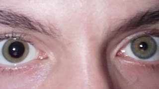
Anisocoria is a condition characterized by an unequal size of the eyes' pupils. Affecting up to 20% of the population, anisocoria is often entirely harmless, but can be a sign of more serious medical problems.

<span class="mw-page-title-main">Parinaud's syndrome</span> Inability to move the eyes up and down

Parinaud's syndrome is a constellation of neurological signs indicating injury to the dorsal midbrain. More specifically, compression of the vertical gaze center at the rostral interstitial nucleus of medial longitudinal fasciculus (riMLF).

<span class="mw-page-title-main">Neurosyphilis</span> Infection of the central nervous system in a patient with syphilis

Neurosyphilis is the infection of the central nervous system in a patient with syphilis. In the era of modern antibiotics, the majority of neurosyphilis cases have been reported in HIV-infected patients. Meningitis is the most common neurological presentation in early syphilis. Tertiary syphilis symptoms are exclusively neurosyphilis, though neurosyphilis may occur at any stage of infection.

<span class="mw-page-title-main">Pinealoma</span> Medical condition

A pinealoma is a tumor of the pineal gland, a part of the brain that produces melatonin. If a pinealoma destroys the cells of the pineal gland in a child, it can cause precocious puberty.

<span class="mw-page-title-main">Relative afferent pupillary defect</span> Medical condition

A relative afferent pupillary defect (RAPD), also known as a Marcus Gunn pupil, is a medical sign observed during the swinging-flashlight test whereupon the patient's pupils dilate when a bright light is swung from the unaffected eye to the affected eye. The affected eye still senses the light and produces pupillary sphincter constriction to some degree, albeit reduced.

Optic neuropathy is damage to the optic nerve from any cause. The optic nerve is a bundle of millions of fibers in the retina that sends visual signals to the brain.

<span class="mw-page-title-main">Douglas Argyll Robertson</span>

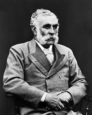
Douglas Moray Cooper Lamb Argyll Robertson FRSE, FRCSEd LLD was a Scottish ophthalmologist and surgeon. He introduced physostigmine into ophthalmic practice and the Argyll Robertson pupil is named after him. He was president of the Royal College of Surgeons of Edinburgh.

<span class="mw-page-title-main">Henri Parinaud</span> French ophthalmologist and neurologist

Henri Parinaud was a French ophthalmologist and neurologist, most noted for his work in the field of neuro-ophthalmology.

<span class="mw-page-title-main">Pupillary response</span> Physiological response that varies the size of the pupil

Pupillary response is a physiological response that varies the size of the pupil, via the optic and oculomotor cranial nerve.

<span class="mw-page-title-main">Meningeal syphilis</span> Medical condition

Meningeal syphilis is a chronic form of syphilis infection that affects the central nervous system. Treponema pallidum, a spirochate bacterium, is the main cause of syphilis, which spreads drastically throughout the body and can infect all its systems if not treated appropriately. Treponema pallidum is the main cause of the onset of meningeal syphilis and other treponemal diseases, and it consists of a cytoplasmic and outer membrane that can cause a diverse array of diseases in the central nervous system and brain.

<span class="mw-page-title-main">Accommodative excess</span> Medical condition

In ophthalmology, accommodative excess occurs when an individual uses more than normal accommodation for performing certain near work. Accommodative excess has traditionally been defined as accommodation that is persistently higher than expected for the patient's age. Modern definitions simply regard it as an inability to relax accommodation readily. Excessive accommodation is seen in association with excessive convergence also.

References

  1. Digre, Kathleen A. (1986). "Light-Near Dissociation". content.lib.utah.edu. Spencer S. Eccles Health Sciences Library, University of Utah. Retrieved 20 October 2016.
  2. Dente, Christopher; Gurwood, Andrew (10 September 1999). "The Argyll Robertson pupil". Optometry Today: 23–25. Retrieved 7 March 2021.
  3. Fletcher WA, Sharpe JA (1986). "Tonic pupils in neurosyphilis". Neurology. 36 (2): 188–92. doi:10.1212/wnl.36.2.188. PMID   3945389. S2CID   11710415.
  4. Thompson HS, Kardon RH (2006). "The Argyll Robertson pupil". Journal of Neuro-Ophthalmology. 26 (2): 134–8. doi: 10.1097/01.wno.0000222971.09745.91 . PMID   16845316.
  5. Digre, Kathleen A. (1986). "Convergence Retraction Nystagmus (Parinaud's Syndrome)". content.lib.utah.edu. Spencer S. Eccles Health Sciences Library, University of Utah. Retrieved 20 October 2016.
  6. Kawasaki, A (December 1999). "Physiology, assessment, and disorders of the pupil". Current Opinion in Ophthalmology. 10 (6): 394–400. doi:10.1097/00055735-199912000-00005. PMID   10662243.
  7. Thompson, HS; Kardon, RH (June 2006). "Irene E. Loewenfeld, PhD Physiologist of the Pupil". Journal of Neuro-Ophthalmology. 26 (2): 139–48. doi: 10.1097/01.wno.0000222970.02122.a0 . PMID   16845317.

Further reading