Glaucoma is a group of eye diseases that lead to damage of the optic nerve, which is important for transmitting visual information from the eye to the brain. This damage is often caused by increased pressure within the eye, known as intraocular pressure (IOP) and may cause vision loss if left untreated. The word glaucoma originated from the Greek word ΓλαύV̇ξ (glaukos), which means "to glow". Glaucoma has been called the "silent thief of sight" because the loss of vision usually occurs slowly over a long period of time. It is associated with old age, a family history of glaucoma, and certain medical conditions or medications.
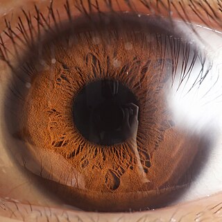
In humans and most mammals and birds, the iris is a thin, annular structure in the eye, responsible for controlling the diameter and size of the pupil, and thus the amount of light reaching the retina. Eye color is defined by the iris. In optical terms, the pupil is the eye's aperture, while the iris is the diaphragm.

Eye surgery, also known as ophthalmic surgery or ocular surgery, is surgery performed on the eye or its adnexa. Eye surgery is part of ophthalmology and is performed by an ophthalmologist or eye surgeon. The eye is a fragile organ, and requires due care before, during, and after a surgical procedure to minimize or prevent further damage. An eye surgeon is responsible for selecting the appropriate surgical procedure for the patient, and for taking the necessary safety precautions. Mentions of eye surgery can be found in several ancient texts dating back as early as 1800 BC, with cataract treatment starting in the fifth century BC. It continues to be a widely practiced class of surgery, with various techniques having been developed for treating eye problems.

The aqueous humour is a transparent water-like fluid similar to blood plasma, but containing low protein concentrations. It is secreted from the ciliary body, a structure supporting the lens of the eyeball. It fills both the anterior and the posterior chambers of the eye, and is not to be confused with the vitreous humour, which is located in the space between the lens and the retina, also known as the posterior cavity or vitreous chamber. Blood cannot normally enter the eyeball.

Phacoemulsification is a cataract surgery method in which the internal lens of the eye which has developed a cataract is emulsified with the tip of an ultrasonic handpiece and aspirated from the eye. Aspirated fluids are replaced with irrigation of balanced salt solution to maintain the volume of the anterior chamber during the procedure. This procedure minimises the incision size and reduces the recovery time and risk of surgery induced astigmatism.

The ciliary body is a part of the eye that includes the ciliary muscle, which controls the shape of the lens, and the ciliary epithelium, which produces the aqueous humor. The aqueous humor is produced in the non-pigmented portion of the ciliary body. The ciliary body is part of the uvea, the layer of tissue that delivers oxygen and nutrients to the eye tissues. The ciliary body joins the ora serrata of the choroid to the root of the iris.
Uveitis is inflammation of the uvea, the pigmented layer of the eye between the inner retina and the outer fibrous layer composed of the sclera and cornea. The uvea consists of the middle layer of pigmented vascular structures of the eye and includes the iris, ciliary body, and choroid. Uveitis is described anatomically, by the part of the eye affected, as anterior, intermediate or posterior, or panuveitic if all parts are involved. Anterior uveitis (iridocyclitis) is the most common, with the incidence of uveitis overall affecting approximately 1:4500, most commonly those between the ages of 20-60. Symptoms include eye pain, eye redness, floaters and blurred vision, and ophthalmic examination may show dilated ciliary blood vessels and the presence of cells in the anterior chamber. Uveitis may arise spontaneously, have a genetic component, or be associated with an autoimmune disease or infection. While the eye is a relatively protected environment, its immune mechanisms may be overcome resulting in inflammation and tissue destruction associated with T-cell activation.

A red eye is an eye that appears red due to illness or injury. It is usually injection and prominence of the superficial blood vessels of the conjunctiva, which may be caused by disorders of these or adjacent structures. Conjunctivitis and subconjunctival hemorrhage are two of the less serious but more common causes.

In ophthalmology, gonioscopy is a routine procedure that measures the angle between the iris and the cornea, using a goniolens together with a slit lamp or operating microscope. Its use is important in diagnosing and monitoring various eye conditions associated with glaucoma.

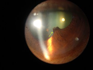
Ectopia lentis is a displacement or malposition of the eye's crystalline lens from its normal location. A partial dislocation of a lens is termed lens subluxation or subluxated lens; a complete dislocation of a lens is termed lens luxation or luxated lens.

Tropicamide, sold under the brand name Mydriacyl among others, is a medication used to dilate the pupil and help with examination of the eye. Specifically it is used to help examine the back of the eye. It is applied as eye drops. Effects occur within 40 minutes and last for up to a day.

An iridectomy, also known as a surgical iridectomy or corectomy, is the surgical removal of part of the iris. These procedures are most frequently performed in the treatment of closed-angle glaucoma and iris melanoma.
The posterior chamber is a narrow space behind the peripheral part of the iris, and in front of the suspensory ligament of the lens and the ciliary processes. The posterior chamber consists of small space directly posterior to the iris but anterior to the lens. The posterior chamber is part of the anterior segment and should not be confused with the vitreous chamber.

Trabeculectomy is a surgical procedure used in the treatment of glaucoma to relieve intraocular pressure by removing part of the eye's trabecular meshwork and adjacent structures. It is the most common glaucoma surgery performed and allows drainage of aqueous humor from within the eye to underneath the conjunctiva where it is absorbed. This outpatient procedure was most commonly performed under monitored anesthesia care using a retrobulbar block or peribulbar block or a combination of topical and subtenon anesthesia. Due to the higher risks associated with bulbar blocks, topical analgesia with mild sedation is becoming more common. Rarely general anesthesia will be used, in patients with an inability to cooperate during surgery.
Glaucoma is a group of diseases affecting the optic nerve that results in vision loss and is frequently characterized by raised intraocular pressure (IOP). There are many glaucoma surgeries, and variations or combinations of those surgeries, that facilitate the escape of excess aqueous humor from the eye to lower intraocular pressure, and a few that lower IOP by decreasing the production of aqueous humor.

Phacolytic glaucoma (PG) is a form of glaucoma which is caused due to a leaking mature or immature cataract. Inflammatory glaucoma which occurs in phacolysis is a condition which is a result of the leakage of protein within the lens into the capsule of a mature or hyper mature cataract and involves a simple procedure to be cured that is referred to as cataract extraction.

Secondary glaucoma is a collection of progressive optic nerve disorders associated with a rise in intraocular pressure (IOP) which results in the loss of vision. In clinical settings, it is defined as the occurrence of IOP above 21 mmHg requiring the prescription of IOP-managing drugs. It can be broadly divided into two subtypes: secondary open-angle glaucoma and secondary angle-closure glaucoma, depending on the closure of the angle between the cornea and the iris. Principal causes of secondary glaucoma include optic nerve trauma or damage, eye disease, surgery, neovascularization, tumours and use of steroid and sulfa drugs. Risk factors for secondary glaucoma include uveitis, cataract surgery and also intraocular tumours. Common treatments are designed according to the type and the underlying causative condition, in addition to the consequent rise in IOP. These include drug therapy, the use of miotics, surgery or laser therapy.
Schwartz–Matsuo syndrome is a human eye disease characterised by rhegmatogenous retinal detachment, elevated intraocular pressure (IOP) and open angle of anterior chamber.
Uveitis–glaucoma–hyphaema (UGH) syndrome, also known as Ellingson syndrome, is a complication of cataract surgery, caused by intraocular lens subluxation or dislocation. The chafing of mispositioned intraocular lens over iris, ciliary body or iridocorneal angle cause elevated intraocular pressure (IOP) anterior uveitis and hyphema. It is most commonly caused by anterior chamber IOLs and sulcus IOLs but, the condition can be seen with any type of IOL, including posterior chamber lenses and cosmetic iris implants.

Uveitic glaucoma is most commonly a progression stage of noninfectious anterior uveitis or iritis.