Env (gene)

Last updated
TLV/ENV coat polyprotein
Identifiers
SymbolTLV_coat
Pfam PF00429
InterPro IPR018154
Available protein structures:
Pfam   structures / ECOD  
PDB RCSB PDB; PDBe; PDBj
PDBsum structure summary

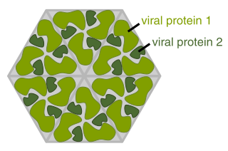
Env is a viral gene that encodes the protein forming the viral envelope. [1] The expression of the env gene enables retroviruses to target and attach to specific cell types, and to infiltrate the target cell membrane. [2]

Contents

Analysis of the structure and sequence of several different env genes suggests that Env proteins are type 1 fusion machines. [3] Type 1 fusion machines initially bind a receptor on the target cell surface, which triggers a conformational change, allowing for binding of the fusion protein. The fusion peptide inserts itself in the host cell membrane and brings the host cell membrane very close to the viral membrane to facilitate membrane fusion. [4]

While there are significant differences in sequence of the env gene between retroviruses, the gene is always located downstream of gag , pro, and pol . The env mRNA must be spliced for expression.

The mature product of the env gene is the viral spike protein, which has two main parts: the surface protein (SU) and the transmembrane protein (TM). The tropism of the virus is determined by the SU protein domain because it is responsible for the receptor-binding function of the virus. The SU domain therefore determines the specificity of the virus for a single receptor molecule. [2]

Physical structure

Oligomerization

The retroviral glycoproteins are oligomeric complexes that are composed of SU-TM heterodimers, which are made in the endoplasmic reticulum after the translation of the glycosylated Env precursor. [5] The arrangement of these heterodimers determines the 3D structure of the knobbed spike on the viral surface. The Env proteins of the Avian Sarcoma and Leukosis virus (ASLV) and the Murine Leukemia Virus (MLV) are both trimers of SU-TM heterodimers. [6] The Env protein of Human Immunodeficiency Virus (HIV) also has a trimeric structure of heterodimers. [7] It is believed that the intracellular transport of the nascent protein depends, to some extent, on the oligomerization of Env precursors, which allows hydrophobic sequences to be buried inside the protein structure. This oligomerization has also been implicated in fusion initiation with the membrane of the target cell. [8]

Post-translational modification

Env can be modified by the addition of mannose-rich oligosaccharides, a process that takes place in the rough endoplasmic reticulum and is carried out by the enzymes of the host cell. Cotranslational glycosylation take place at the asparagine in the Asn-X-Ser or Asn-X-Thr motifs. Different retroviruses vary widely in N-linked glycosylation sites: HIV-1 can have as many as 30 sites glycosylated, 25 of which reside in gp120. At the other end of the spectrum, MMTV (Mouse Mammary Tumor Virus has only 4 sites for oligosaccharide addition (two on gp52 and two on gp37). The addition of oligosaccharides is believed to play a role in the proper folding of Env, presumably by stabilizing the protein structure. Without proper folding, protein transport and function can be severely compromised. [2] The importance of glycosylation of Env in HIV-1 was ascertained by synthesizing the glycoprotein in the presence of a glycosylation inhibitor, tunicamycin. The synthesized protein was incorrectly folded and incapable of binding CD4. Receptor binding was only minimally affected, however, when the secreted env product was enzymatically deglycosylated. [9]

In HIV

Diagram of HIV virion HI-virion-structure en.svg
Diagram of HIV virion

The env gene codes for the gp160 protein which forms a homotrimer, and is cleaved into gp120 and gp41 by the host cell protease, furin. To form an active fusion protein, SU gp120 and TM gp41 polypeptides remain non-covalently bound together, but this interaction is often not stable, leading to shed, soluble gp120 and membrane-bound, gp41 'stumps'. Separately, cleavage by furin is inefficient, and virions often are released with inactive, uncleaved gp160. Because of the high prevalence of these inactive forms, the immune system often produces antibodies which target inactive gp160, rather than active forms of the envelope protein. See Replication cycle of HIV.

Env expression is regulated by the gene product of rev . Experimental deletion of rev resulted in the inability to detect the Env protein and levels of env mRNA in the cell cytoplasm were significantly diminished. However, when total cellular RNA was analyzed, env RNA totals were not significantly different in the presence and absence of rev coexpression. It was found that without rev expression, there was a marked increase in nuclear env RNA, which suggests that rev plays an important role in the nuclear export of env mRNA. [10] The role of rev was further elucidated when it was found that rev acts in trans to target a specific sequence present in the env gene of HIV-1 to initiate export of incompletely spliced HIV-1 RNA from the nucleus. [11]

env gp160; envelope glycoprotein
Identifiers
Organism HIV 1
Symbolenv
Entrez 155971
RefSeq (Prot) NP_057856.1
UniProt P04578
Other data
Chromosome viral genome: 0.01 - 0.01 Mb
Search for
Structures Swiss-model
Domains InterPro

gp120

Exposed on the surface of the viral envelope, the glycoprotein gp120 binds to the CD4 receptor on any target cell that has such a receptor, particularly the helper T-cell. Strains of HIV-1 have been isolated that are able to enter host cells that are CD4 negative. This CD4-independence is associated with spontaneous mutation in the env gene. The presence of a co-receptor, CXCR4, is sufficient for this mutant strain to infect human cells. The strain with this phenotype was found to have seven mutations in the sequence coding for gp120 and it is proposed that these mutations induce conformational changes in gp120 that allow the virus to directly interact with the co-receptor. [12]

Since CD4 receptor binding is the most obvious step in HIV infection, gp120 was among the first targets of HIV vaccine research. These efforts have been hampered by the fusion mechanism used by HIV, which makes neutralization by antibodies extremely difficult. Prior to binding the host cell, gp120 remains effectively hidden from antibodies because it is buried in the protein and shielded by sugars. Gp120 is only exposed when in close proximity to a host cell and the space between the viral and host cell membranes is small enough to sterically hinder the binding of antibodies. [13]

gp41

The glycoprotein gp41 is non-covalently bound to gp120, and provides the second step by which HIV enters the cell. It is originally buried within the viral envelope, but when gp120 binds to a CD4 receptor, gp120 changes its conformation causing gp41 to become exposed, where it can assist in fusion with the host cell.

Fusion inhibitor drugs such as enfuvirtide block the fusion process by binding to gp41. [14]

Env in MMTV

The Mouse Mammary Tumor Virus (MMTV) env gene codes for a polyprotein gp70 ( P10259 ) that is cleaved to yield the surface (SU) and transmembrane (TM) Env products. Gp52 is the SU subunit in MMTV and gp36 is the TM subunit. Gp52 is a 52,000-dalton glycoprotein and gp36 is a 36,000-dalton glycoprotein. [15] [16]

MMTV Env is of particular interest to researchers because of the discovery that it encodes an immunoreceptor tyrosine-based activation motif (ITAM) that has been shown to transform human and murine mammary cell in culture. This ITAM depolarizes epithelial acinar structures, thereby changing the phenotype of the cells and causing them to become cancerous. [16]

Env in ASLV

Subgroup A

Avian Sarcoma and Leukosis Viruses (ASLV) have ten subgroups (A through J). The envelope glycoprotein of subgroup A is called EnvA and its env gene codes for precursor protein known as Pr95. This precursor is cleaved by host cell enzymes to yield the surface protein subunit, gp85, and the transmembrane protein subunit, gp37, which heterodimerize and then form a trimer. The virus cannot infect cells before the processing of the envelope precursor protein is completed. [17] For the virus to penetrate the cytosol of a host cell, a low pH is necessary. [18]

Env in MLV

The env gene of Murine Leukemia Virus (MLV) codes for the 71,000-dalton glycoprotein, gp71. This membrane receptor was isolated from Rauscher murine leukemia virus (R-MuLV). [19]

Env in mammalian evolution

The retroviral protein env has been captured multiple times during mammalian evolution and is expressed in placental tissue, where it facilitates fusion of fetal and maternal cells. The protein is called syncytin in mammals. [20] [21]

See also


Related Research Articles

<span class="mw-page-title-main">Viral protein</span>

The term viral protein refers to both the products of the genome of a virus and any host proteins incorporated into the viral particle. Viral proteins are grouped according to their functions, and groups of viral proteins include structural proteins, nonstructural proteins, regulatory proteins, and accessory proteins. Viruses are non-living and do not have the means to reproduce on their own, instead depending on their host cell's machinery to do this. Thus, viruses do not code for most of the proteins required for their replication and the translation of their mRNA into viral proteins, but use proteins encoded by the host cell for this purpose.

The genome and proteins of HIV (human immunodeficiency virus) have been the subject of extensive research since the discovery of the virus in 1983. "In the search for the causative agent, it was initially believed that the virus was a form of the Human T-cell leukemia virus (HTLV), which was known at the time to affect the human immune system and cause certain leukemias. However, researchers at the Pasteur Institute in Paris isolated a previously unknown and genetically distinct retrovirus in patients with AIDS which was later named HIV." Each virion comprises a viral envelope and associated matrix enclosing a capsid, which itself encloses two copies of the single-stranded RNA genome and several enzymes. The discovery of the virus itself occurred two years following the report of the first major cases of AIDS-associated illnesses.

<span class="mw-page-title-main">Envelope glycoprotein GP120</span> Glycoprotein exposed on the surface of the HIV virus

Envelope glycoprotein GP120 is a glycoprotein exposed on the surface of the HIV envelope. It was discovered by Professors Tun-Hou Lee and Myron "Max" Essex of the Harvard School of Public Health in 1984. The 120 in its name comes from its molecular weight of 120 kDa. Gp120 is essential for virus entry into cells as it plays a vital role in attachment to specific cell surface receptors. These receptors are DC-SIGN, Heparan Sulfate Proteoglycan and a specific interaction with the CD4 receptor, particularly on helper T-cells. Binding to CD4 induces the start of a cascade of conformational changes in gp120 and gp41 that lead to the fusion of the viral membrane with the host cell membrane. Binding to CD4 is mainly electrostatic although there are van der Waals interactions and hydrogen bonds.

<span class="mw-page-title-main">Gp41</span> Subunit of the envelope protein complex of retroviruses

Gp41 also known as glycoprotein 41 is a subunit of the envelope protein complex of retroviruses, including human immunodeficiency virus (HIV). Gp41 is a transmembrane protein that contains several sites within its ectodomain that are required for infection of host cells. As a result of its importance in host cell infection, it has also received much attention as a potential target for HIV vaccines.

Simian foamy virus (SFV) is a species of the genus Spumavirus that belongs to the family of Retroviridae. It has been identified in a wide variety of primates, including prosimians, New World and Old World monkeys, as well as apes, and each species has been shown to harbor a unique (species-specific) strain of SFV, including African green monkeys, baboons, macaques, and chimpanzees. As it is related to the more well-known retrovirus human immunodeficiency virus (HIV), its discovery in primates has led to some speculation that HIV may have been spread to the human species in Africa through contact with blood from apes, monkeys, and other primates, most likely through bushmeat-hunting practices.

Entry inhibitors, also known as fusion inhibitors, are a class of antiviral drugs that prevent a virus from entering a cell, for example, by blocking a receptor. Entry inhibitors are used to treat conditions such as HIV and hepatitis D.

<span class="mw-page-title-main">Spike protein</span> Glycoprotein spike on a viral capsid or viral envelope

In virology, a spike protein or peplomer protein is a protein that forms a large structure known as a spike or peplomer projecting from the surface of an enveloped virus. The proteins are usually glycoproteins that form dimers or trimers.

Visna-maedi virus from the genus Lentivirus and subfamily Orthoretrovirinae, is a retrovirus that causes encephalitis and chronic pneumonitis in sheep. It is known as visna when found in the brain, and maedi when infecting the lungs. Lifelong, persistent infections in sheep occur in the lungs, lymph nodes, spleen, joints, central nervous system, and mammary glands; The condition is sometimes known as ovine progressive pneumonia (OPP), particularly in the United States, or Montana sheep disease. White blood cells of the monocyte/macrophage lineage are the main target of the virus.

CD4 immunoadhesin is a recombinant fusion protein consisting of a combination of CD4 and the fragment crystallizable region, similarly known as immunoglobulin. It belongs to the antibody (Ig) gene family. CD4 is a surface receptor for human immunodeficiency virus (HIV). The CD4 immunoadhesin molecular fusion allow the protein to possess key functions from each independent subunit. The CD4 specific properties include the gp120-binding and HIV-blocking capabilities. Properties specific to immunoglobulin are the long plasma half-life and Fc receptor binding. The properties of the protein means that it has potential to be used in AIDS therapy as of 2017. Specifically, CD4 immunoadhesin plays a role in antibody-dependent cell-mediated cytotoxicity (ADCC) towards HIV-infected cells. While natural anti-gp120 antibodies exhibit a response towards uninfected CD4-expressing cells that have a soluble gp120 bound to the CD4 on the cell surface, CD4 immunoadhesin, however, will not exhibit a response. One of the most relevant of these possibilities is its ability to cross the placenta.

<span class="mw-page-title-main">CLEC4M</span> Protein-coding gene in the species Homo sapiens

C-type lectin domain family 4 member M is a protein that in humans is encoded by the CLEC4M gene. CLEC4M has also been designated as CD299.

<span class="mw-page-title-main">PRKCH</span> Protein-coding gene in the species Homo sapiens

Protein kinase C eta type is an enzyme that in humans is encoded by the PRKCH gene.

<span class="mw-page-title-main">Syncytin-1</span> Protein-coding gene in the species Homo sapiens

Syncytin-1 also known as enverin is a protein found in humans and other primates that is encoded by the ERVW-1 gene. Syncytin-1 is a cell-cell fusion protein whose function is best characterized in placental development. The placenta in turn aids in embryo attachment to the uterus and establishment of a nutrient supply.

<span class="mw-page-title-main">IFNA7</span> Protein-coding gene in the species Homo sapiens

Interferon alpha-7 is a protein that in humans is encoded by the IFNA7 gene.

<span class="mw-page-title-main">HLA-DQB2</span> Protein-coding gene in the species Homo sapiens

HLA class II histocompatibility antigen, DX beta chain is a protein that in humans is encoded by the HLA-DQB2 gene.

<span class="mw-page-title-main">MAN1A1</span> Protein-coding gene in the species Homo sapiens

Mannosyl-oligosaccharide 1,2-alpha-mannosidase IA is an enzyme that in humans is encoded by the MAN1A1 gene.

<span class="mw-page-title-main">GCS1</span> Protein-coding gene in the species Homo sapiens

Mannosyl-oligosaccharide glucosidase is an enzyme that in humans is encoded by the MOGS gene.

Avian sarcoma leukosis virus (ASLV) is an endogenous retrovirus that infects and can lead to cancer in chickens; experimentally it can infect other species of birds and mammals. ASLV replicates in chicken embryo fibroblasts, the cells that contribute to the formation of connective tissues. Different forms of the disease exist, including lymphoblastic, erythroblastic, and osteopetrotic.

<span class="mw-page-title-main">Vpu protein</span>

Vpu is an accessory protein that in HIV is encoded by the vpu gene. Vpu stands for "Viral Protein U". The Vpu protein acts in the degradation of CD4 in the endoplasmic reticulum and in the enhancement of virion release from the plasma membrane of infected cells. Vpu induces the degradation of the CD4 viral receptor and therefore participates in the general downregulation of CD4 expression during the course of HIV infection. Vpu-mediated CD4 degradation is thought to prevent CD4-Env binding in the endoplasmic reticulum to facilitate proper Env assembly into virions. It is found in the membranes of infected cells, but not the virus particles themselves.

2F5 is a broadly neutralizing human monoclonal antibody (mAb) that has been shown to bind to and neutralize HIV-1 in vitro, making it a potential candidate for use in vaccine synthesis. 2F5 recognizes an epitope in the membrane-proximal external region (MPER) of HIV-1 gp41. 2F5 then binds to this epitope and its constant region interacts with the viral lipid membrane, which neutralizes the virus.

Bovine immunodeficiency virus (BIV) is a retrovirus belonging to the genus Lentivirus. It is similar to the human immunodeficiency virus (HIV) and infects cattle. The cells primarily infected are lymphocytes and monocytes/macrophages.

References

  1. Gene+Products,+env at the U.S. National Library of Medicine Medical Subject Headings (MeSH)
  2. 1 2 3 Coffin JM, Hughes SH, Vamus HE (1997). Retroviruses. Plainview, N.Y: Cold Spring Harbor Laboratory Press. ISBN   978-0-87969-497-5.
  3. Caffrey M, Cai M, Kaufman J, Stahl SJ, Wingfield PT, Covell DG, Gronenborn AM, Clore GM (August 1998). "Three-dimensional solution structure of the 44 kDa ectodomain of SIV gp41". EMBO J. 17 (16): 4572–84. doi:10.1093/emboj/17.16.4572. PMC   1170787 . PMID   9707417.
  4. Colman PM, Lawrence MC (April 2003). "The structural biology of type I viral membrane fusion". Nat. Rev. Mol. Cell Biol. 4 (4): 309–19. doi:10.1038/nrm1076. PMID   12671653. S2CID   31703688.
  5. Einfeld D, Hunter E (November 1988). "Oligomeric structure of a prototype retrovirus glycoprotein". Proc. Natl. Acad. Sci. U.S.A. 85 (22): 8688–92. Bibcode:1988PNAS...85.8688E. doi: 10.1073/pnas.85.22.8688 . PMC   282525 . PMID   2847170.
  6. Kamps CA, Lin YC, Wong PK (October 1991). "Oligomerization and transport of the envelope protein of Moloney murine leukemia virus-TB and of ts1, a neurovirulent temperature-sensitive mutant of MoMuLV-TB". Virology. 184 (2): 687–94. doi:10.1016/0042-6822(91)90438-H. PMID   1887590.
  7. Tran EE, Borgnia MJ, Kuybeda O, Schauder DM, Bartesaghi A, Frank GA, Sapiro G, Milne JL, Subramaniam S (2012). "Structural mechanism of trimeric HIV-1 envelope glycoprotein activation". PLOS Pathog. 8 (7): e1002797. doi: 10.1371/journal.ppat.1002797 . PMC   3395603 . PMID   22807678.
  8. Earl PL, Doms RW, Moss B (September 1992). "Multimeric CD4 binding exhibited by human and simian immunodeficiency virus envelope protein dimers". J. Virol. 66 (9): 5610–4. doi:10.1128/JVI.66.9.5610-5614.1992. PMC   289124 . PMID   1501294.
  9. Li Y, Luo L, Rasool N, Kang CY (January 1993). "Glycosylation is necessary for the correct folding of human immunodeficiency virus gp120 in CD4 binding". J. Virol. 67 (1): 584–8. doi:10.1128/JVI.67.1.584-588.1993. PMC   237399 . PMID   8416385.
  10. Hammarskjöld ML, Heimer J, Hammarskjöld B, Sangwan I, Albert L, Rekosh D (May 1989). "Regulation of human immunodeficiency virus env expression by the rev gene product". J. Virol. 63 (5): 1959–66. doi:10.1128/JVI.63.5.1959-1966.1989. PMC   250609 . PMID   2704072.
  11. Malim MH, Hauber J, Le SY, Maizel JV, Cullen BR (March 1989). "The HIV-1 rev trans-activator acts through a structured target sequence to activate nuclear export of unspliced viral mRNA". Nature. 338 (6212): 254–7. Bibcode:1989Natur.338..254M. doi:10.1038/338254a0. PMID   2784194. S2CID   4367958.
  12. Dumonceaux J, Nisole S, Chanel C, Quivet L, Amara A, Baleux F, Briand P, Hazan U (January 1998). "Spontaneous mutations in the env gene of the human immunodeficiency virus type 1 NDK isolate are associated with a CD4-independent entry phenotype". J. Virol. 72 (1): 512–9. doi:10.1128/JVI.72.1.512-519.1998. PMC   109402 . PMID   9420253.
  13. Labrijn AF, Poignard P, Raja A, Zwick MB, Delgado K, Franti M, Binley J, Vivona V, Grundner C, Huang CC, Venturi M, Petropoulos CJ, Wrin T, Dimitrov DS, Robinson J, Kwong PD, Wyatt RT, Sodroski J, Burton DR (October 2003). "Access of antibody molecules to the conserved coreceptor binding site on glycoprotein gp120 is sterically restricted on primary human immunodeficiency virus type 1". J. Virol. 77 (19): 10557–65. doi:10.1128/JVI.77.19.10557-10565.2003. PMC   228502 . PMID   12970440.
  14. Lalezari JP, Henry K, O'Hearn M, Montaner JS, Piliero PJ, Trottier B, Walmsley S, Cohen C, Kuritzkes DR, Eron JJ, Chung J, DeMasi R, Donatacci L, Drobnes C, Delehanty J, Salgo M (May 2003). "Enfuvirtide, an HIV-1 fusion inhibitor, for drug-resistant HIV infection in North and South America". N. Engl. J. Med. 348 (22): 2175–85. doi: 10.1056/NEJMoa035026 . PMID   12637625.
  15. Callis AH, Ritzi EM (September 1980). "Detection and characterization of mouse mammary tumor virus cell surface antigens: estimation of antigen abundance by protein A assay". J. Virol. 35 (3): 876–87. doi:10.1128/JVI.35.3.876-887.1980. PMC   288881 . PMID   6252344.
  16. 1 2 Katz E, Lareef MH, Rassa JC, Grande SM, King LB, Russo J, Ross SR, Monroe JG (February 2005). "MMTV Env encodes an ITAM responsible for transformation of mammary epithelial cells in three-dimensional culture". J. Exp. Med. 201 (3): 431–9. doi:10.1084/jem.20041471. PMC   2213037 . PMID   15684322.
  17. Balliet JW, Gendron K, Bates P (April 2000). "Mutational analysis of the subgroup A avian sarcoma and leukosis virus putative fusion peptide domain". J. Virol. 74 (8): 3731–9. doi:10.1128/JVI.74.8.3731-3739.2000. PMC   111882 . PMID   10729148.
  18. Barnard RJ, Narayan S, Dornadula G, Miller MD, Young JA (October 2004). "Low pH is required for avian sarcoma and leukosis virus Env-dependent viral penetration into the cytosol and not for viral uncoating". J. Virol. 78 (19): 10433–41. doi:10.1128/JVI.78.19.10433-10441.2004. PMC   516436 . PMID   15367609.
  19. DeLarco J, Todaro GJ (July 1976). "Membrane receptors for murine leukemia viruses: characterization using the purified viral envelope glycoprotein, gp71". Cell. 8 (3): 365–71. doi:10.1016/0092-8674(76)90148-3. PMID   8213. S2CID   45192638.
  20. Nova. 2016. Endogenous retroviruses. Retrieved from https://www.pbs.org/wgbh/nova/next/evolution/endogenous-retroviruses
  21. Lavialle C, Cornelis G, Dupressoir A, Esnault C, Heidmann O, Vernochet C, Heidmann T (September 2013). "Paleovirology of 'syncytins', retroviral env genes exapted for a role in placentation". Philosophical Transactions of the Royal Society of London. Series B, Biological Sciences. 368 (1626): 20120507. doi:10.1098/rstb.2012.0507. PMC   3758191 . PMID   23938756.