Guillardia

Last updated

Guillardia
41598 2017 2668 Fig2d HTML.jpg
Guillardia theta . DAPI-staining images showing the representative cells of cell cycle stages based on the localization of the nucleus and the shape of the chloroplast. DIC, images of differential interference contact; Chl, chloroplast autofluorescence; Chl/DAPI, merged images of Chl and DAPI. The double arrowhead indicates constriction of the chloroplast division site. Scale bar = 5 µm
Scientific classification OOjs UI icon edit-ltr.svg
Kingdom: Chromista
Phylum: Cryptophyta
Class: Cryptophyceae
Order: Pyrenomonadales
Family: Geminigeraceae
Genus: Guillardia
D. R. A. Hill & R. Wetherbee
Species:
G. theta
Binomial name
Guillardia theta
D. R. A. Hill & R. Wetherbee

Guillardia is a genus of marine biflagellate cryptomonad algae with a plastid obtained through secondary endosymbiosis of a red alga. [1]

Contents

Originally identified in Connecticut by Richard Guillard in the 1960s, Guillardia only has one described species. [2] The genus is rare in the wild, but cultures well and has been frequently studied since its original discovery. The general morphology of the small cell is well described, and shares many similarities with other cryptomonads, though it contains a unique organization of periplasm. [2] Guillardia is the only cryptomonad to have its entire nucleus, nucleomorph, and plastid genome sequenced. [2] [3] [4] This knowledge has prompted further studies on gene transfer between chloroplast, the ancestral red alga l nucleomorph, and the nucleus, [5] as well as regulation of photosynthetic [6] and cell cycle gene expression within the plastid. [7] The genus is also important in research across biological disciplines; Guillardia serves as a model organism in the study of secondary endosymbiosis and photosynthesis in cryptomonads due to its ease of culture and sequenced genome. [8] Two anion channelrhodopsins have also been isolated from Guillardiatheta for neurobiological research applications as optogenetic inhibitors. [9]

Etymology

Originally this genus was referred to as “Cryptomonas species theta” or “Flagellate theta’. [2] [4] It was then named Guillardia by Hill and Weatherbee after Dr. Robert Guillard, the researcher who originally isolated the genus. [2]

Type Species: Guillardia theta. [10]

History of Knowledge

The cryptomonad Guillardia theta was first isolated by Dr. Robert Guillard in Connecticut in 1963, where he defined it as “flagellate theta” in a symposium on the organic sources of nitrogen in marine diatoms. [2] Since then, the organism has been successfully cultured many times. When it was referred to as Cryptomonas theta in the early 1980s, the flagella and a unique periplast were described. [11] [12] Following these studies, the plastid genome was mapped [13] and Hill and Weatherbee named and characterized the genus in 1990, [2] before the plastid genome was fully sequenced in 1999, [3] confirming the plastid’s common ancestry with red algae. Since these original studies, many other aspects of this organism have been identified, including the mechanisms of nucleomorph and plastid division and their regulation, [14] and photosynthetic pigments, [15] mechanisms, and regulation. [6] Because it grows so well in culture, Guillardia theta is also frequently used as a model organism in modern day studies investigating cryptomonads characteristics.

Habitat and Ecology

In the wild, Guillardia theta is a rare planktonic marine organism, and the majority of studies have been completed from cultures. The genus was originally isolated from Milford Harbor in Connecticut, and has only been found in one other location in Denmark since its original discovery. [2] Milford Harbor includes many discrete areas such as estuaries, mud flats, marine basins, marinas, beaches, marshes, and coastal shores that provide habitats to a variety of different organisms. [16] Though the precise isolation location was not recorded, the genus is thought to proliferate as phytoplankton in the still water of the marine basin. [1]

A. Larsen is the only other researcher to have identified Guillardia in the wild within the Wadden Sea in Denmark. However, this was only revealed through a personal communique with Hill and Weatherbee, and never published. [2]   Another northern marine habitat, the Wadden Sea consists of an agglomeration of sandbanks that provide estuary, open water, marsh, and sandy beach habitats to the local ecosystems. [17]

As the genus is relatively rare in the wild, its role in ecosystems is not well understood. Guillardia is a photosynthetic phytoplankton with two plastids, indicating a role in primary production within the system. [1] Additionally, ciliates are known predators of the genus in culture, suggesting the role of Guillardia as prey within aquatic systems. Mesodinium pulex , a well studied phagotrophic ciliate common to marine, brackish, and freshwater environments ingested and grew on Guillardiatheta cultures. [18]

Description

Morphology

The morphology of the genus Guillardia is well described. The cell is dorso-ventrally flattened and approximately 7-11μm long. [2] As a member of the cryptomonads, it has an anterior gullet and contains a nucleus with nucleolus, a double lobed, four membraned plastid with pyrenoid, a nucleomorph closely associated with the plastid, mitochondria, Golgi apparatus associated with two flagella, starch deposits, and ejectosomes on the gullet and periplast. [1] The structure of the periplasm, a layer of thin sheets composed of irregular plates made from crystalline subunits, is a defining characteristic of the genus. Guillardia’s inner periplasm consists of a single sheet adjacent to the plasma membrane while the inner periplasm of other cryptomonads is made of from uniformly shaped plates. [1] In both types of periplasm, the peripheral ejectosomes lay beneath the periplasm in vesicles and a non crystalline material separates the periplasm from the plasma membrane. [1] [2]

Ejectosomes in Guillardia and other cryptomonads are primarily used for defense and evasion, lining the gullet of the cell. In Guillardia, uneven elongated strands lay within vesicles, while strand length varies across other cryptomonad genera. Strand length ranges from 200 nm to 3.6 μm. In response to external stressors like rapid pH change, osmolarity change, or light intensity changes, coils shoot out from their vesicles in the surrounding environment. The impact of the ejectosome strands with an object such as another organism causes the uneven backwards motion of Guillardia for predator evasion. [8]

Plastid

The plastid in Guillardia arose from a secondary endosymbiosis event of a red alga l cell. Like other cryptomonads, Guillardia is key for understanding secondary endosymbiosis as it retains the nucleus of the algal endosymbiont in the form of a nucleomorph within a periplastidial compartment and four membranes surrounding the plastidial complex. [1] The outermost membrane is hypothesized to be a remnant of the ancestral phagocytic vesicle and is continuous with the Guillardia endoplasmic reticulum. The small periplastidial cytoplasm is also hypothesized to retain components of its cytoskeleton, due to tubulin genes localized to the nucleomorph. [19] While many plastidial proteins remain in the nucleomorph, those that underwent endosymbiotic gene transfer to the host nucleus are targeted back through the outermost membrane through co-translational translocation with a bipartite N-terminal signal sequence. [2] [7] Each subsequent membrane the protein passes retains unique translocation mechanisms. Also contained within the plastid are eukaryotic ribosomes and a starch granule filled pyrenoid, where CO2 fixation occurs through the enzyme RUBISCO. [1]

Like other cryptomonads, the light harvesting pigments of the plastidial chloroplasts are phycobiliproteins and chlorophyll a/c-binding proteins, homologous to those found in red algal lineages. Unlike in red algae, the phycobiliprotein antenna in Guillardia are localized in the thylakoid lumens as small soluble protein complexes, instead of the large antenna associated with the thylakoid membranes characteristic of algal photosynthesis. [15] Mechanisms used to control photosynthetic pigments in Guillardia vary depending on the growth stage. In logarithmic growth stages, Guillardia uses state transitions to modulate energy inputs, while in the stationary growth phase, the cell uses non photochemical quenching, a mechanism to protect plants and algae from high light intensity. [7] It is unclear why the two mechanisms of regulating energy input are differentiated in the different growth phases of Guillardia.

Motility

Motility occurs primarily through two asymmetric flagella, the longest protruding anteriorly from the gullet, while the shorter flagellum points to the back of the cell. [1] A rhizostyle and rootlet system also contribute to the motility of Guillardia. [11] Interestingly, the photaxis mechanisms of Guillardiatheta which incorporate anion channelrhodopsins to initiate a motion response have been used in neuroscience applications as optogenetic inhibitors. [9]

Cell Division

In order to properly divide asexually, Guillardia must replicate its cell, as well as the nucleomorph and chloroplast of the plastid. [1] The genus divides through mitosis, and has never been observed dividing sexually; however, meiosis related genes have been found in the nucleus, suggesting it has the capability to do so. [5] Mitosis in Guillardia begins after plastid division, with the formation of mitotic spindles and initiation of basal body and flagella division. Like many other flagellated protists, both preexisting flagella become the daughter locomotion flagellum, while new basal bodies develop into trailing flagellum through flagellar transformation. Through metaphase, a chromatin plate with tunnels results from the dissolution of the nuclear membrane. During anaphase, microtubule spindles thread through the plate tunnels and attach to chromatin, splitting the plate in two. [1] Remnants of the nuclear membrane also appear to border the mitotic spindle remaining in contact with the endoplasmic reticulum throughout mitosis. [14] Cytokinesis occurs in Guillardia during metaphase and anaphase, with a thin layer of amorphous materials instead of microtubule structures. [1]

Plastidial division occurs prior to flagellar division in preprophase, and both nucleomorph and chloroplast division occur once per Guillardia cell cycle. [1] Plastid division occurs via the constriction of the dorsal bridge that connects the two lobes of the plastid. Before completion of chloroplast division, the nucleomorph divides by invaginating the inner and outer nucleomorph membranes. [14] Synchronization of chloroplast, nucleomorph, and host cell division is vital for the evolution of the red algal endosymbiont into an organelle. Nucleus encoded nucleomorph HISTONE H2A mRNA accumulates during S phase, while nucleomorph encoded genes that regulate nucleomorph replication and division are constantly expressed. [6] This suggests that the endosymbiont lost the ability to regulate replication cycle dependent transcription, but the control of host nucleus cell-cycle dependent genes regulates nucleomorph and chloroplast replication and division.

Characteristics of the Genome

Guillardiatheta was the first cryptomonad with a complete sequenced genome. Its nucleus is haploid, with a tetraploid nucleomorph and mitochondria and plastids with high copy numbers. [20] Since the original sequencing, the plastid and nucleomorph genome have also been sequenced and mapped to better understand the algal ancestry of the plastid and taxonomic history of the genus. [1]   The nuclear genome is approximately 87 mega base pairs in size encoding 21,000 predicted proteins, 57% being completely unique with no known homologs in other organisms. [1] [5] The genome contains almost all eukaryotic complexity hallmarks including endomembrane system, transcription, RNA processing and translation, post translational modification, protein turnover, and cytoskeletal genes. The Guillardia nuclear genome was also found to have many spliceosomal introns, and a large family of putative tyrosine kinases. Of 7451 genes in the Guillardia nucleus, 508 were determined to originate in algal lineages. [5] Despite data suggesting that the majority of these genes originate from green algal lineages, this comparison is not reliable as many genome databases tend to be biased towards green algal genes. [1] [5]

Nucleomorph sequencing reveals a relatively small genome with 487 protein genes, few housekeeping genes, with only 31 genes being targeted to the plastid. [5] It is clear that the nucleomorph has been significantly reduced in size and almost entirely relies on protein targeting to the periplastidial complex. The host nuclear genome codes for transcription associated proteins that presumably act to regulate gene expression in the nucleomorph, as well as DNA replication proteins and protein kinases associated with cell replication. [1] [5] [8] The transfer of these genes to the host genome clearly depicts the loss of self sufficiency of the plastid endosymbiont.  However, complete sequencing of both nuclear and nucleomorph genomes indicate that throughout endosymbiont gene transfer, proteins often take on new functions and occupy different compartments, so function cannot be determined based on evolutionary history. Guillardia’s genome contains a high level of mosaicism with genes derived from host nucleus, nucleomorph, plastid, and other foreign alga derived proteins. [5]

The plastid genome of Guillardia theta was also the first plastid from a nucleomorph containing organism to be physically mapped and sequenced to elucidate the endosymbiotic origin of the plastid. [5] [1] The genome consists of 121 kilo base pairs, of that 4kbp encode the two rRNA cistrons for ribosomal production. In the coding regions, 46 genes are for photosynthesis, 10 genes are biosynthetic, replication, and division genes, 44 encode  ribosomal proteins, and 7 are involved in transcription and translation. [3] Some genes overlap and there are no introns contained within the genome, making it quite compact. [13] Additionally there are many polycistronic genes that are identical to those identified in the plastid of a red alga, Porphyra purpurea. [3] This suggests the common ancestry of the plastid in both Guillardia and Porphyra.

Practical Importance

Guillardia has been frequently used as a model cryptomonad for algal endosymbiont genomics and many other cryptomonad studies. Because the genus cultures so well, it was the first cryptomonad to have its entire nuclear, nucleomorph, and plastid genome sequenced. [1] The information gleaned from this data helped to elucidate mechanisms of secondary endosymbiosis present in protist lineages containing endosymbiotic red algal plastids. [3] In addition to molecular sequencing, the replication mechanisms, and photosynthetic mechanisms incorporated in the plastid of Guillardia organisms have been well studied as a model for other cryptophyte species. The genus was also used as a model to study periplast starch synthesis in cryptophytes, demonstrating that the Guillardia theta periplast uses a UDP-glucose based pathway to synthesize starch. [21]

Additionally, anion channelrhodopsin proteins from Guillardia theta have been found to induce neuron hyperpolarization in optogenetic assays. [9] Channelrhodopsins are light gated anion channels that induce flagellar movement towards light sources in algae. In human studies, anion channelrhodopsins can be deployed to induce chloride driven hyperpolarization, silencing targeted neurons at specific timepoints. [22] This allows neuroscientists to study neuronal circuitry by photo-suppressing specific neurons. Before the discovery of anion channelrhodopsins in Guillardia, methods for optogenetic silencing of neurons were less effective and precise. While this discovery continues to be incredibly useful for neuroscience research, further research demonstrated a Rhodomonas salina anion channelrhodopsin to have a decreased response time between stimulation and channel opening. [22] As such, Guillardia channelrhodopsins are no longer used as frequently for neuroscience assays.

Furthermore, it has been used as prey for culturing and study of other organisms, like Mesodinium ciliates. [8] [18]

Related Research Articles

<span class="mw-page-title-main">Chloroplast</span> Plant organelle that conducts photosynthesis

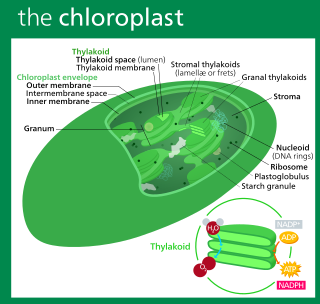
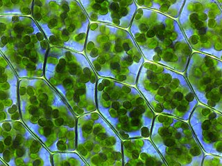
A chloroplast is a type of membrane-bound organelle known as a plastid that conducts photosynthesis mostly in plant and algal cells. The photosynthetic pigment chlorophyll captures the energy from sunlight, converts it, and stores it in the energy-storage molecules ATP and NADPH while freeing oxygen from water in the cells. The ATP and NADPH is then used to make organic molecules from carbon dioxide in a process known as the Calvin cycle. Chloroplasts carry out a number of other functions, including fatty acid synthesis, amino acid synthesis, and the immune response in plants. The number of chloroplasts per cell varies from one, in unicellular algae, up to 100 in plants like Arabidopsis and wheat.

<span class="mw-page-title-main">Symbiogenesis</span> Evolutionary theory holding that eukaryotic organelles evolved through symbiosis with prokaryotes

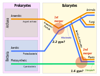
Symbiogenesis is the leading evolutionary theory of the origin of eukaryotic cells from prokaryotic organisms. The theory holds that mitochondria, plastids such as chloroplasts, and possibly other organelles of eukaryotic cells are descended from formerly free-living prokaryotes taken one inside the other in endosymbiosis. Mitochondria appear to be phylogenetically related to Rickettsiales bacteria, while chloroplasts are thought to be related to cyanobacteria.

<span class="mw-page-title-main">Cryptomonad</span> Subphylum of algae

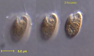
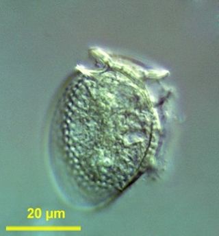
The cryptomonads are a group of algae, most of which have plastids. They are common in freshwater, and also occur in marine and brackish habitats. Each cell is around 10–50 μm in size and flattened in shape, with an anterior groove or pocket. At the edge of the pocket there are typically two slightly unequal flagella.

<span class="mw-page-title-main">Plastid</span> Plant cell organelles that perform photosynthesis and store starch

The plastid is a membrane-bound organelle found in the cells of plants, algae, and some other eukaryotic organisms. They are considered to be intracellular endosymbiotic cyanobacteria. Examples include chloroplasts, chromoplasts, and leucoplasts.

<span class="mw-page-title-main">Chlorarachniophyte</span> Group of algae

The chlorarachniophytes are a small group of exclusively marine algae widely distributed in tropical and temperate waters. They are typically mixotrophic, ingesting bacteria and smaller protists as well as conducting photosynthesis. Normally they have the form of small amoebae, with branching cytoplasmic extensions that capture prey and connect the cells together, forming a net. They may also form flagellate zoospores, which characteristically have a single subapical flagellum that spirals backwards around the cell body, and walled coccoid cells.

<span class="mw-page-title-main">Chromista</span> Eukaryotic biological kingdom

Chromista is a proposed but polyphyletic biological kingdom consisting of single-celled and multicellular eukaryotic species that share similar features in their photosynthetic organelles (plastids). It includes all eukaryotes whose plastids contain chlorophyll c and are surrounded by four membranes. If the ancestor already possessed chloroplasts derived by endosymbiosis from red algae, all non-photosynthetic Chromista have secondarily lost the ability to photosynthesise. Its members might have arisen independently as separate evolutionary groups from the last eukaryotic common ancestor.

Cryptomonas is the name-giving genus of the Cryptomonads established by German biologist Christian Gottfried Ehrenberg in 1831. The algae are common in freshwater habitats and brackish water worldwide and often form blooms in greater depths of lakes. The cells are usually brownish or greenish in color and are characteristic of having a slit-like furrow at the anterior. They are not known to produce any toxins. They are used to feed small zooplankton, which is the food source for small fish in fish farms. Many species of Cryptomonas can only be identified by DNA sequencing. Cryptomonas can be found in several marine ecosystems in Australia and South Korea.

<span class="mw-page-title-main">Nucleomorph</span>

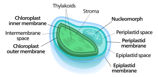
Nucleomorphs are small, vestigial eukaryotic nuclei found between the inner and outer pairs of membranes in certain plastids. They are thought to be vestiges of primitive red and green algal nuclei that were engulfed by a larger eukaryote. Because the nucleomorph lies between two sets of membranes, nucleomorphs support the endosymbiotic theory and are evidence that the plastids containing them are complex plastids. Having two sets of membranes indicate that the plastid, a prokaryote, was engulfed by a eukaryote, an alga, which was then engulfed by another eukaryote, the host cell, making the plastid an example of secondary endosymbiosis.

<span class="mw-page-title-main">Kleptoplasty</span> Form of algae symbiosis

Kleptoplasty or kleptoplastidy is a process in symbiotic relationships whereby plastids, notably chloroplasts from algae, are sequestered by the host. The word is derived from Kleptes (κλέπτης) which is Greek for thief. The alga is eaten normally and partially digested, leaving the plastid intact. The plastids are maintained within the host, temporarily continuing photosynthesis and benefiting the host.

<span class="mw-page-title-main">Archaeplastida</span> Clade of eukaryotes containing land plants and some algae

The Archaeplastida are a major group of eukaryotes, comprising the photoautotrophic red algae (Rhodophyta), green algae, land plants, and the minor group glaucophytes. It also includes the non-photosynthetic lineage Rhodelphidia, a predatorial (eukaryotrophic) flagellate that is sister to the Rhodophyta, and probably the microscopic picozoans. The Archaeplastida have chloroplasts that are surrounded by two membranes, suggesting that they were acquired directly through a single endosymbiosis event by feeding on a cyanobacterium. All other groups which have chloroplasts, besides the amoeboid genus Paulinella, have chloroplasts surrounded by three or four membranes, suggesting they were acquired secondarily from red or green algae. Unlike red and green algae, glaucophytes have never been involved in secondary endosymbiosis events.

<span class="mw-page-title-main">Cryptophyceae</span> Class of single-celled organisms

The cryptophyceae are a class of algae, most of which have plastids. About 220 species are known, and they are common in freshwater, and also occur in marine and brackish habitats. Each cell is around 10–50 μm in size and flattened in shape, with an anterior groove or pocket. At the edge of the pocket there are typically two slightly unequal flagella.

An apicoplast is a derived non-photosynthetic plastid found in most Apicomplexa, including Toxoplasma gondii, and Plasmodium falciparum and other Plasmodium spp., but not in others such as Cryptosporidium. It originated from algae through secondary endosymbiosis; there is debate as to whether this was a green or red alga. The apicoplast is surrounded by four membranes within the outermost part of the endomembrane system. The apicoplast hosts important metabolic pathways like fatty acid synthesis, isoprenoid precursor synthesis and parts of the heme biosynthetic pathway.

<span class="mw-page-title-main">Red algae</span> Division of archaeplastids

Red algae, or Rhodophyta, are one of the oldest groups of eukaryotic algae. The Rhodophyta comprises one of the largest phyla of algae, containing over 7,000 currently recognized species with taxonomic revisions ongoing. The majority of species (6,793) are found in the Florideophyceae (class), and mostly consist of multicellular, marine algae, including many notable seaweeds. Red algae are abundant in marine habitats but relatively rare in freshwaters. Approximately 5% of red algae species occur in freshwater environments, with greater concentrations found in warmer areas. Except for two coastal cave dwelling species in the asexual class Cyanidiophyceae, there are no terrestrial species, which may be due to an evolutionary bottleneck in which the last common ancestor lost about 25% of its core genes and much of its evolutionary plasticity.

<i>Rhodomonas</i> Genus of single-celled organisms

Rhodomonas is a genus of cryptomonads. It is characterized by its red colour, the square-shaped plates of its inner periplast, its short furrow ending in a gullet, and a distinctly shaped chloroplast closely associated with its nucleomorph. Historically, Rhodomonas was characterized by its red chloroplast alone, but this no longer occurs as its taxonomy has become increasingly based on molecular and cellular data. Currently, there is some debate about the taxonomic validity of Rhodomonas as a genus and further research is needed to verify its taxonomic status. Rhodomonas is typically found in marine environments, although freshwater reports exist. It is commonly used as a live feed for various aquaculture species.

<i>Hemiselmis</i> Genus of single-celled organisms

Hemiselmis is a genus of cryptomonads.

<i>Dinophysis</i> Genus of single-celled organisms

Dinophysis is a genus of dinoflagellates common in tropical, temperate, coastal and oceanic waters. It was first described in 1839 by Christian Gottfried Ehrenberg.

<span class="mw-page-title-main">Chloroplast DNA</span> DNA located in cellular organelles called chloroplasts

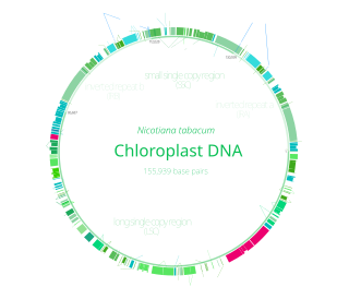
Chloroplast DNA (cpDNA) is the DNA located in chloroplasts, which are photosynthetic organelles located within the cells of some eukaryotic organisms. Chloroplasts, like other types of plastid, contain a genome separate from that in the cell nucleus. The existence of chloroplast DNA was identified biochemically in 1959, and confirmed by electron microscopy in 1962. The discoveries that the chloroplast contains ribosomes and performs protein synthesis revealed that the chloroplast is genetically semi-autonomous. The first complete chloroplast genome sequences were published in 1986, Nicotiana tabacum (tobacco) by Sugiura and colleagues and Marchantia polymorpha (liverwort) by Ozeki et al. Since then, a great number of chloroplast DNAs from various species have been sequenced.

A plastid is a membrane-bound organelle found in plants, algae and other eukaryotic organisms that contribute to the production of pigment molecules. Most plastids are photosynthetic, thus leading to color production and energy storage or production. There are many types of plastids in plants alone, but all plastids can be separated based on the number of times they have undergone endosymbiotic events. Currently there are three types of plastids; primary, secondary and tertiary. Endosymbiosis is reputed to have led to the evolution of eukaryotic organisms today, although the timeline is highly debated.

Lepidodinium is a genus of dinoflagellates belonging to the family Gymnodiniaceae.

References

  1. 1 2 3 4 5 6 7 8 9 10 11 12 13 14 15 16 17 18 19 Hoef-Emden, Kerstin; Archibald, John M. (2016), Archibald, John M.; Simpson, Alastair G.B.; Slamovits, Claudio H.; Margulis, Lynn (eds.), "Cryptophyta (Cryptomonads)", Handbook of the Protists, Cham: Springer International Publishing, pp. 1–41, doi:10.1007/978-3-319-32669-6_35-1, ISBN   978-3-319-32669-6 , retrieved 28 April 2023
  2. 1 2 3 4 5 6 7 8 9 10 11 12 Hill, David R. A.; Wetherbee, Richard (1 September 1990). "Guillardia theta gen. et sp.nov. (Cryptophyceae)". Canadian Journal of Botany (in French). 68 (9): 1873–1876. doi:10.1139/b90-245. ISSN   0008-4026.
  3. 1 2 3 4 5 Douglas, Susan E.; Penny, Susanne L. (1 February 1999). "The Plastid Genome of the Cryptophyte Alga, Guillardia theta: Complete Sequence and Conserved Synteny Groups Confirm Its Common Ancestry with Red Algae". Journal of Molecular Evolution. 48 (2): 236–244. Bibcode:1999JMolE..48..236D. doi:10.1007/PL00006462. ISSN   1432-1432. PMID   9929392. S2CID   2005223.
  4. 1 2 Gillott, Marcelle A.; Gibbs, Sarah P. (December 1980). "THE CRYPTOMONAD NUCLEOMORPH: ITS ULTRASTRUCTURE AND EVOLUTIONARY SIGNIFICANCE1". Journal of Phycology. 16 (4): 558–568. doi:10.1111/j.1529-8817.1980.tb03074.x. S2CID   84702891.
  5. 1 2 3 4 5 6 7 8 9 Curtis, Bruce A.; Tanifuji, Goro; Burki, Fabien; Gruber, Ansgar; Irimia, Manuel; Maruyama, Shinichiro; Arias, Maria C.; Ball, Steven G.; Gile, Gillian H.; Hirakawa, Yoshihisa; Hopkins, Julia F.; Kuo, Alan; Rensing, Stefan A.; Schmutz, Jeremy; Symeonidi, Aikaterini (December 2012). "Algal genomes reveal evolutionary mosaicism and the fate of nucleomorphs". Nature. 492 (7427): 59–65. Bibcode:2012Natur.492...59C. doi:10.1038/nature11681. ISSN   1476-4687. PMID   23201678. S2CID   4380094.
  6. 1 2 3 Onuma, Ryo; Mishra, Neha; Miyagishima, Shin-ya (24 May 2017). "Regulation of chloroplast and nucleomorph replication by the cell cycle in the cryptophyte Guillardia theta". Scientific Reports. 7 (1): 2345. Bibcode:2017NatSR...7.2345O. doi:10.1038/s41598-017-02668-2. ISSN   2045-2322. PMC   5443833 . PMID   28539635.
  7. 1 2 3 Cheregi, Otilia; Kotabová, Eva; Prášil, Ondřej; Schröder, Wolfgang P.; Kaňa, Radek; Funk, Christiane (6 August 2015). "Presence of state transitions in the cryptophyte algaGuillardia theta". Journal of Experimental Botany. 66 (20): 6461–6470. doi:10.1093/jxb/erv362. ISSN   0022-0957. PMC   4588893 . PMID   26254328.
  8. 1 2 3 4 Vilchis, María Concepción Lora (6 September 2022). "Cryptophyte: Biology, Culture, and Biotechnological Applications". Progress in Microalgae Research – A Path for Shaping Sustainable Futures. IntechOpen. doi:10.5772/intechopen.107009. ISBN   978-1-80356-024-3.
  9. 1 2 3 Govorunova, Elena G.; Sineshchekov, Oleg A.; Hemmati, Raheleh; Janz, Roger; Morelle, Olivier; Melkonian, Michael; Wong, Gane K.-S.; Spudich, John L. (1 May 2018). "Extending the Time Domain of Neuronal Silencing with Cryptophyte Anion Channelrhodopsins". eNeuro. 5 (3). doi:10.1523/ENEURO.0174-18.2018. ISSN   2373-2822. PMC   6051594 . PMID   30027111.
  10. "AlgaeBase :: Listing the World's Algae". www.algaebase.org. Retrieved 28 April 2023.
  11. 1 2 Gillott, Marcelle A.; Gibbs, Sarah P. (1 July 1983). "Comparison of the flagellar rootlets and periplast in two marine cryptomonads". Canadian Journal of Botany. 61 (7): 1964–1978. doi:10.1139/b83-212. ISSN   0008-4026.
  12. Wetherbee, Richard; Hill, David R. A.; Brett, Steven J. (1 May 1987). "The structure of the periplast components and their association with the plasma membrane in a cryptomonad flagellate". Canadian Journal of Botany (in French). 65 (5): 1019–1026. doi:10.1139/b87-141. ISSN   0008-4026.
  13. 1 2 Douglas, Susan E. (1 December 1988). "Physical mapping of the plastid genome from the chlorophyll c-containing alga, Cryptomonas Φ". Current Genetics. 14 (6): 591–598. doi:10.1007/BF00434085. ISSN   1432-0983. S2CID   33335861.
  14. 1 2 3 McKerracher, Lisa; Gibbs, Sarah P. (1 December 1982). "Cell and nucleomorph division in the alga Cryptomonas". Canadian Journal of Botany. 60 (11): 2440–2452. doi:10.1139/b82-296. ISSN   0008-4026.
  15. 1 2 Kieselbach, Thomas; Cheregi, Otilia; Green, Beverley R.; Funk, Christiane (1 March 2018). "Proteomic analysis of the phycobiliprotein antenna of the cryptophyte alga Guillardia theta cultured under different light intensities". Photosynthesis Research. 135 (1): 149–163. doi:10.1007/s11120-017-0400-0. ISSN   1573-5079. PMC   5784005 . PMID   28540588.
  16. "Milford Point". Audubon Connecticut. 2 July 2015. Retrieved 28 April 2023.
  17. "Denmark | Wadden Sea". www.waddensea-worldheritage.org. Retrieved 28 April 2023.
  18. 1 2 Tarangkoon, Woraporn; Hansen, Per Juel (4 January 2011). "Prey selection, ingestion and growth responses of the common marine ciliate Mesodinium pulex in the light and in the dark". Aquatic Microbial Ecology. 62 (1): 25–38. doi: 10.3354/ame01455 . ISSN   0948-3055.
  19. Keeling, P. J.; Deane, J. A.; Hink-Schauer, C.; Douglas, S. E.; Maier, U. G.; McFadden, G. I. (1 September 1999). "The secondary endosymbiont of the cryptomonad Guillardia theta contains alpha-, beta-, and gamma-tubulin genes". Molecular Biology and Evolution. 16 (9): 1308–1313. doi:10.1093/oxfordjournals.molbev.a026221. ISSN   0737-4038. PMID   10486984.
  20. Hirakawa, Yoshihisa; Ishida, Ken-Ichiro (April 2014). "Polyploidy of Endosymbiotically Derived Genomes in Complex Algae". Genome Biology and Evolution. 6 (4): 974–980. doi:10.1093/gbe/evu071. ISSN   1759-6653. PMC   4007541 . PMID   24709562.
  21. Deschamps, Philippe; Haferkamp, Ilka; Dauvillée, David; Haebel, Sophie; Steup, Martin; Buléon, Alain; Putaux, Jean-Luc; Colleoni, Christophe; d'Hulst, Christophe; Plancke, Charlotte; Gould, Sven; Maier, Uwe; Neuhaus, H. Ekkehard; Ball, Steven (June 2006). "Nature of the Periplastidial Pathway of Starch Synthesis in the Cryptophyte Guillardia theta". Eukaryotic Cell. 5 (6): 954–963. doi:10.1128/EC.00380-05. ISSN   1535-9778. PMC   1489276 . PMID   16757743.
  22. 1 2 Deisseroth, Karl; Hegemann, Peter (15 September 2017). "The form and function of channelrhodopsin". Science. 357 (6356): eaan5544. doi:10.1126/science.aan5544. ISSN   0036-8075. PMC   5723383 . PMID   28912215.