Histone H2A

Last updated

Histone H2A is one of the five main histone proteins involved in the structure of chromatin in eukaryotic cells.

Contents

The other histone proteins are: H1, H2B, H3 and H4.

The crystal structure of the nucleosome core particle consisting of H2A, H2B, H3 and H4 core histones, and DNA. The view is from the top through the superhelical axis. Nucleosome 1KX5 colour coded.png
The crystal structure of the nucleosome core particle consisting of H2A,H2B, H3 andH4core histones, and DNA. The view is from the top through the superhelical axis.
Structure of the H2AFJ protein Structure of the H2AFJ protein.jpg
Structure of the H2AFJ protein

Background

Histones are proteins that package DNA into nucleosomes. [1] Histones are responsible for maintaining the shape and structure of a nucleosome. One chromatin molecule is composed of at least one of each core histones per 100 base pairs of DNA. [2] There are five families of histones known to date; these histones are termed H1/H5, H2A, H2B, H3, and H4. [3] H2A is considered a core histone, along with H2B, H3 and H4. Core formation first occurs through the interaction of two H2A molecules. [3] Then, H2A forms a dimer with H2B; the core molecule is complete when H3-H4 also attaches to form a tetramer.

Sequence variants

Histone H2A is composed of non-allelic variants. [4] The term "Histone H2A" is intentionally non-specific and refers to a variety of closely related proteins that vary often by only a few amino acids. Apart from the canonical form, notable variants include H2A.1, H2A.2, H2A.X, and H2A.Z. H2A variants can be explored using "HistoneDB with Variants" database

Changes in variant composition occur in differentiating cells. This was observed in differentiating neurons during synthesis and turnover; changes in variant composition were seen among the H2A.1 histone. The only variant that remained constant in the neural differentiation was variant H2A.Z. [4] H2A.Z is a variant that exchanges with conventional H2A core protein; this variant is important for gene silencing. [5]

Physically, there are small changes on the surface area of the nucleosome that make the histone differ from H2A. Recent research suggests that H2AZ is incorporated into the nucleosome using a Swr1, a Swi2/Snf2- related adenosine triphosphatase. [6]

Another H2A variant that has been identified is H2AX. This variant has a C-terminal extension that is utilized for DNA repair. The method of repair this variant employs is non-homologous end joining. Direct DNA damage can induce changes to the sequence variants. Experiments performed with ionizing radiation linked γ- phosphorylation of H2AX to DNA double-strand break. [7] A large amount of chromatin is involved with each DNA double-strand break; a response to DNA damage is the formation of γ- H2AX.

Lastly, MacroH2A variant is a variant that is similar to H2A; it is encoded by the H2AFY gene. This variant differs from H2A because of the addition of a fold domain in its C-terminal tail. MacroH2A is expressed in the inactive X chromosome in females. [8]

Structure

Histone tails and their function in chromatin formation Histone tails and their function in chromatin formation.svg
Histone tails and their function in chromatin formation

H2A consists of a main globular domain, an N-terminal tail and a C-terminal tail. [9] Both tails are the location of post-translational modification. Thus far, researchers have not identified any secondary structures that arise in the tails. H2A utilizes a protein fold known as the ‘histone fold’. The histone fold is a three-helix core domain that is connected by two loops. This connection forms a ‘handshake arrangement.’ Most notably, this is termed the helix-turn-helix motif, which allows for dimerization with H2B. The ‘histone fold’ is conserved among H2A at the structural level; however the genetic sequence that encodes for this structure differs between variants. [10]

The structure of macroH2A variant was exposed through X-ray crystallography. The conserved domain contains a DNA binding structure and a peptidase fold. [11] The function of this conserved domain remains unknown. Research suggests that this conserved domain may function as an anchor site for Xist DNA or it may also function as a modifying enzyme.

Function

Basic units of chromatin structure Basic units of chromatin structure.svg
Basic units of chromatin structure

DNA Folding: H2A is important for packaging DNA into chromatin. Since H2A packages DNA molecules into chromatin, the packaging process will affect gene expression. H2A has been correlated with DNA modification and epigenetics. H2A plays a major role in determining the overall structure of chromatin. Inadvertently, H2A has been found to regulate gene expression. [10]

DNA modification by H2A occurs in the cell nucleus. Proteins responsible for nuclear import of H2A protein are karyopherin and importin. [12] Recent studies also show that nucleosome assembly protein 1 is also used to transport of H2A into the nucleus so it can wrap DNA. Other functions of H2A have been seen in the histone variant H2A.Z. This variant is associated with gene activation, silencing and suppression of antisense RNA. In addition, when H2A.Z was studied in human and yeast cells, it was used to promote RNA polymerase II recruitment. [13]

Antimicrobial peptide: Histones are conserved eukaryotic cationic proteins present in the cells and are involved in the antimicrobial activities. In vertebrates and invertebrates, Histone H2A variant is reported to be involved in host immune response by acting as antimicrobial peptides (AMPs). H2A are α-helical molecule, amphipathic protein with hydrophobic and hydrophilic residues on opposing sides that enhances the antimicrobial activity of H2A. [14]

DNA damage response

Site specific ubiquitination of histone H2A has a role in the recruitment of DNA repair proteins to DNA double strand breaks which then may be repaired by either homologous recombination or non-homologous end joining. [15] In the DNA damage response, it is thought that ubiquitination of H2A by the BRCA1/BARD1 heterodimer promotes homologous recombination, and that ubiquitination of H2A by RNF168 protein promotes non-homologous end joining. [15]

Genetics

H2A is coded by many genes in the human genome, including: H2AFB1, H2AFB2, H2AFB3, H2AFJ, H2AFV, H2AFX, H2AFY, H2AFY2, and H2AFZ. Genetic patterns among the different H2A molecules are mostly conserved among variants. The variability in gene expression exists among the regulatory machinery that manages H2A expression. Researchers studied eukaryotic evolutionary lineages of histone proteins and found diversification among the regulatory genes. The greatest differences were observed in core histone gene cis-regulatory sequence motifs and associated protein factors. Variability in gene sequence was seen in bacterial, fungi, plant, and mammalian genes. [10]

One variant of H2A protein is H2ABbd (Barr body deficient) variant. This variant is composed of a different genetic sequence compared to H2A. The variant functions with transcriptionally active domains. [10] Other variations associated with H2ABbd are located within its C-terminus. H2ABbd has a shorter C-terminal domain compared to the large C-terminal found on H2A. The two C terminals are about 48% identical. H2ABbd functions with active chromosomes. Thus far, it is missing from Xi chromosomes in fibroblast cells. Lastly, it found to be associated with acetylated H4. [16]

Different functions of H2A.Z compared to H2A are correlated with genetic differences between H2A and the variant. Resistance to nucleosomes occurs in H2A.Z by binding to H1 factor. H2A.Z gene is an essential gene in yeast and it is denoted as Htz1. Comparatively, vertebrates have two H2A.Z genes. [10] These genes, H2A.Z1 and H2A.Z2 encode for proteins that differ from H2A.Z by three residues. At first researchers figured that these genes were redundant; however, when a mutant H2A.Z1 was created, it resulted in lethality during mammalian tests. [16] Therefore, H2A.Z1 is an essential gene. On the other hand, researchers have not identified the function of H2A.Z2 variant. It is known that it is transcribed in mammals and this gene expression is conserved among mammalian species. This conservation suggests that the gene is functional. [16] When studying H2A.Z in plants species, the protein different among residues from species to species. These differences contribute to differences in cell-cycle regulation. [16] This phenomenon was only observed in plants.

Phylogenetic trees were created to show the divergence of variants from their ancestors. The divergence of variant, H2A.X, from H2A occurred at multiple origins in a phylogenetic tree. Acquisition of the phosphorylation motif was consistent with the many origins of H2A that arose from an ancestral H2A.X. Finally, the presence of H2A.X and absence of H2A in fungi leads researchers to believe that H2A.X was the original ancestor of the histone protein H2A [10]

Modification of H2A

H2A modification is under current research. However, modification of H2A does occur. Serine phosphorylation sites have been identified on H2A. Threonine O-GlcNAc has also been identified on H2A. Large differences exist between the modified residues of H2A variants. For example, H2ABbd lacks modified residues that exist in H2A. [16] The differences in modification change the function of H2ABbd compared to H2A. As previously mentioned, variant H2AX was found to function in DNA repair. This function is dependent upon the phosphorylation of H2AX C-terminal. [7] Once H2AX becomes phosphorylated, it can function in DNA repair. The H2A.X variant differs from H2A through modification. The C-terminal of H2A.X contains an additional motif compared to H2A. The motif that is added is Ser-Gln-(Glu/Asp)- (hydrophobic residue). [16] The motif becomes heavily phosphorylated at the serine residue; if this phosphorylation occurs the variant becomes γH2A.X. Phosphorylation occurs due to dsDNA breaks. [16] Modification on histone proteins can sometimes result in a change in function. Different H2A variants were exploited to have different functions, genetic sequences, and modifications.

See also

Related Research Articles

<span class="mw-page-title-main">Histone</span> Protein family around which DNA winds to form nucleosomes

In biology, histones are highly basic proteins abundant in lysine and arginine residues that are found in eukaryotic cell nuclei and in most Archaeal phyla. They act as spools around which DNA winds to create structural units called nucleosomes. Nucleosomes in turn are wrapped into 30-nanometer fibers that form tightly packed chromatin. Histones prevent DNA from becoming tangled and protect it from DNA damage. In addition, histones play important roles in gene regulation and DNA replication. Without histones, unwound DNA in chromosomes would be very long. For example, each human cell has about 1.8 meters of DNA if completely stretched out; however, when wound about histones, this length is reduced to about 90 micrometers (0.09 mm) of 30 nm diameter chromatin fibers.

<span class="mw-page-title-main">Nucleosome</span> Basic structural unit of DNA packaging in eukaryotes

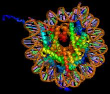
A nucleosome is the basic structural unit of DNA packaging in eukaryotes. The structure of a nucleosome consists of a segment of DNA wound around eight histone proteins and resembles thread wrapped around a spool. The nucleosome is the fundamental subunit of chromatin. Each nucleosome is composed of a little less than two turns of DNA wrapped around a set of eight proteins called histones, which are known as a histone octamer. Each histone octamer is composed of two copies each of the histone proteins H2A, H2B, H3, and H4.

<span class="mw-page-title-main">Histone acetyltransferase</span> Enzymes that catalyze acyl group transfer from acetyl-CoA to histones

Histone acetyltransferases (HATs) are enzymes that acetylate conserved lysine amino acids on histone proteins by transferring an acetyl group from acetyl-CoA to form ε-N-acetyllysine. DNA is wrapped around histones, and, by transferring an acetyl group to the histones, genes can be turned on and off. In general, histone acetylation increases gene expression.

<span class="mw-page-title-main">Histone octamer</span> 8-protein complex forming the core of nucleosomes

In molecular biology, a histone octamer is the eight-protein complex found at the center of a nucleosome core particle. It consists of two copies of each of the four core histone proteins. The octamer assembles when a tetramer, containing two copies of H3 and two of H4, complexes with two H2A/H2B dimers. Each histone has both an N-terminal tail and a C-terminal histone-fold. Each of these key components interacts with DNA in its own way through a series of weak interactions, including hydrogen bonds and salt bridges. These interactions keep the DNA and the histone octamer loosely associated, and ultimately allow the two to re-position or to separate entirely.

<span class="mw-page-title-main">S phase</span> DNA replication phase of the cell cycle, between G1 and G2 phase

S phase (Synthesis phase) is the phase of the cell cycle in which DNA is replicated, occurring between G1 phase and G2 phase. Since accurate duplication of the genome is critical to successful cell division, the processes that occur during S-phase are tightly regulated and widely conserved.

<span class="mw-page-title-main">Histone H4</span> One of the five main histone proteins involved in the structure of chromatin

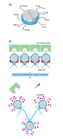
Histone H4 is one of the five main histone proteins involved in the structure of chromatin in eukaryotic cells. Featuring a main globular domain and a long N-terminal tail, H4 is involved with the structure of the nucleosome of the 'beads on a string' organization. Histone proteins are highly post-translationally modified. Covalently bonded modifications include acetylation and methylation of the N-terminal tails. These modifications may alter expression of genes located on DNA associated with its parent histone octamer. Histone H4 is an important protein in the structure and function of chromatin, where its sequence variants and variable modification states are thought to play a role in the dynamic and long term regulation of genes.

Histone H2B is one of the 5 main histone proteins involved in the structure of chromatin in eukaryotic cells. Featuring a main globular domain and long N-terminal and C-terminal tails, H2B is involved with the structure of the nucleosomes.

<span class="mw-page-title-main">Histone acetylation and deacetylation</span> Biological processes used in gene regulation

Histone acetylation and deacetylation are the processes by which the lysine residues within the N-terminal tail protruding from the histone core of the nucleosome are acetylated and deacetylated as part of gene regulation.

<span class="mw-page-title-main">H2AFX</span> Histone protein from the H2A family

H2A histone family member X is a type of histone protein from the H2A family encoded by the H2AFX gene. An important phosphorylated form is γH2AX (S139), which forms when double-strand breaks appear.

<span class="mw-page-title-main">Histone-modifying enzymes</span> Type of enzymes

Histone-modifying enzymes are enzymes involved in the modification of histone substrates after protein translation and affect cellular processes including gene expression. To safely store the eukaryotic genome, DNA is wrapped around four core histone proteins, which then join to form nucleosomes. These nucleosomes further fold together into highly condensed chromatin, which renders the organism's genetic material far less accessible to the factors required for gene transcription, DNA replication, recombination and repair. Subsequently, eukaryotic organisms have developed intricate mechanisms to overcome this repressive barrier imposed by the chromatin through histone modification, a type of post-translational modification which typically involves covalently attaching certain groups to histone residues. Once added to the histone, these groups elicit either a loose and open histone conformation, euchromatin, or a tight and closed histone conformation, heterochromatin. Euchromatin marks active transcription and gene expression, as the light packing of histones in this way allows entry for proteins involved in the transcription process. As such, the tightly packed heterochromatin marks the absence of current gene expression.

<span class="mw-page-title-main">HIST2H2BE</span> Protein-coding gene in the species Homo sapiens

Histone H2B type 2-E is a protein that in humans is encoded by the HIST2H2BE gene.

<span class="mw-page-title-main">Histone H2A.Z</span> Protein-coding gene in the species Homo sapiens

Histone H2A.Z is a protein that in humans is encoded by the H2AZ1 gene.

Histones are basic nuclear proteins that are responsible for the nucleosome structure of the chromosomal fiber in eukaryotes. Nucleosomes consist of approximately 146 bp of DNA wrapped around a histone octamer composed of pairs of each of the four core histones. The chromatin fiber is further compacted through the interaction of a linker histone, H1, with the DNA between the nucleosomes to form higher order chromatin structures. The H2AFZ gene encodes a replication-independent member of the histone H2A family that is distinct from other members of the family. Studies in mice have shown that this particular histone is required for embryonic development and indicate that lack of functional histone H2A leads to embryonic lethality.

Histone variants are proteins that substitute for the core canonical histones in nucleosomes in eukaryotes and often confer specific structural and functional features. The term might also include a set of linker histone (H1) variants, which lack a distinct canonical isoform. The differences between the core canonical histones and their variants can be summarized as follows: (1) canonical histones are replication-dependent and are expressed during the S-phase of cell cycle whereas histone variants are replication-independent and are expressed during the whole cell cycle; (2) in animals, the genes encoding canonical histones are typically clustered along the chromosome, are present in multiple copies and are among the most conserved proteins known, whereas histone variants are often single-copy genes and show high degree of variation among species; (3) canonical histone genes lack introns and use a stem loop structure at the 3’ end of their mRNA, whereas histone variant genes may have introns and their mRNA tail is usually polyadenylated. Complex multicellular organisms typically have a large number of histone variants providing a variety of different functions. Recent data are accumulating about the roles of diverse histone variants highlighting the functional links between variants and the delicate regulation of organism development.

H2BK5ac is an epigenetic modification to the DNA packaging protein Histone H2B. It is a mark that indicates the acetylation at the 5th lysine residue of the histone H2B protein. H2BK5ac is involved in maintaining stem cells and colon cancer.

H4K20me is an epigenetic modification to the DNA packaging protein Histone H4. It is a mark that indicates the mono-methylation at the 20th lysine residue of the histone H4 protein. This mark can be di- and tri-methylated. It is critical for genome integrity including DNA damage repair, DNA replication and chromatin compaction.

H4K16ac is an epigenetic modification to the DNA packaging protein Histone H4. It is a mark that indicates the acetylation at the 16th lysine residue of the histone H4 protein.

H4K5ac is an epigenetic modification to the DNA packaging protein histone H4. It is a mark that indicates the acetylation at the 5th lysine residue of the histone H4 protein. H4K5 is the closest lysine residue to the N-terminal tail of histone H4. It is enriched at the transcription start site (TSS) and along gene bodies. Acetylation of histone H4K5 and H4K12ac is enriched at centromeres.

H4K8ac, representing an epigenetic modification to the DNA packaging protein histone H4, is a mark indicating the acetylation at the 8th lysine residue of the histone H4 protein. It has been implicated in the prevalence of malaria.

H4K91ac is an epigenetic modification to the DNA packaging protein histone H4. It is a mark that indicates the acetylation at the 91st lysine residue of the histone H4 protein. No known diseases are attributed to this mark but it might be implicated in melanoma.

H3K36ac is an epigenetic modification to the DNA packaging protein Histone H3. It is a mark that indicates the acetylation at the 36th lysine residue of the histone H3 protein.

References

  1. Youngson RM (2006). Collins dictionary of human biology. Glasgow [Scotland]: Collins. ISBN   978-0-00-722134-9.
  2. Khorasanizadeh S (January 2004). "The nucleosome: from genomic organization to genomic regulation". Cell. 116 (2): 259–72. doi: 10.1016/s0092-8674(04)00044-3 . PMID   14744436. S2CID   15504162.
  3. 1 2 Cox MM, Lehninger AL, Nelson DL (2005). Lehninger principles of biochemistry (4th ed.). New York: W.H. Freeman. ISBN   978-0-7167-4339-2.
  4. 1 2 Bosch A, Suau P (November 1995). "Changes in core histone variant composition in differentiating neurons: the roles of differential turnover and synthesis rates". European Journal of Cell Biology. 68 (3): 220–5. PMID   8603674.
  5. Suto RK, Clarkson MJ, Tremethick DJ, Luger K (December 2000). "Crystal structure of a nucleosome core particle containing the variant histone H2A.Z". Nature Structural Biology. 7 (12): 1121–4. doi:10.1038/81971. PMID   11101893. S2CID   5966635.
  6. Mizuguchi G, Shen X, Landry J, Wu WH, Sen S, Wu C (January 2004). "ATP-driven exchange of histone H2AZ variant catalyzed by SWR1 chromatin remodeling complex". Science. 303 (5656): 343–8. Bibcode:2004Sci...303..343M. doi: 10.1126/science.1090701 . PMID   14645854. S2CID   9881829.
  7. 1 2 Jakob B, Splinter J, Conrad S, Voss KO, Zink D, Durante M, Löbrich M, Taucher-Scholz G (August 2011). "DNA double-strand breaks in heterochromatin elicit fast repair protein recruitment, histone H2AX phosphorylation and relocation to euchromatin". Nucleic Acids Research. 39 (15): 6489–99. doi:10.1093/nar/gkr230. PMC   3159438 . PMID   21511815.
  8. Costanzi C, Pehrson JR (June 1998). "Histone macroH2A1 is concentrated in the inactive X chromosome of female mammals". Nature. 393 (6685): 599–601. Bibcode:1998Natur.393..599C. doi:10.1038/31275. PMID   9634239. S2CID   205001095.
  9. Ghoneim M, Fuchs HA, Musselman CA (2021). "Histone Tail Conformations: A Fuzzy Affair with DNA". Trends Biochem Sci. 46 (7): 564–578. doi: 10.1016/j.tibs.2020.12.012 . PMC   8195839 . PMID   33551235.{{cite journal}}: CS1 maint: multiple names: authors list (link)
  10. 1 2 3 4 5 6 Mariño-Ramírez L, Jordan IK, Landsman D (2006). "Multiple independent evolutionary solutions to core histone gene regulation". Genome Biology. 7 (12): R122. doi: 10.1186/gb-2006-7-12-r122 . PMC   1794435 . PMID   17184543.
  11. Allen MD, Buckle AM, Cordell SC, Löwe J, Bycroft M (July 2003). "The crystal structure of AF1521 a protein from Archaeoglobus fulgidus with homology to the non-histone domain of macroH2A". Journal of Molecular Biology. 330 (3): 503–11. doi:10.1016/s0022-2836(03)00473-x. PMID   12842467.
  12. Mosammaparast N, Ewart CS, Pemberton LF (December 2002). "A role for nucleosome assembly protein 1 in the nuclear transport of histones H2A and H2B". The EMBO Journal. 21 (23): 6527–38. doi:10.1093/emboj/cdf647. PMC   136951 . PMID   12456659.
  13. Mariño-Ramírez L, Levine KM, Morales M, Zhang S, Moreland RT, Baxevanis AD, Landsman D (2011). "The Histone Database: an integrated resource for histones and histone fold-containing proteins". Database. 2011: bar048. doi:10.1093/database/bar048. PMC   3199919 . PMID   22025671.
  14. Arockiaraj J, Gnanam AJ, Kumaresan V, Palanisamy R, Bhatt P, Thirumalai MK, Roy A, Pasupuleti M, Kasi M (November 2013). "An unconventional antimicrobial protein histone from freshwater prawn Macrobrachium rosenbergii: analysis of immune properties". Fish & Shellfish Immunology. 35 (5): 1511–22. doi:10.1016/j.fsi.2013.08.018. PMID   23994279.
  15. 1 2 Uckelmann M, Sixma TK. Histone ubiquitination in the DNA damage response. DNA Repair (Amst). 2017 Aug;56:92-101. doi: 10.1016/j.dnarep.2017.06.011. Epub 2017 Jun 9. PMID 28624371
  16. 1 2 3 4 5 6 7 Talbert PB, Henikoff S (April 2010). "Histone variants--ancient wrap artists of the epigenome". Nature Reviews. Molecular Cell Biology. 11 (4): 264–75. doi:10.1038/nrm2861. PMID   20197778. S2CID   10934412.