Histone H2B

Last updated

Histone H2B is one of the 5 main histone proteins involved in the structure of chromatin in eukaryotic cells. Featuring a main globular domain and long N-terminal and C-terminal tails, H2B is involved with the structure of the nucleosomes. [1]

Contents

Structure

Histone tails and their function in chromatin formation Histone tails and their function in chromatin formation.svg
Histone tails and their function in chromatin formation

Histone H2B is a lightweight structural protein made of 126 amino acids. [2] Many of these amino acids have a positive charge at cellular pH, which allows them to interact with the negatively charged phosphate groups in DNA. [3] Along with a central globular domain, histone H2B has two[ verification needed ] flexible histone tails that extend outwards – one at the N-terminal end and one at C-terminal end. These are highly involved in condensing chromatin from the beads-on-a-string conformation to a 30-nm fiber. [3] Similar to other histone proteins, histone H2B has a distinct histone fold that is optimized for histone-histone as well as histone-DNA interactions. [1]

Two copies of histone H2B come together with two copies each of histone H2A, histone H3, and histone H4 to form the octamer core of the nucleosome [2] to give structure to DNA. [3] To facilitate this formation, histone H2B first binds to histone H2A to form a heterodimer. [2] Two of these heterodimers then bind together with a heterotetramer made of histone H3 and histone H4, giving the nucleosome its characteristic disk shape. [3] DNA is then wrapped around the entire nucleosome in groups of approximately 160 base pairs of DNA. [1] The wrapping continues until all chromatin has been packaged with the nucleosomes. [4]

Function

Basic units of chromatin structure Basic units of chromatin structure.svg
Basic units of chromatin structure

Histone H2B is a structural protein that helps organize eukaryotic DNA. [5] It plays an important role in the biology of the nucleus where it is involved in the packaging and maintaining of chromosomes, [5] regulation of transcription, and replication and repair of DNA. [2] Histone H2B helps regulate chromatin structure and function through post-translational modifications and specialized histone variants. [4]

Acetylation and ubiquitination are examples of two post-translational modifications that affect the function of histone H2B in particular ways. Hyperacetylation of histone tails helps DNA-binding proteins access chromatin by weakening histone-DNA and nucleosome-nucleosome interactions. [6] Furthermore, acetylation of a specific lysine residue binds to bromine-containing domains of certain transcription and chromatin regulatory proteins. This docking facilitates the recruitment of these proteins to the correct region of the chromosome. Ubiquitinated histone H2B is often found in regions of active transcription. [6] Through the facilitation of chromatin remodeling, it stimulates transcriptional elongation and sets the stage for further modifications that regulate multiple elements of transcription. [6] Specifically, the ubiquitin on histone H2B opens up and unfolds regions of chromatin allowing transcription machinery access to the promoter and coding regions of DNA. [7]

While only a few isoforms of histone H2B have been studied in depth, researchers have found that histone H2B variants serve important roles. If certain variants stopped functioning, centromeres would not form correctly, genome integrity would be lost, and the DNA damage response would be silenced. [4] Specifically, in some lower eukaryotes, a histone H2B variant binds to a histone H2A variant called H2AZ, localizes to active genes, and supports transcription in those regions. In mice, a variant called H2BE helps control the expression of olfactory genes. This supports the idea that isoforms of histone H2B may have specialized functions in different tissues. [4]

DNA damage response

Ubiquitination of histone H2B in response to DNA damage is important for timely initiation of DNA repair [8] . Ubiquitinase RNF20/RNF40 specifically modifies histone H2B at position K120 and this modification is need for recruitment to damaged DNA of the factors necessary for repair by the pathways of homologous recombination and non-homologous end joining [8] .

Isoforms

There are sixteen variants of histone H2B found in humans, thirteen of which are expressed in regular body cells and three of which are only expressed in the testes. These variants, also called isoforms, are proteins that are structurally very similar to the regular histone H2B but feature some specific variations in their amino acid sequence. [4] All variants of histone H2B contain the same number of amino acids, and the variations in sequence are few in number. Only two to five amino acids are changed, but even these small differences can alter the isoform’s, higher level structure. [4]

Histone H2B isoforms interact differently with other proteins, are found in specific regions of chromatin, have different types and numbers of post-translational modifications, and are more or less stable than regular histone H2B. All of these differences accumulate and cause the isoforms to have unique functions and even function differently in different tissues. [4]

Many histone H2B isoforms are expressed in a DNA replication independent manner. They are produced at the same level during all phases of the cell cycle. Regular histone H2B is only added to nucleosomes during the S-phase of the cell cycle when DNA is replicated; histone H2B isoforms can be added to nucleosomes at other times during the cell cycle. [4] Histone variants of H2B can be explored using "HistoneDB with Variants" database.

Post-Translational Modifications

Histone H2B is modified by a combination of several types of post-translational modifications. [1] These modifications affect the structural and functional organization of chromatin, [9] and the majority of them are found outside the globular domain of the nucleosome where amino acid residues are more accessible. [7] Possible modifications include acetylation, methylation, phosphorylation, ubiquitination, and sumoylation. [9] Acetylation, phosphorylation, and ubiquitination are the most common and most studied modifications of histone H2B.

Acetylation

Histone H2B proteins found both in the promoter and coding regions of genes contain specific patterns of hyperacetylation and hypoacetylation on certain lysine residues found in the N-terminal tail. [9] Acetylation relies on specific histone acetyltransferases that work at gene promoters during transcriptional activation. [1] Adding an acetyl group to lysine residues in one of several positions in the amino acid tail contributes to the activation of transcription. [3] In fact, scientists consider acetylation of histone H2B’s N-terminal tails, such as H2BK5ac, to be an extremely important part of regulating gene transcription. [9]

O-GlcNAcylation

Modification of H2B S112 with O-GlcNAc is thought to facilitate monoubiquitination of K112, which in turn is associated with transcriptionally activated regions. [10]

Phosphorylation

In histone H2B, a phosphorylated serine or threonine residue activates transcription. [3] When a cell experiences metabolic stress, an AMP-activated protein kinase phosphorylates the lysine at position 36 in histone H2B of the promoter and coding regions on DNA, which helps regulate transcriptional elongation. [2] If cells receive multiple apoptotic stimuli, caspase-3 activates the Mst1 kinase, which phosphorylates the serine at position 14 in all histone H2B proteins, which helps facilitate chromatin condensation. DNA damage can induce this same response on a more localized scale very quickly to help facilitate DNA repair. [2]

Ubiquitination

Ubiquitin residues are usually added to the lysine at position 120 on histone H2B. Ubiquitinating this lysine residue activates transcription. [3] Scientists have discovered other ubiquitination sites in recent years, but they are not well studied or understood at this time. [4] Ubiquitin-conjugating enzymes and ubiquitin ligases regulate the ubiquitination of histone H2B. These enzymes use co-transcription to conjugate ubiquitin to histone H2B. Histone H2B’s level of ubiquitination varies throughout the cell cycle. All ubiquitin moieties are removed from histone H2B during metaphase and re-conjugated during anaphase. [7]

Genetics

Histone H2B’s amino acid sequence is highly evolutionarily conserved. Even distantly related species have extremely similar histone H2B proteins. [3] The histone H2B family contains 214 members from many different and diverse species. In humans, histone H2B is coded for by twenty-three different genes, [11] none of which contain introns. [2] All of these genes are located in histone cluster 1 on chromosome 6 and cluster 2 and cluster 3 on chromosome 1. In each gene cluster, histone H2B genes share a promoter region with sequences that code for histone H2A. While all genes in the histone cluster are transcribed at high levels during S-phase, individual histone H2B genes are also expressed at other times during the cell cycle. They are dually regulated by the cluster promoter sequences and their specific promoter sequences. [4]

See also

Related Research Articles

<span class="mw-page-title-main">Histone</span> Family proteins package and order the DNA into structural units called nucleosomes.

In biology, histones are highly basic proteins abundant in lysine and arginine residues that are found in eukaryotic cell nuclei. They act as spools around which DNA winds to create structural units called nucleosomes. Nucleosomes in turn are wrapped into 30-nanometer fibers that form tightly packed chromatin. Histones prevent DNA from becoming tangled and protect it from DNA damage. In addition, histones play important roles in gene regulation and DNA replication. Without histones, unwound DNA in chromosomes would be very long. For example, each human cell has about 1.8 meters of DNA if completely stretched out; however, when wound about histones, this length is reduced to about 90 micrometers (0.09 mm) of 30 nm diameter chromatin fibers.

<span class="mw-page-title-main">Nucleosome</span> Basic structural unit of DNA packaging in eukaryotes

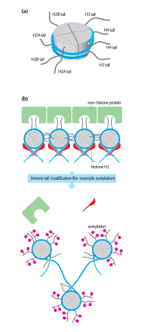
A nucleosome is the basic structural unit of DNA packaging in eukaryotes. The structure of a nucleosome consists of a segment of DNA wound around eight histone proteins and resembles thread wrapped around a spool. The nucleosome is the fundamental subunit of chromatin. Each nucleosome is composed of a little less than two turns of DNA wrapped around a set of eight proteins called histones, which are known as a histone octamer. Each histone octamer is composed of two copies each of the histone proteins H2A, H2B, H3, and H4.

<span class="mw-page-title-main">Histone acetyltransferase</span> Enzymes that catalyze acyl group transfer from acetyl-CoA to histones

Histone acetyltransferases (HATs) are enzymes that acetylate conserved lysine amino acids on histone proteins by transferring an acetyl group from acetyl-CoA to form ε-N-acetyllysine. DNA is wrapped around histones, and, by transferring an acetyl group to the histones, genes can be turned on and off. In general, histone acetylation increases gene expression.

<span class="mw-page-title-main">Histone octamer</span> 8-protein complex forming the core of nucleosomes

In molecular biology, a histone octamer is the eight-protein complex found at the center of a nucleosome core particle. It consists of two copies of each of the four core histone proteins. The octamer assembles when a tetramer, containing two copies of H3 and two of H4, complexes with two H2A/H2B dimers. Each histone has both an N-terminal tail and a C-terminal histone-fold. Each of these key components interacts with DNA in its own way through a series of weak interactions, including hydrogen bonds and salt bridges. These interactions keep the DNA and the histone octamer loosely associated, and ultimately allow the two to re-position or to separate entirely.

<span class="mw-page-title-main">Histone H4</span> One of the five main histone proteins involved in the structure of chromatin

Histone H4 is one of the five main histone proteins involved in the structure of chromatin in eukaryotic cells. Featuring a main globular domain and a long N-terminal tail, H4 is involved with the structure of the nucleosome of the 'beads on a string' organization. Histone proteins are highly post-translationally modified. Covalently bonded modifications include acetylation and methylation of the N-terminal tails. These modifications may alter expression of genes located on DNA associated with its parent histone octamer. Histone H4 is an important protein in the structure and function of chromatin, where its sequence variants and variable modification states are thought to play a role in the dynamic and long term regulation of genes.

The histone code is a hypothesis that the transcription of genetic information encoded in DNA is in part regulated by chemical modifications to histone proteins, primarily on their unstructured ends. Together with similar modifications such as DNA methylation it is part of the epigenetic code. Histones associate with DNA to form nucleosomes, which themselves bundle to form chromatin fibers, which in turn make up the more familiar chromosome. Histones are globular proteins with a flexible N-terminus that protrudes from the nucleosome. Many of the histone tail modifications correlate very well to chromatin structure and both histone modification state and chromatin structure correlate well to gene expression levels. The critical concept of the histone code hypothesis is that the histone modifications serve to recruit other proteins by specific recognition of the modified histone via protein domains specialized for such purposes, rather than through simply stabilizing or destabilizing the interaction between histone and the underlying DNA. These recruited proteins then act to alter chromatin structure actively or to promote transcription. For details of gene expression regulation by histone modifications see table below.

<span class="mw-page-title-main">Histone acetylation and deacetylation</span>

Histone acetylation and deacetylation are the processes by which the lysine residues within the N-terminal tail protruding from the histone core of the nucleosome are acetylated and deacetylated as part of gene regulation.

<span class="mw-page-title-main">Histone-modifying enzymes</span> Type of enzymes

Histone-modifying enzymes are enzymes involved in the modification of histone substrates after protein translation and affect cellular processes including gene expression. To safely store the eukaryotic genome, DNA is wrapped around four core histone proteins, which then join to form nucleosomes. These nucleosomes further fold together into highly condensed chromatin, which renders the organism's genetic material far less accessible to the factors required for gene transcription, DNA replication, recombination and repair. Subsequently, eukaryotic organisms have developed intricate mechanisms to overcome this repressive barrier imposed by the chromatin through histone modification, a type of post-translational modification which typically involves covalently attaching certain groups to histone residues. Once added to the histone, these groups elicit either a loose and open histone conformation, euchromatin, or a tight and closed histone conformation, heterochromatin. Euchromatin marks active transcription and gene expression, as the light packing of histones in this way allows entry for proteins involved in the transcription process. As such, the tightly packed heterochromatin marks the absence of current gene expression.

H3K27ac is an epigenetic modification to the DNA packaging protein histone H3. It is a mark that indicates acetylation of the lysine residue at N-terminal position 27 of the histone H3 protein.

H4K16ac is an epigenetic modification to the DNA packaging protein Histone H4. It is a mark that indicates the acetylation at the 16th lysine residue of the histone H4 protein.

H4K8ac, representing an epigenetic modification to the DNA packaging protein histone H4, is a mark indicating the acetylation at the 8th lysine residue of the histone H4 protein. It has been implicated in the prevalence of malaria.

H4K12ac is an epigenetic modification to the DNA packaging protein histone H4. It is a mark that indicates the acetylation at the 12th lysine residue of the histone H4 protein. H4K12ac is involved in learning and memory. It is possible that restoring this modification could reduce age-related decline in memory.

H4K91ac is an epigenetic modification to the DNA packaging protein histone H4. It is a mark that indicates the acetylation at the 91st lysine residue of the histone H4 protein. No known diseases are attributed to this mark but it might be implicated in melanoma.

H3K23ac is an epigenetic modification to the DNA packaging protein Histone H3. It is a mark that indicates the acetylation at the 23rd lysine residue of the histone H3 protein.

H3K14ac is an epigenetic modification to the DNA packaging protein Histone H3. It is a mark that indicates the acetylation at the 14th lysine residue of the histone H3 protein.

H3K9ac is an epigenetic modification to the DNA packaging protein Histone H3. It is a mark that indicates the acetylation at the 9th lysine residue of the histone H3 protein.

H3K36ac is an epigenetic modification to the DNA packaging protein Histone H3. It is a mark that indicates the acetylation at the 36th lysine residue of the histone H3 protein.

H3K56ac is an epigenetic modification to the DNA packaging protein Histone H3. It is a mark that indicates the acetylation at the 56th lysine residue of the histone H3 protein.

H3S10P is an epigenetic modification to the DNA packaging protein histone H3. It is a mark that indicates the phosphorylation the 10th serine residue of the histone H3 protein.

H3S28P is an epigenetic modification to the DNA packaging protein histone H3. It is a mark that indicates the phosphorylation the 28th serine residue of the histone H3 protein.

References

  1. 1 2 3 4 5 Bhasin M, Reinherz EL, Reche PA (2006). "Recognition and classification of histones using support vector machine" (PDF). Journal of Computational Biology. 13 (1): 102–12. doi:10.1089/cmb.2006.13.102. PMID   16472024.
  2. 1 2 3 4 5 6 7 Rønningen T, Shah A, Oldenburg AR, Vekterud K, Delbarre E, Moskaug JØ, Collas P (December 2015). "Prepatterning of differentiation-driven nuclear lamin A/C-associated chromatin domains by GlcNAcylated histone H2B". Genome Research. 25 (12): 1825–35. doi:10.1101/gr.193748.115. PMC   4665004 . PMID   26359231.
  3. 1 2 3 4 5 6 7 8 Lodish HF, Berk A, Kaiser C, Krieger M, Bretscher A, Ploegh HL, Amon A, Scott MP (2013). Molecular Cell Biology (Seventh ed.). New York: Freeman. pp. 258–328. ISBN   978-1-4292-3413-9.
  4. 1 2 3 4 5 6 7 8 9 10 Molden RC, Bhanu NV, LeRoy G, Arnaudo AM, Garcia BA (2015). "Multi-faceted quantitative proteomics analysis of histone H2B isoforms and their modifications". Epigenetics & Chromatin. 8 (15): 15. doi:10.1186/s13072-015-0006-8. PMC   4411797 . PMID   25922622.
  5. 1 2 Wu J, Mu S, Guo M, Chen T, Zhang Z, Li Z, Li Y, Kang X (January 2016). "Histone H2B gene cloning, with implication for its function during nuclear shaping in the Chinese mitten crab, Eriocheir sinensis". Gene. 575 (2 Pt 1): 276–84. doi:10.1016/j.gene.2015.09.005. PMID   26343795.
  6. 1 2 3 "Histone H2B (53H3) Mouse mAb #2934". Cell Signaling Technology. Retrieved 24 November 2015.
  7. 1 2 3 Osley MA (September 2006). "Regulation of histone H2A and H2B ubiquitylation". Briefings in Functional Genomics & Proteomics. 5 (3): 179–89. doi: 10.1093/bfgp/ell022 . PMID   16772277.
  8. 1 2 Uckelmann M, Sixma TK. Histone ubiquitination in the DNA damage response. DNA Repair (Amst). 2017 Aug;56:92-101. doi: 10.1016/j.dnarep.2017.06.011. Epub 2017 Jun 9. PMID: 28624371
  9. 1 2 3 4 Golebiowski F, Kasprzak KS (November 2005). "Inhibition of core histones acetylation by carcinogenic nickel(II)". Molecular and Cellular Biochemistry. 279 (1–2): 133–9. doi:10.1007/s11010-005-8285-1. PMID   16283522. S2CID   25071586.
  10. R, Fujiki; W, Hashiba; H, Sekine; A, Yokoyama; T, Chikanishi; S, Ito; Y, Imai; J, Kim; Hh, He (2011-11-27). "GlcNAcylation of Histone H2B Facilitates Its Monoubiquitination". Nature. 480 (7378): 557–60. Bibcode:2011Natur.480..557F. doi:10.1038/nature10656. PMC   7289526 . PMID   22121020.
  11. Khare SP, Habib F, Sharma R, Gadewal N, Gupta S, Galande S. "Histone H2B". HIstome: A Relational Knowledgebase of Human Proteins and Histone Modifying Enzymes. Nucleic Acids Research. Archived from the original on 16 October 2016. Retrieved 24 November 2015.