Nephrotic syndrome

Last updated
Nephrotic syndrome
Diabetic glomerulosclerosis (1) HE.jpg
Microscopic image of diabetic glomerulosclerosis, the main cause of nephrotic syndrome in adults.
Specialty Nephrology
Symptoms Swelling, weight gain, feeling tired, foamy urine [1]
Complications Blood clots, infections, high blood pressure [1]
Causes Focal segmental glomerulosclerosis, membranous nephropathy, minimal change disease, diabetes, lupus [1] [2]
Diagnostic method Urine testing, kidney biopsy [1]
Differential diagnosis Nephritic syndrome, cirrhosis, severe malnutrition [2]
TreatmentDirected at underlying cause [1]
Frequency5 per 100,000 per year [3] [4]

Nephrotic syndrome is a collection of symptoms due to kidney damage. [1] This includes protein in the urine, low blood albumin levels, high blood lipids, and significant swelling. [1] Other symptoms may include weight gain, feeling tired, and foamy urine. [1] Complications may include blood clots, infections, and high blood pressure. [1]

Contents

Causes include a number of kidney diseases such as focal segmental glomerulosclerosis, membranous nephropathy, and minimal change disease. [1] [2] It may also occur as a complication of diabetes or lupus. [1] The underlying mechanism typically involves damage to the glomeruli of the kidney. [1] Diagnosis is typically based on urine testing and sometimes a kidney biopsy. [1] It differs from nephritic syndrome in that there are no red blood cells in the urine. [2]

Treatment is directed at the underlying cause. [1] Other efforts include managing high blood pressure, high blood cholesterol, and infection risk. [1] A low-salt diet and limiting fluids are often recommended. [1] About 5 per 100,000 people are affected per year. [3] [4] The usual underlying cause varies between children and adults. [4]

Signs and symptoms

Nephrotic syndrome is usually accompanied by retention of water and sodium. The degree to which this occurs can vary between slight edema in the eyelids that decreases during the day, to affecting the lower limbs, to generalized swelling, to full blown anasarca. Gezichtsoedeem.jpg
Nephrotic syndrome is usually accompanied by retention of water and sodium. The degree to which this occurs can vary between slight edema in the eyelids that decreases during the day, to affecting the lower limbs, to generalized swelling, to full blown anasarca.

Nephrotic syndrome is characterized by large amounts of proteinuria (>3.5 g per 1.73 m2 body surface area per day, [6] or > 40 mg per square meter body surface area per hour in children), hypoalbuminemia (< 3.5 g/dl), hyperlipidaemia, and edema that begins in the face. Lipiduria (lipids in urine) can also occur, but is not essential for the diagnosis of nephrotic syndrome.

Hyperlipidaemia is caused by two factors: [7]

A few other characteristics seen in nephrotic syndrome are:

The main signs of nephrotic syndrome are: [10]

Complications

Nephrotic syndrome can be associated with a series of complications that can affect an individual's health and quality of life: [15]

Causes

Histological image of a normal kidney glomerulus. It is possible to see a glomerulus in the centre of the image surrounded by kidney tubules. Glomerulus pas.JPG
Histological image of a normal kidney glomerulus. It is possible to see a glomerulus in the centre of the image surrounded by kidney tubules.

Nephrotic syndrome has many causes and may either be the result of a glomerular disease that can be either limited to the kidney, called primary nephrotic syndrome (primary glomerulonephrosis), or a condition that affects the kidney and other parts of the body, called secondary nephrotic syndrome. [20]

Primary glomerulonephrosis

Primary causes of nephrotic syndrome are usually described by their histology: [21]

They are considered to be "diagnoses of exclusion", i.e. they are diagnosed only after secondary causes have been excluded.

Secondary glomerulonephrosis

Diabetic glomerulonephritis in a person with nephrotic syndrome. Diabetic glomerulosclerosis (8) PAM.jpg
Diabetic glomerulonephritis in a person with nephrotic syndrome.

Secondary causes of nephrotic syndrome have the same histologic patterns as the primary causes, though they may exhibit some differences suggesting a secondary cause, such as inclusion bodies. [24] They are usually described by the underlying cause, such as:[ citation needed ]

By histologic pattern

Membranous nephropathy (MN)

Focal segmental glomerulosclerosis (FSGS) [26]

Minimal change disease (MCD) [26]

  • Drugs, especially NSAIDs in the elderly
  • Malignancy, especially Hodgkin's lymphoma
  • Allergy
  • Bee sting

Membranoproliferative Glomerulonephritis

Genetics

Over 50 mutations are known to be associated with this condition. [28] [29]

Non-Genetic

There is no known genetic cause for idiopathic nephrotic syndrome. This is thought to be caused by a hitherto unknown circulating permeability factor that travels in the circulation to the podocyte within the glomerulus of the kidney. This circulating factor damages the podocyte which changes its structure. The podocytes are now less able to restrict urinary protein loss. Despite not knowing the specific indentity of the circulating factor, scientists are learning more about it. It is thought to be either T cell or B cell derived, [30] [31] hence why staroid treatment can be effective for some patients. There is also evidence that the circulating factor could be signalling via the PAR-1 receptro on the podocytes. [32]

Pathophysiology

Drawing of the kidney glomerulus. Gray1130.svg
Drawing of the kidney glomerulus.

The kidney glomerulus filters the blood that arrives at the kidney. It is formed of capillaries with small pores that allow small molecules to pass through that have a molecular weight of less than 40,000 daltons, [33] but not larger macromolecules such as proteins.

In nephrotic syndrome, the glomeruli are affected by an inflammation or a hyalinization (the formation of a homogenous crystalline material within cells) that allows proteins such as albumin, antithrombin or the immunoglobulins to pass through the cell membrane and appear in urine. [15]

Albumin is the main protein in the blood that is able to maintain an oncotic pressure, which prevents the leakage of fluid into the extracellular medium and the subsequent formation of edemas.[ citation needed ]

As a response to hypoproteinemia the liver commences a compensatory mechanism involving the synthesis of proteins, such as alpha-2 macroglobulin and lipoproteins. [15] An increase in the latter can cause the hyperlipidemia associated with this syndrome.

Diagnosis

Urinalysis will be able to detect high levels of proteins and occasionally microscopic haematuria. Urinbecher.jpg
Urinalysis will be able to detect high levels of proteins and occasionally microscopic haematuria.
Ultrasound of a kidney with nephrotic syndrome. There is a hyperechoic kidney without demarcation of the cortex and medulla. Ultrasonography of kidney with nephrotic syndrome.jpg
Ultrasound of a kidney with nephrotic syndrome. There is a hyperechoic kidney without demarcation of the cortex and medulla.

Along with obtaining a complete medical history, a series of biochemical tests are required in order to arrive at an accurate diagnosis that verifies the presence of the illness. In addition, imaging of the kidneys (for structure and presence of two kidneys) is sometimes carried out, and/or a biopsy of the kidneys. The first test will be a urinalysis to test for high levels of proteins, [35] as a healthy subject excretes an insignificant amount of protein in their urine. The test will involve a 24-hour bedside urinary total protein estimation. The urine sample is tested for proteinuria (>3.5 g per 1.73 m2 per 24 hours). It is also examined for urinary casts, which are more a feature of active nephritis. Next a blood screen, comprehensive metabolic panel (CMP) will look for hypoalbuminemia: albumin levels of ≤2.5 g/dL (normal=3.5-5 g/dL). Then a Creatinine Clearance CCr test will evaluate kidney function particularly the glomerular filtration capacity. [36] Creatinine formation is a result of the breakdown of muscular tissue, it is transported in the blood and eliminated in urine. Measuring the concentration of organic compounds in both liquids evaluates the capacity of the glomeruli to filter blood. Electrolytes and urea levels may also be analysed at the same time as creatinine (EUC test) in order to evaluate kidney function. A lipid profile will also be carried out as high levels of cholesterol (hypercholesterolemia), specifically elevated LDL, usually with concomitantly elevated VLDL, is indicative of nephrotic syndrome.[ citation needed ]

A kidney biopsy may also be used as a more specific and invasive test method. A study of a sample's anatomical pathology may then allow the identification of the type of glomerulonephritis involved. [35] However, this procedure is usually reserved for adults as the majority of children experience minimal change disease that has a remission rate of 95% with corticosteroids. [37] A biopsy is usually only indicated for children that are corticosteroid resistant as the majority have focal and segmental glomeruloesclerosis. [37]

Further investigations are indicated if the cause is not clear including analysis of auto-immune markers (ANA, ASOT, C3, cryoglobulins, serum electrophoresis), or ultrasound of the whole abdomen.

Classification

A broad classification of nephrotic syndrome based on underlying cause:

Nephrotic
syndrome
PrimarySecondary

Nephrotic syndrome is often classified histologically:

Nephrotic syndrome
MCD FSGS MGN MPGN

Differential diagnosis

Some symptoms that are present in nephrotic syndrome, such as edema and proteinuria, also appear in other illnesses. Therefore, other pathologies need to be excluded in order to arrive at a definitive diagnosis. [38]

Acute fluid overload can cause edema in someone with kidney failure. These people are known to have kidney failure, and have either drunk too much or missed their dialysis. In addition, when Metastatic cancer spreads to the lungs or abdomen it causes effusions and fluid accumulation due to obstruction of lymphatic vessels and veins, as well as serous exudation.

Treatment

The treatment of nephrotic syndrome can be symptomatic or can directly address the injuries caused to the kidney.[ citation needed ]

Symptomatic

The objective of this treatment is to treat the imbalances brought about by the illness: [44] edema, hypoalbuminemia, hyperlipidaemia, hypercoagulability and infectious complications.

  1. Analyse haemoglobin and haematocrit levels.
  2. A solution of 25% albumin is used that is administered for only 4 hours in order to avoid pulmonary edema.
  3. Haemoglobin and haematocrit levels are analysed again: if the haematocrit value is less than the initial value (a sign of correct expansion) the diuretics are administered for at least 30 minutes. If the haematocrit level is greater than the initial one this is a contraindication for the use of diuretics as they would increase said value.
It may be necessary to give a person potassium or require a change in dietary habits if the diuretic drug causes hypokalaemia as a side effect.

In addition to these key imbalances, vitamin D and calcium are also taken orally in case the alteration of vitamin D causes severe hypocalcaemia, this treatment has the goal of restoring physiological levels of calcium in the person. [56]

Kidney damage

The treatment of kidney damage may reverse or delay the progression of the disease. [44] Kidney damage is treated by prescribing drugs:

The susceptibility testing in vitro to glucocorticoids on the person's peripheral blood mononuclear cells is associated with the number of new cases of not optimal clinical responses: the most sensitive people in vitro have shown a higher number of cases of corticodependence, while the most resistant people in vitro showed a higher number of cases of ineffective therapy. [59]

Prognosis

The prognosis for nephrotic syndrome under treatment is generally good although this depends on the underlying cause, the age of the person and their response to treatment. It is usually good in children, because minimal change disease responds very well to steroids and does not cause chronic kidney failure. Any relapses that occur become less frequent over time; [60] the opposite occurs with mesangiocapillary glomerulonephritis, in which the kidney fails within three years of the disease developing, making dialysis necessary and subsequent kidney transplant. [60] In addition children under the age of 5 generally have a poorer prognosis than prepubescents, as do adults older than 30 years of age as they have a greater risk of kidney failure. [61]

Other causes such as focal segmental glomerulosclerosis frequently lead to end stage kidney disease. Factors associated with a poorer prognosis in these cases include level of proteinuria, blood pressure control and kidney function (GFR).[ citation needed ]

Without treatment nephrotic syndrome has a very bad prognosis especially rapidly progressing glomerulonephritis, which leads to acute kidney failure after a few months.[ citation needed ]

Epidemiology

Nephrotic syndrome can affect any age, although it is mainly found in adults with a ratio of adults to children of 26 to 1. [62]

The syndrome presents in different ways in the two groups: the most frequent glomerulopathy in children is minimal change disease (66% of cases), followed by focal segmental glomerulosclerosis (8%) and mesangiocapillary glomerulonephritis (6%). [24] In adults the most common disease is mesangiocapillary glomerulonephritis (30-40%), followed by focal and segmental glomeruloesclerosis (15-25%) and minimal change disease (20%). The latter usually presents as secondary and not primary as occurs in children. Its main cause is diabetic nephropathy. [24] It usually presents in a person from their 40s or 50s. Of the glomerulonephritis cases, approximately 60% to 80% are primary, while the remainder are secondary. [62]

There are also differences in epidemiology between the sexes, the disease is more common in men than in women by a ratio of 2 to 1. [62]

The epidemiological data also reveals information regarding the most common way that symptoms develop in people with nephrotic syndrome: [62] spontaneous remission occurs in up to 20% or 30% of cases during the first year of the illness. However, this improvement is not definitive as some 50% to 60% of people with Nephrotic syndrome die and/or develop chronic kidney failure 6 to 14 years after this remission. On the other hand, between 10% and 20% of people have continuous episodes of remissions and relapses without dying or jeopardizing their kidney. The main causes of death are cardiovascular, as a result of the chronicity of the syndrome, and thromboembolic accidents.

Related Research Articles

Albuminuria is a pathological condition wherein the protein albumin is abnormally present in the urine. It is a type of proteinuria. Albumin is a major plasma protein ; in healthy people, only trace amounts of it are present in urine, whereas larger amounts occur in the urine of patients with kidney disease. For a number of reasons, clinical terminology is changing to focus on albuminuria more than proteinuria.

<span class="mw-page-title-main">Edema</span> Accumulation of excess fluid in body tissue

Edema, also spelled oedema, and also known as fluid retention, dropsy, hydropsy and swelling, is the build-up of fluid in the body's tissue. Most commonly, the legs or arms are affected. Symptoms may include skin which feels tight, the area may feel heavy, and joint stiffness. Other symptoms depend on the underlying cause.

<span class="mw-page-title-main">Proteinuria</span> Presence of an excess of serum proteins in the urine

Proteinuria is the presence of excess proteins in the urine. In healthy persons, urine contains very little protein, less than 150 mg/day; an excess is suggestive of illness. Excess protein in the urine often causes the urine to become foamy. Severe proteinuria can cause nephrotic syndrome in which there is worsening swelling of the body.

<span class="mw-page-title-main">Nephritis</span> Inflammation of the kidneys

Nephritis is inflammation of the kidneys and may involve the glomeruli, tubules, or interstitial tissue surrounding the glomeruli and tubules. It is one of several different types of nephropathy.

<span class="mw-page-title-main">Hematuria</span> Medical condition

Hematuria or haematuria is defined as the presence of blood or red blood cells in the urine. "Gross hematuria" occurs when urine appears red, brown, or tea-colored due to the presence of blood. Hematuria may also be subtle and only detectable with a microscope or laboratory test. Blood that enters and mixes with the urine can come from any location within the urinary system, including the kidney, ureter, urinary bladder, urethra, and in men, the prostate. Common causes of hematuria include urinary tract infection (UTI), kidney stones, viral illness, trauma, bladder cancer, and exercise. These causes are grouped into glomerular and non-glomerular causes, depending on the involvement of the glomerulus of the kidney. But not all red urine is hematuria. Other substances such as certain medications and foods can cause urine to appear red. Menstruation in women may also cause the appearance of hematuria and may result in a positive urine dipstick test for hematuria. A urine dipstick test may also give an incorrect positive result for hematuria if there are other substances in the urine such as myoglobin, a protein excreted into urine during rhabdomyolysis. A positive urine dipstick test should be confirmed with microscopy, where hematuria is defined by three or more red blood cells per high power field. When hematuria is detected, a thorough history and physical examination with appropriate further evaluation can help determine the underlying cause.

<span class="mw-page-title-main">Glomerulonephritis</span> Term for several kidney diseases

Glomerulonephritis (GN) is a term used to refer to several kidney diseases. Many of the diseases are characterised by inflammation either of the glomeruli or of the small blood vessels in the kidneys, hence the name, but not all diseases necessarily have an inflammatory component.

<span class="mw-page-title-main">Diabetic nephropathy</span> Chronic loss of kidney function

Diabetic nephropathy, also known as diabetic kidney disease, is the chronic loss of kidney function occurring in those with diabetes mellitus. Diabetic nephropathy is the leading causes of chronic kidney disease (CKD) and end-stage renal disease (ESRD) globally. The triad of protein leaking into the urine, rising blood pressure with hypertension and then falling renal function is common to many forms of CKD. Protein loss in the urine due to damage of the glomeruli may become massive, and cause a low serum albumin with resulting generalized body swelling (edema) so called nephrotic syndrome. Likewise, the estimated glomerular filtration rate (eGFR) may progressively fall from a normal of over 90 ml/min/1.73m2 to less than 15, at which point the patient is said to have end-stage renal disease. It usually is slowly progressive over years.

<span class="mw-page-title-main">Membranous glomerulonephritis</span> Medical condition

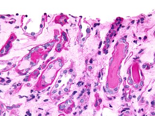
Membranous glomerulonephritis (MGN) is a slowly progressive disease of the kidney affecting mostly people between ages of 30 and 50 years, usually white people.

<span class="mw-page-title-main">Minimal change disease</span> Medical condition

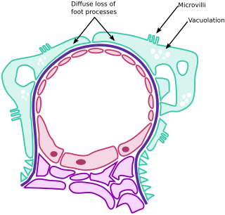
Minimal change disease is a disease affecting the kidneys which causes nephrotic syndrome. Nephrotic syndrome leads to the loss of significant amounts of protein in the urine, which causes the widespread edema and impaired kidney function commonly experienced by those affected by the disease. It is most common in children and has a peak incidence at 2 to 6 years of age. MCD is responsible for 10–25% of nephrotic syndrome cases in adults. It is also the most common cause of nephrotic syndrome of unclear cause (idiopathic) in children.

<span class="mw-page-title-main">Nephritic syndrome</span> Medical condition

Nephritic syndrome is a syndrome comprising signs of nephritis, which is kidney disease involving inflammation. It often occurs in the glomerulus, where it is called glomerulonephritis. Glomerulonephritis is characterized by inflammation and thinning of the glomerular basement membrane and the occurrence of small pores in the podocytes of the glomerulus. These pores become large enough to permit both proteins and red blood cells to pass into the urine. By contrast, nephrotic syndrome is characterized by proteinuria and a constellation of other symptoms that specifically do not include hematuria. Nephritic syndrome, like nephrotic syndrome, may involve low level of albumin in the blood due to the protein albumin moving from the blood to the urine.

<span class="mw-page-title-main">Focal segmental glomerulosclerosis</span> Kidney disease

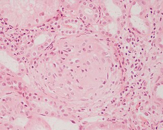
Focal segmental glomerulosclerosis (FSGS) is a histopathologic finding of scarring (sclerosis) of glomeruli and damage to renal podocytes. This process damages the filtration function of the kidney, resulting in protein presence in the urine due to protein loss. FSGS is a leading cause of excess protein loss—nephrotic syndrome—in children and adults. Signs and symptoms include proteinuria and edema. Kidney failure is a common long-term complication of the disease. FSGS can be classified as primary, secondary, or genetic, depending on whether a particular toxic or pathologic stressor or genetic predisposition can be identified as the cause. Diagnosis is established by renal biopsy, and treatment consists of glucocorticoids and other immune-modulatory drugs. Response to therapy is variable, with a significant portion of patients progressing to end-stage kidney failure. An American epidemiological study 20 years ago demonstrated that FSGS is estimated to occur in 7 persons per million, with males and African-Americans at higher risk.

<span class="mw-page-title-main">Urinary cast</span> Cylindrical protein structure in urine in certain disease states

Urinary casts are microscopic cylindrical structures produced by the kidney and present in the urine in certain disease states. They form in the distal convoluted tubule and collecting ducts of nephrons, then dislodge and pass into the urine, where they can be detected by microscopy.

<span class="mw-page-title-main">Hypoalbuminemia</span> Medical condition

Hypoalbuminemia is a medical sign in which the level of albumin in the blood is low. This can be due to decreased production in the liver, increased loss in the gastrointestinal tract or kidneys, increased use in the body, or abnormal distribution between body compartments. Patients often present with hypoalbuminemia as a result of another disease process such as malnutrition as a result of severe anorexia nervosa, sepsis, cirrhosis in the liver, nephrotic syndrome in the kidneys, or protein-losing enteropathy in the gastrointestinal tract. One of the roles of albumin is being the major driver of oncotic pressure in the bloodstream and the body. Thus, hypoalbuminemia leads to abnormal distributions of fluids within the body and its compartments. As a result, associated symptoms include edema in the lower legs, ascites in the abdomen, and effusions around internal organs. Laboratory tests aimed at assessing liver function diagnose hypoalbuminemia. Once identified, it is a poor prognostic indicator for patients with a variety of different diseases. Yet, it is only treated in very specific indications in patients with cirrhosis and nephrotic syndrome. Treatment instead focuses on the underlying cause of the hypoalbuminemia. Albumin is an acute negative phase respondent and not a reliable indicator of nutrition status.

Congenital nephrotic syndrome is a rare kidney disease which manifests in infants during the first 3 months of life, and is characterized by high levels of protein in the urine (proteinuria), low levels of protein in the blood, and swelling. This disease is primarily caused by genetic mutations which result in damage to components of the glomerular filtration barrier and allow for leakage of plasma proteins into the urinary space.

<span class="mw-page-title-main">Rapidly progressive glomerulonephritis</span> Medical condition

Rapidly progressive glomerulonephritis (RPGN) is a syndrome of the kidney that is characterized by a rapid loss of kidney function, with glomerular crescent formation seen in at least 50% or 75% of glomeruli seen on kidney biopsies. If left untreated, it rapidly progresses into acute kidney failure and death within months. In 50% of cases, RPGN is associated with an underlying disease such as Goodpasture syndrome, systemic lupus erythematosus or granulomatosis with polyangiitis; the remaining cases are idiopathic. Regardless of the underlying cause, RPGN involves severe injury to the kidneys' glomeruli, with many of the glomeruli containing characteristic glomerular crescents.

<span class="mw-page-title-main">Mesangial proliferative glomerulonephritis</span> Medical condition

Mesangial proliferative glomerulonephritis (MesPGN) is a morphological pattern characterized by a numerical increase in mesangial cells and expansion of the extracellular matrix within the mesangium of the glomerulus. The increase in the number of mesangial cells can be diffuse or local and immunoglobulin and/or complement deposition can also occur. MesPGN is associated with a variety of disease processes affecting the glomerulus, though can be idiopathic. The clinical presentation of MesPGN usually consists of hematuria or nephrotic syndrome. Treatment is often consistent with the histologic pattern of and/or disease process contributing to mesangial proliferative glomerulonephritis, and usually involves some form of immunosuppressant.

Glomerulonephrosis is a non-inflammatory disease of the kidney (nephrosis) presenting primarily in the glomerulus as nephrotic syndrome. The nephron is the functional unit of the kidney and it contains the glomerulus, which acts as a filter for blood to retain proteins and blood lipids. Damage to these filtration units results in important blood contents being released as waste in urine. This disease can be characterized by symptoms such as fatigue, swelling, and foamy urine, and can lead to chronic kidney disease and ultimately end-stage renal disease, as well as cardiovascular diseases. Glomerulonephrosis can present as either primary glomerulonephrosis or secondary glomerulonephrosis.

Diffuse proliferative glomerulonephritis (DPGN) is a type of glomerulonephritis that is the most serious form of renal lesions in SLE and is also the most common, occurring in 35% to 60% of patients. In absence of SLE, DPGN pathology looks more like Membranoproliferative glomerulonephritis

Sickle cell nephropathy is a type of nephropathy associated with sickle cell disease which causes kidney complications as a result of sickling of red blood cells in the small blood vessels. The hypertonic and relatively hypoxic environment of the renal medulla, coupled with the slow blood flow in the vasa recta, favors sickling of red blood cells, with resultant local infarction. Functional tubule defects in patients with sickle cell disease are likely the result of partial ischemic injury to the renal tubules.

Monoclonal gammopathy of renal significance (MGRS) are a group of kidney disorders that present with kidney damage due to nephrotoxic monoclonal immunoglobulins secreted by clonal plasma cells or B cells. By definition, people with MGRS do not meet criteria for multiple myeloma or other hematologic malignancies. The term MGRS was introduced in 2012 by the International Kidney and Monoclonal Gammopathy Research Group (IKMG). MGRS is associated with monoclonal gammopathy of undetermined significance (MGUS). People with MGUS have a monoclonal gammopathy but does not meet the criteria for the clonal burden nor the presence of end organ damage seen in hematologic malignancies. In a population based study based on the NHANES III health survey; 6% of patients with MGUS were subsequently classified as having MGRS. The prevalence and incidence of MGRS in the general population or in specific populations is not known but it is more prevalent in those over the age of 50 as there is a monoclonal protein (M-protein) present in 3% of those 50 and years older and 5% of those 70 years and older, placing those 50 and older at increased risk of MGRS.

References

  1. 1 2 3 4 5 6 7 8 9 10 11 12 13 14 15 16 "Nephrotic Syndrome in Adults". National Institute of Diabetes and Digestive and Kidney Diseases. February 2014. Retrieved 9 November 2017.
  2. 1 2 3 4 Ferri, Fred F. (2017). Ferri's Clinical Advisor 2018 E-Book: 5 Books in 1. Elsevier Health Sciences. p. 889. ISBN   9780323529570.
  3. 1 2 Kher, Kanwal; Schnaper, H. William; Greenbaum, Larry A. (2016). Clinical Pediatric Nephrology, Third Edition. CRC Press. p. 307. ISBN   9781482214635.
  4. 1 2 3 Kelly, Christopher R.; Landman, Jaime (2012). The Netter Collection of Medical Illustrations - Urinary System e-Book. Elsevier Health Sciences. p. 101. ISBN   978-1455726561.
  5. Behrman, Richard E.; Robert M Kliegman; Hal B. Jenson (2008). Nelson Tratado de Pediatria (in Spanish). Elsevier, España. p. 1755. ISBN   978-8481747478.
  6. "Electronic Learning Module for Kidney and Urinary Tract Diseases". Archived from the original on 2008-12-20. Retrieved 2015-12-25.
  7. Davis, K; Kohli, P (15 July 2019). "What to know about hyperlipidemia". Medical News Today. Retrieved 1 July 2021.
  8. "Case Based Pediatrics Chapter". Hawaii.edu. Retrieved 23 August 2018.
  9. Freedberg, Irwin M.; et al., eds. (2003). Fitzpatrick's dermatology in general medicine (6th ed.). New York, NY [u.a.]: McGraw-Hill. p. 659. ISBN   0-07-138076-0.
  10. "Manifestaciones clínicas del síndrome nefrótico" (PDF). See table 4.2. Archived from the original (PDF) on 24 September 2015. Retrieved 12 Sep 2008.
  11. 1 2 García - Conde, J.; Merino Sánchez, J.; González Macías, J. (1995). "Fisiopatología glomerular". Patología General. Semiología Clínica y Fisiopatología. McGraw - Hill Interamericana. ISBN   8448600932.
  12. Parra Herrán, Carlos Eduardo; Castillo Londoño, Juan Sebastián; López Panqueva, Rocío del Pilar; Andrade Pérez, Rafael Enrique. "Síndrome nefrótico y proteinuria en rango no nefrótico" . Retrieved 2008-09-14.
  13. "Valores normales de proteína en orina de 24 horas" . Retrieved 24 August 2012.
  14. Klahr S, Tripathy K, Bolanos O (1967). "Qualitative and Quantitative Analysis of Urinary Lipids in the Nephrotic Syndrome". J. Clin. Invest. 46 (9): 1475–81. doi:10.1172/JCI105639. PMC   292893 . PMID   6036540.
  15. 1 2 3 Álvarez, Sandalio Durán (1999). "Complicaciones agudas del síndrome nefrótico" [Acute complications of nephrotic syndrome]. Revista Cubana de Pediatría (in Spanish). 7 (4).
  16. Ruiz, S.; Soto, S.; Rodado, R.; Alcaraz, F.; López Guillén, E. (September 2007). "Peritonitis bacteriana espontánea como forma de presentación de síndrome nefrótico idiopático en un adulto de raza negra" [Spontaneous bacterial peritonitis as form of presentation of idiophatic nephrotic syndrome in an adult black race]. Anales de Medicina Interna (in Spanish). 24 (9): 442–4. doi: 10.4321/s0212-71992007000900008 . PMID   18198954.
  17. Zollo, Anthony J (2005). "Nefrología". Medicina interna. Secretos (Cuarta ed.). Elsevier España. p. 283. ISBN   8481748862.
  18. 1 2 "Balance de nitrógeno y equilibrio nitrogenado" . Retrieved 8 Sep 2008. It occurs during renal pathologies, during fasting, in eating disorders or during heavy physical exercise.
  19. "Cushing's Syndrome". The Lecturio Medical Concept Library. Retrieved 11 July 2021.
  20. "Ascites". The Lecturio Medical Concept Library. Retrieved 1 July 2021.
  21. "Descripción histológica de las glomerulonefritis ideopáticas" . Retrieved 8 Sep 2008.
  22. 1 2 "Patient information: The nephrotic syndrome (Beyond the Basics)" . Retrieved 2013-06-28.
  23. James W Lohr, MD. "Rapidly progressive glomerulonephritis" . Retrieved 2013-06-28.
  24. 1 2 3 "Frecuencia de las glomerulonefritis y causas de las glomerulonefritis secundarias" . Retrieved 8 Sep 2008.[ permanent dead link ]
  25. "Fármacos que pueden producir síndrome nefrótico" . Retrieved 8 Sep 2008.
  26. 1 2 Fogo AB, Bruijn JA. Cohen AH, Colvin RB, Jennette JC. Fundamentals of Renal Pathology. Springer. ISBN   978-0-387-31126-5.
  27. 1 2 "Nephrotic syndrome". Archived from the original on 2013-04-23. Retrieved 2016-05-21.
  28. Bierzynska, Agnieszka; McCarthy, Hugh J.; Soderquest, Katrina; Sen, Ethan S.; Colby, Elizabeth; Ding, Wen Y.; Nabhan, Marwa M.; Kerecuk, Larissa; Hegde, Shivram; Hughes, David; Marks, Stephen; Feather, Sally; Jones, Caroline; Webb, Nicholas J.A.; Ognjanovic, Milos (April 2017). "Genomic and clinical profiling of a national nephrotic syndrome cohort advocates a precision medicine approach to disease management". Kidney International. 91 (4): 937–947. doi:10.1016/j.kint.2016.10.013. hdl: 1983/c730c0d6-5527-435a-8c27-a99fd990a0e8 . ISSN   0085-2538. PMID   28117080. S2CID   4768411.
  29. Braun, Daniela A.; Lovric, Svjetlana; Schapiro, David; Schneider, Ronen; Marquez, Jonathan; Asif, Maria; Hussain, Muhammad Sajid; Daga, Ankana; Widmeier, Eugen; Rao, Jia; Ashraf, Shazia; Tan, Weizhen; Lusk, C. Patrick; Kolb, Amy; Jobst-Schwan, Tilman; Schmidt, Johanna Magdalena; Hoogstraten, Charlotte A.; Eddy, Kaitlyn; Kitzler, Thomas M.; Shril, Shirlee; Moawia, Abubakar; Schrage, Kathrin; Khayyat, Arwa Ishaq A.; Lawson, Jennifer A.; Gee, Heon Yung; Warejko, Jillian K.; Hermle, Tobias; Majmundar, Amar J.; Hugo, Hannah; Budde, Birgit; Motameny, Susanne; Altmüller, Janine; Noegel, Angelika Anna; Fathy, Hanan M.; Gale, Daniel P.; Waseem, Syeda Seema; Khan, Ayaz; Kerecuk, Larissa; Hashmi, Seema; Mohebbi, Nilufar; Ettenger, Robert; Serdaroğlu, Erkin; Alhasan, Khalid A.; Hashem, Mais; Goncalves, Sara; Ariceta, Gema; Ubetagoyena, Mercedes; Antonin, Wolfram; Baig, Shahid Mahmood; Alkuraya, Fowzan S.; Shen, Qian; Xu, Hong; Antignac, Corinne; Lifton, Richard P.; Mane, Shrikant; Nürnberg, Peter; Khokha, Mustafa K.; Hildebrandt, Friedhelm (4 September 2018). "Mutations in multiple components of the nuclear pore complex cause nephrotic syndrome". Journal of Clinical Investigation. 128 (10): 4313–4328. doi:10.1172/JCI98688. PMC   6159964 . PMID   30179222.
  30. van den Berg, José G.; Weening, Jan J. (2004-07-27). "Role of the immune system in the pathogenesis of idiopathic nephrotic syndrome". Clinical Science. 107 (2): 125–136. doi:10.1042/cs20040095. ISSN   0143-5221. PMID   15157184.
  31. May, Carl J.; Welsh, Gavin I.; Chesor, Musleeha; Lait, Phillipa J.; Schewitz-Bowers, Lauren P.; Lee, Richard W. J.; Saleem, Moin A. (2019-10-01). "Human Th17 cells produce a soluble mediator that increases podocyte motility via signaling pathways that mimic PAR-1 activation". American Journal of Physiology. Renal Physiology. 317 (4): F913–F921. doi:10.1152/ajprenal.00093.2019. ISSN   1931-857X. PMC   6843047 . PMID   31339775.
  32. May, Carl J.; Chesor, Musleeha; Hunter, Sarah E.; Hayes, Bryony; Barr, Rachel; Roberts, Tim; Barrington, Fern A.; Farmer, Louise; Ni, Lan; Jackson, Maisie; Snethen, Heidi; Tavakolidakhrabadi, Nadia; Goldstone, Max; Gilbert, Rodney; Beesley, Matt (March 2023). "Podocyte protease activated receptor 1 stimulation in mice produces focal segmental glomerulosclerosis mirroring human disease signaling events". Kidney International. 104 (2): 265–278. doi: 10.1016/j.kint.2023.02.031 . ISSN   0085-2538. PMID   36940798. S2CID   257639270.
  33. "Apuntes de fisiopatología de sistemas". Archived from the original on 2008-09-08. Retrieved 8 Sep 2008.
  34. Hansen, Kristoffer; Nielsen, Michael; Ewertsen, Caroline (23 December 2015). "Ultrasonography of the Kidney: A Pictorial Review". Diagnostics. 6 (1): 2. doi: 10.3390/diagnostics6010002 . PMC   4808817 . PMID   26838799.
  35. 1 2 "Nefrología y urología" . Retrieved 12 Sep 2008.
  36. "El diagnóstico del síndrome nefrótico" . Retrieved 12 Sep 2008.
  37. 1 2 Voguel S, Andrea; Azócar P, Marta; Nazal Ch, Vilma; Salas del C, Paulina (June 2006). "Indicaciones de la biospsia renal en niños". Revista Chilena de Pediatría. 77 (3): 295–303. doi: 10.4067/S0370-41062006000300011 . Retrieved 2008-09-14.
  38. "Diagnóstico diferencial en el síndrome nefrótico". Archived from the original on 2009-03-06. Retrieved 2008-09-14.{{cite web}}: CS1 maint: bot: original URL status unknown (link)
  39. Harold Friedman, H (2001). "General problems". Problem-oriented Medical Diagnosis (Seventh ed.). Lippincott Williams & Wilkins. pp. 3 and 4. ISBN   0-7817-2909-2.
  40. "El edema en la insuficiencia cardíaca" . Retrieved 2008-09-14.
  41. 1 2 Goldman, Lee; Braunwald, Eugene (2000). "Edemas". Cardiología en atención primaria. Harcourt. pp. 114–117. ISBN   8481744328.
  42. Rivera, F; Egea, J.J; Jiménez del Cerro, L.A; Olivares, J. "La proteinuria en el mieloma múltiple" . Retrieved 2008-09-14.[ permanent dead link ]
  43. Bustillo Solano, Emilio. "Relación de la proteinuria con el nivel de hemoglobina glicosilada en los diabéticos". Archived from the original on 2008-09-14. Retrieved 2008-09-14.
  44. 1 2 Curtis, Michael J.; Page, Clive P.; Walker, Michael J.A; Hoffman, Brian B. (1998). "Fisiopatología y enfermedades renales". Farmacología integrada. Harcourt. ISBN   8481743402.
  45. Saz Peiro, Pablo. "El reposo prolongado" (PDF). Archived from the original (PDF) on 24 January 2009. Retrieved 8 Sep 2008.
  46. 1 2 3 "Dietoterapia del síndrome nefrótico". Archived from the original on 2009-01-22. Retrieved 8 Sep 2008.
  47. "Lista de alimentos ricos en sodio" . Retrieved 8 Sep 2008.
  48. "Fluidoterapia: tipos de expansores" (PDF). Archived from the original (PDF) on 2008-09-20. Retrieved 8 Sep 2008. Plasma expanders are natural or synthetic substances (dextran, albumin...), that are able to retain liquid in the vascular space.
  49. "Lista de alimentos ricos en proteínas". Archived from the original on 7 September 2008. Retrieved 8 Sep 2008. Expressed as grams per 100 g of food.
  50. "Sustitución de los alimentos ricos en grasas de la dieta". Archived from the original on February 12, 2008. Retrieved 8 Sep 2008. Organizations in the US recommend that no more than 30% of total daily calorie intake is from fats.
  51. Martín Zurro, Armando (2005). "Hipolipemiante, diuréticos, estatina.". Compendio de atención primaria: Conceptos, organización y práctica clínica (Segunda ed.). Elsevier España. p. 794. ISBN   8481748161.
  52. Jiménez Alonso, Juan. "Profilaxis de los fenómenos tromboembólicos" (PDF). Retrieved 2008-09-14.[ permanent dead link ]
  53. Glassock RJ (August 2007). "Prophylactic anticoagulation in nephrotic syndrome: a clinical conundrum". J. Am. Soc. Nephrol. 18 (8): 2221–5. doi: 10.1681/ASN.2006111300 . PMID   17599972.
  54. "Rango Internacional Normalizado (INR)" . Retrieved 2008-09-14.
  55. "Tratamiento de la hipercoagulabilidad". Archived from the original on 2008-09-15. Retrieved 2008-09-14.
  56. "Tratamiento de la hipocalcemia". Archived from the original on 2008-09-15. Retrieved 2008-09-14.
  57. Hahn D, Hodson EM, Willis NS, Craig JC (2015). "Corticosteroid therapy for nephrotic syndrome in children". Cochrane Database of Systematic Reviews. 2015 (3): CD001533. doi:10.1002/14651858.CD001533.pub5. PMC   7025788 . PMID   25785660.
  58. According to MedlinePlus, avascular necrosis is the death of the bone caused by insufficient blood supply to the bone.
  59. Cuzzoni, E; De Iudicibus, S; Stocco, G; Favretto, D (2016). "In Vitro sensitivity to methyl-prednisolone is associated with clinical response in pediatric idiopathic nephrotic syndrome". Clin Pharmacol Ther. 100 (3): 268–74. doi:10.1002/cpt.372. hdl: 11368/2878528 . PMID   27007551. S2CID   37671642.
  60. 1 2 Guerrero Fernández, J. "Pronóstico de la enfermedad" . Retrieved 2016-05-21.
  61. "Síndrome nefrótico idiopático: diagnóstico histológico por biopsia renal percutanea". 1995. Archived from the original on 2016-03-25. Retrieved 2016-05-21.
  62. 1 2 3 4 Borrego R., Jaime; Montero C., Orlando (2003). Nefrología: Fundamentos de medicina (Cuarta ed.). Corporación para investigaciones biológicas. p. 340. ISBN   9589400639.