Nuclear pore

Last updated

Nuclear Pore
Diagram human cell nucleus.svg
Diagram of the human cell nucleus with nuclear pores.
NuclearPore crop.png
Schematic diagram of a nuclear pore complex within the nuclear envelope (1) with the outer ring (2), spokes (3), basket (4), and filaments (5).
Details
Identifiers
Latin porus nuclearis
MeSH D022022
TH H1.00.01.2.01005
FMA 63148
Anatomical terminology

A nuclear pore is a channel as part of the nuclear pore complex (NPC), a large protein complex found in the nuclear envelope of eukaryotic cells. The nuclear envelope (NE) surrounds the cell nucleus containing DNA and facilitates the selective membrane transport of various molecules.

Contents

The nuclear pore complex predominantly consists of proteins known as nucleoporins (Nups). Each human NPC comprises at least 456 individual protein molecules, with 34 distinct nucleoporin proteins, [1] in 2022 90% of the structure was known, [2] then in 2024 the nuclear basket was decoded. About half of the nucleoporins encompass solenoid protein domains, such as alpha solenoids or beta-propeller folds, and occasionally both as separate structural domains. Conversely, the remaining nucleoporins exhibit characteristics of "natively unfolded" or intrinsically disordered proteins, characterized by high flexibility and a lack of ordered tertiary structure. These disordered proteins, referred to as FG nucleoporins (FG-Nups), contain multiple phenylalanineglycine repeats (FG repeats) in their amino acid sequences. [3] FG-Nups is one of three main types of nucleoporins found in the NPC. The other two are the transmembrane Nups and the scaffold Nups. The transmembrane Nups are made up of transmembrane α-helices and play a vital part in anchoring the NPC to the nuclear envelope. The scaffold Nups are made up of α-solenoid and β-propeller folds, and create the structural framework of NPCs. [4]

The principal function of nuclear pore complexes is to facilitate selective membrane transport of various molecules across the nuclear envelope. This includes the transportation of RNA and ribosomal proteins from the nucleus to the cytoplasm, as well as proteins (such as DNA polymerase and lamins), carbohydrates, signaling molecules, and lipids moving into the nucleus. Notably, the nuclear pore complex (NPC) can actively mediate up to 1000 translocations per complex per second. While smaller molecules can passively diffuse through the pores, larger molecules are often identified by specific signal sequences and are facilitated by nucleoporins to traverse the nuclear envelope.

Evolutionary conserved features in sequences that code for nucleoporins regulate molecular transport through the nuclear pore. [5] [6] Nucleoporin-mediated transport does not entail direct energy expenditure but instead relies on concentration gradients associated with the RAN cycle (Ras-related nuclear protein cycle).

The count of nuclear pore complexes varies across cell types and different stages of the cell's life cycle, with approximately 1,000 NPCs typically found in vertebrate cells. [7] The human nuclear pore complex (hNPC) is a substantial structure, with a molecular weight of 120 megadaltons (MDa). [8] Each NPC comprises eight protein subunits encircling the actual pore, forming the outer ring. Additionally, these subunits project a spoke-shaped protein over the pore channel. The central region of the pore may exhibit a plug-like structure; however, its precise nature remains unknown, and it is yet undetermined whether it represents an actual plug or merely cargo transiently caught in transit.

Nuclear Pore Complex: Size and Composition

The nuclear pore complex (NPC) is a crucial cellular structure with a diameter of approximately 120 nanometers in vertebrates. Its channel varies from 5.2 nanometers in humans [9] to 10.7 nm in the frog Xenopus laevis , with a depth of roughly 45 nm. [10] Additionally, mRNA, being single-stranded, has a thickness ranging from 0.5 to 1 nm. The mammalian NPC has a molecular mass of about 124 megadaltons (MDa), comprising approximately 30 distinct protein components, each in multiple copies. The mammalian NPCs contain about 800 nucleoporins each that are organized into distinct NPC subcomplexes. [11] Conversely, the yeast Saccharomyces cerevisiae possesses a smaller mass, estimated at only 66 MDa. [12]

Transport through the nuclear pore complex

The Ran-GTP cycle, which drives the import and export of RNA and proteins through the nuclear protein complex. Rancycle nuclearimport nuclearexport.png
The Ran-GTP cycle, which drives the import and export of RNA and proteins through the nuclear protein complex.
Scanning and illumination microscopy of nuclear pores, lamina, and chromatin. 3D-SIM-1 NPC Confocal vs 3D-SIM detail.jpg
Scanning and illumination microscopy of nuclear pores, lamina, and chromatin.

Nuclear pore complex (NPC) serves highly regulated gateway for the transport of molecules between the nucleus and the cytoplasm. This intricate system enables the selective passage for molecules including proteins, RNA, and signaling molecules, ensuring proper cellular function and homeostasis. Small molecules such as proteins water and ions can diffuse through NPCs, but cargoes (>40 KDa) such as RNA and protein require the participation of soluble transport receptors. [13]

The largest family of nuclear transport receptors are karyopherin's, these are also knowing as importins or exportins. These are a superfamily of nuclear transport receptors that facilitate the translocation of proteins, RNAs, and ribonuclear particles across the NPC in a Ran GTP hydrolase-dependent process. [14] This family is further subdivided to the karyopherin-α and the karyopherin-β subfamilies. Other nuclear transport receptors include NTF2 and some NTF2-like proteins.

Three models have been suggested to explain the translocation mechanism:

Import of proteins

Nuclear proteins are synthesized in the cytoplasm and need to be imported through the NPCs into the nucleus. Import can be directed by various signals, of which nuclear localization signal (NLS) are best characterized. [15] Several NLS sequences are known, generally containing a conserved sequence with basic residues such as PKKKRKV. Any material with an NLS will be taken up by importins to the nucleus.[ citation needed ]

Importation begins with Importin-α binding to the NLS sequence of cargo proteins, forming a complex. Importin-β then attaches to Importin-α, facilitating transport towards the NPC.[ citation needed ]

As the complex reaches the NPC, it diffuses through the pore without the need for additional energy. Upon entry into nucleus, RanGTP binds to Importin-β and displaces it from the complex. Then the cellular apoptosis susceptibility protein (CAS), an exportin which in the nucleus is bound to RanGTP, displaces Importin-α from the cargo. The NLS-protein is thus free in the nucleoplasm. The Importinβ-RanGTP and Importinα-CAS-RanGTP complex diffuses back to the cytoplasm where GTPs are hydrolyzed to GDP leading to the release of Importinβ and Importinα which become available for a new NLS-protein import round.[ citation needed ]

While translocation through the NPC is not energy-dependent, the overall import cycle needs the hydrolysis of two GTPs molecules, making it an active transport process. The import cycle is powered by the nucleo-cytoplasmic RanGTP gradient. This gradient arises from the exclusive nuclear localization of RanGEFs, proteins that exchange GDP to GTP on Ran molecules. Thus, there is an elevated RanGTP concentration in the nucleus compared to the cytoplasm.[ citation needed ]

Export of proteins

In addition to nuclear import, certain molecules and macromolecular complexes, such as ribosome subunits and messenger RNAs, require export from the nucleus to the cytoplasm. This export process mirrors the import mechanism in complexity and importance.[ citation needed ]

In a classical export scenario, proteins with a nuclear export sequence (NES) form a heterotrimeric complex with an exportin and RanGTP within the nucleus. Example of such an exportin is CRM1. This complex subsequently translocate to the cytoplasm, where GTP hydrolysis occurs, releasing the NES-containing protein. The resulting CRM1-RanGDP complex returns to the nucleus, where RanGEFs catalyze the exchange of GDP for GTP on Ran, replenishing the system's energy source. This entire process is energy-dependent and consumes one GTP molecule. Notably, the export activity mediated by CRM1 can be inhibited by compounds like Leptomycin B

Export of RNA

Different export pathways through the NPC for various RNA classes. RNA export is signal-mediated, with nuclear export signals (NES) present in RNA-binding proteins, except for tRNA which lacks an adapter. It is notable that all viral RNAs and cellular RNAs (tRNA, rRNA, U snRNA, microRNA) except mRNA are dependent on RanGTP. Conserved mRNA export factors are necessary for mRNA nuclear export. Export factors are Mex67/Tap (large subunit) and Mtr2/p15 (small subunit).[ citation needed ]

In highest eukaryotes, mRNA export is believed to be spicling-dependent. Splicing recruits the TREX protein complex to spliced messages, serving as an adapter for TAP, a low-affinity RNA-binding protein However, there are alternative mRNA export pathways that do not rely on splicing for specialized messages such as histones. Recent work also suggest an interplay between splicing-dependent export and one of these alternative mRNA export pathways for secretory and mitochondrial transcripts. [16]

Assembly of the NPC

Cell nucleus containing nuclear pores. Blausen 0212 CellNucleus.png
Cell nucleus containing nuclear pores.

Since the NPC regulates genome access, its presence in significant quantities during cell cycle stages characterized by high transcription rates is crucial. For example, cycling mammalian and yeast cells double the amount of NPC in the nucleus between the G1 and G2 phase. Similarly, oocytes accumulate abundant NPCs in anticipation of the rapid mitotic activity during early development. Moreover, interphase cells must maintain NPC generation to sustain consistent NPC levels, as some may incur damage. Furthermore, certain cells can even increase the NPC numbers due to increased transcriptional demand. [17]

Theories of assembly

There are several theories as to how NPCs are assembled. As the immunodepletion of certain protein complexes, such as the Nup 107–160 complex, leads to the formation of poreless nuclei, it seems likely that the Nup complexes are involved in fusing the outer membrane of the nuclear envelope with the inner and not that the fusing of the membrane begins the formation of the pore.[ citation needed ] There are several ways that this could lead to the formation of the full NPC.

Disassembly

During mitosis the NPC appears to disassemble in stages, except in lower eukaryotes like yeast, where NPC disassembly does not happen during mitosis. [20] Peripheral nucleoporins such as the Nup 153 Nup 98 and Nup 214 disassociate from the NPC. The rest, which can be considered a scaffold proteins remain stable, as cylindrical ring complexes within the nuclear envelope. This disassembly of the NPC peripheral groups is largely thought to be phosphate driven, as several of these nucleoporins are phosphorylated during the stages of mitosis. However, the enzyme involved in the phosphorylation is unknown in vivo. In metazoans (which undergo open mitosis) the NE degrades quickly after the loss of the peripheral Nups. The reason for this may be due to the change in the NPC's architecture. This change may make the NPC more permeable to enzymes involved in the degradation of the NE such as cytoplasmic tubulin, as well as allowing the entry of key mitotic regulator proteins. In organisms that undergo a semi-open mitosis such as the filamentous fungus Aspergillus nidulans , 14 out of the 30 nucleoporins disassemble from the core scaffold structure, driven by the activation of the NIMA and Cdk1 kinases that phosphorylate nucleoporins and open nuclear pores [21] [22] thereby widening the nuclear pore and allowing the entry of mitotic regulators. [23]

Preservation of integrity

In fungi undergoing closed mitosis, where the nucleus remains intact, changes in the permeability barrier of the nuclear envelope (NE) are attributed to alterations within the NPC. These changes facilitate the entry of mitotic regulators into the nucleus. Studies in Aspergillys nidulans suggest that the NPC composition appears to be effeveted by the mitotiv kinase NIMA. NIMA potentially phosphorylates nucleoporins Nup98 and Gle2/Rae1, leading to NPC remodeling. [24] This remodeling allows the nuclear entry of the protein complex cdc2/cyclinB and various other proteins, including soluble tubulin. The NPC scaffold remains intact throughout the whole closed mitosis. This seems to preserver the integrity of NE.[ citation needed ]

Related Research Articles

<span class="mw-page-title-main">Cell nucleus</span> Eukaryotic membrane-bounded organelle containing DNA

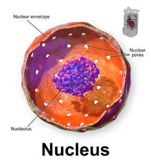
The cell nucleus is a membrane-bound organelle found in eukaryotic cells. Eukaryotic cells usually have a single nucleus, but a few cell types, such as mammalian red blood cells, have no nuclei, and a few others including osteoclasts have many. The main structures making up the nucleus are the nuclear envelope, a double membrane that encloses the entire organelle and isolates its contents from the cellular cytoplasm; and the nuclear matrix, a network within the nucleus that adds mechanical support.

<span class="mw-page-title-main">Nucleoplasm</span> Protoplasm that permeates a cells nucleus

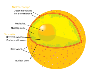
The nucleoplasm, also known as karyoplasm, is the type of protoplasm that makes up the cell nucleus, the most prominent organelle of the eukaryotic cell. It is enclosed by the nuclear envelope, also known as the nuclear membrane. The nucleoplasm resembles the cytoplasm of a eukaryotic cell in that it is a gel-like substance found within a membrane, although the nucleoplasm only fills out the space in the nucleus and has its own unique functions. The nucleoplasm suspends structures within the nucleus that are not membrane-bound and is responsible for maintaining the shape of the nucleus. The structures suspended in the nucleoplasm include chromosomes, various proteins, nuclear bodies, the nucleolus, nucleoporins, nucleotides, and nuclear speckles.

<span class="mw-page-title-main">Telophase</span> Final stage of a cell division for eukaryotic cells both in mitosis and meiosis

Telophase is the final stage in both meiosis and mitosis in a eukaryotic cell. During telophase, the effects of prophase and prometaphase are reversed. As chromosomes reach the cell poles, a nuclear envelope is re-assembled around each set of chromatids, the nucleoli reappear, and chromosomes begin to decondense back into the expanded chromatin that is present during interphase. The mitotic spindle is disassembled and remaining spindle microtubules are depolymerized. Telophase accounts for approximately 2% of the cell cycle's duration.

A nuclear localization signalorsequence (NLS) is an amino acid sequence that 'tags' a protein for import into the cell nucleus by nuclear transport. Typically, this signal consists of one or more short sequences of positively charged lysines or arginines exposed on the protein surface. Different nuclear localized proteins may share the same NLS. An NLS has the opposite function of a nuclear export signal (NES), which targets proteins out of the nucleus.

<span class="mw-page-title-main">Nuclear dimorphism</span>

Nuclear dimorphism is a term referred to the special characteristic of having two different kinds of nuclei in a cell. There are many differences between the types of nuclei. This feature is observed in protozoan ciliates, like Tetrahymena, and some foraminifera. Ciliates contain two nucleus types: a macronucleus that is primarily used to control metabolism, and a micronucleus which performs reproductive functions and generates the macronucleus. The compositions of the nuclear pore complexes help determine the properties of the macronucleus and micronucleus. Nuclear dimorphism is subject to complex epigenetic controls. Nuclear dimorphism is continuously being studied to understand exactly how the mechanism works and how it is beneficial to cells. Learning about nuclear dimorphism is beneficial to understanding old eukaryotic mechanisms that have been preserved within these unicellular organisms but did not evolve into multicellular eukaryotes.

Karyopherins are proteins involved in transporting molecules between the cytoplasm and the nucleus of a eukaryotic cell. The inside of the nucleus is called the karyoplasm. Generally, karyopherin-mediated transport occurs through nuclear pores which act as a gateway into and out of the nucleus. Most proteins require karyopherins to traverse the nuclear pore.

Importin is a type of karyopherin that transports protein molecules from the cell's cytoplasm to the nucleus. It does so by binding to specific recognition sequences, called nuclear localization sequences (NLS).

<span class="mw-page-title-main">Ran (protein)</span> GTPase functioning in nuclear transport

Ran also known as GTP-binding nuclear protein Ran is a protein that in humans is encoded by the RAN gene. Ran is a small 25 kDa protein that is involved in transport into and out of the cell nucleus during interphase and also involved in mitosis. It is a member of the Ras superfamily.

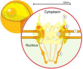
Nuclear transport refers to the mechanisms by which molecules move across the nuclear membrane of a cell. The entry and exit of large molecules from the cell nucleus is tightly controlled by the nuclear pore complexes (NPCs). Although small molecules can enter the nucleus without regulation, macromolecules such as RNA and proteins require association with transport factors known as nuclear transport receptors, like karyopherins called importins to enter the nucleus and exportins to exit.

<span class="mw-page-title-main">Nucleoporin 210kDa</span> Protein-coding gene in the species Homo sapiens

Nuclear pore glycoprotein-210 (gp210) is an essential trafficking regulator in the eukaryotic nuclear pore complex. Gp-210 anchors the pore complex to the nuclear membrane. and protein tagging reveals its primarily located on the luminal side of double layer membrane at the pore. A single polypeptide motif of gp210 is responsible for sorting to nuclear membrane, and indicate the carboxyl tail of the protein is oriented toward the cytoplasmic side of the membrane.

<span class="mw-page-title-main">Nuclear pore glycoprotein p62</span> Protein-coding gene in the species Homo sapiens

Nuclear pore glycoprotein p62 is a protein complex associated with the nuclear envelope. The p62 protein remains associated with the nuclear pore complex-lamina fraction. p62 is synthesized as a soluble cytoplasmic precursor of 61 kDa followed by modification that involve addition of N-acetylglucosamine residues, followed by association with other complex proteins. In humans it is encoded by the NUP62 gene.

<span class="mw-page-title-main">Nucleoporin</span> Family of proteins that form the nuclear pore complex

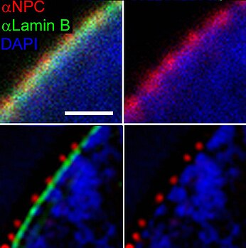
Nucleoporins are a family of proteins which are the constituent building blocks of the nuclear pore complex (NPC). The nuclear pore complex is a massive structure embedded in the nuclear envelope at sites where the inner and outer nuclear membranes fuse, forming a gateway that regulates the flow of macromolecules between the cell nucleus and the cytoplasm. Nuclear pores enable the passive and facilitated transport of molecules across the nuclear envelope. Nucleoporins, a family of around 30 proteins, are the main components of the nuclear pore complex in eukaryotic cells. Nucleoporin 62 is the most abundant member of this family. Nucleoporins are able to transport molecules across the nuclear envelope at a very high rate. A single NPC is able to transport 60,000 protein molecules across the nuclear envelope every minute.

<span class="mw-page-title-main">KPNB1</span> Protein-coding gene in the species Homo sapiens

Importin subunit beta-1 is a protein that in humans is encoded by the KPNB1 gene.

<span class="mw-page-title-main">Nucleoporin 153</span> Protein-coding gene in the species Homo sapiens

Nucleoporin 153 (Nup153) is a protein which in humans is encoded by the NUP153 gene. It is an essential component of the basket of nuclear pore complexes (NPCs) in vertebrates, and is required for the anchoring of NPCs. It also acts as the docking site of an importing karyopherin. On the cytoplasmic side of the NPC, Nup358 fulfills an analogous role.

<span class="mw-page-title-main">Transportin 1</span> Protein-coding gene in the species Homo sapiens

Transportin-1 is a protein that in humans is encoded by the TNPO1 gene.

<span class="mw-page-title-main">Nucleoporin 107</span> Protein-coding gene in the species Homo sapiens

Nucleoporin 107 (Nup107) is a protein that in humans is encoded by the NUP107 gene.

<span class="mw-page-title-main">Nuclear pore complex protein Nup133</span> Protein-coding gene in the species Homo sapiens

Nuclear pore complex protein Nup133, or Nucleoporin Nup133, is a protein that in humans is encoded by the NUP133 gene.

A nuclear export signal (NES) is a short target peptide containing 4 hydrophobic residues in a protein that targets it for export from the cell nucleus to the cytoplasm through the nuclear pore complex using nuclear transport. It has the opposite effect of a nuclear localization signal, which targets a protein located in the cytoplasm for import to the nucleus. The NES is recognized and bound by exportins.

Importin alpha, or karyopherin alpha refers to a class of adaptor proteins that are involved in the import of proteins into the cell nucleus. They are a sub-family of karyopherin proteins.

Gene gating is a phenomenon by which transcriptionally active genes are brought next to nuclear pore complexes (NPCs) so that nascent transcripts can quickly form mature mRNA associated with export factors. Gene gating was first hypothesised by Günter Blobel in 1985. It has been shown to occur in Saccharomyces cerevisiae, Caenorhabditis elegans, Drosophila melanogaster as well as mammalian model systems.

References

  1. Lin, D. H., Stuwe, T., Schilbach, S., Rundlet, E. J., Perriches, T., Mobbs, G., ... Hoelz, A. (2016). Architecture of the nuclear pore complex symmetric core. Science, 352(6283), aaf1015. http://doi.org/10.1126/science.aaf1015
  2. Mosalaganti S, Obarska-Kosinska A, Siggel M, Taniguchi R, Turoňová B, Zimmerli CE, et al. (June 10, 2022). "AI-based structure prediction empowers integrative structural analysis of human nuclear pores". Science. 376 (6598). doi:10.1126/science.abm9506. ISSN   0036-8075.
  3. Field MC, Rout MP (April 3, 2019). "Pore timing: the evolutionary origins of the nucleus and nuclear pore complex". F1000Research. 8: 369. doi: 10.12688/f1000research.16402.1 . PMC   6449795 . PMID   31001417.
  4. Nag N, Sasidharan S, Uversky VN, Saudagar P, Tripathi T (April 2022). "Phase separation of FG-nucleoporins in nuclear pore complexes". Biochimica et Biophysica Acta. Molecular Cell Research. 1869 (4): 119205. doi: 10.1016/j.bbamcr.2021.119205 . PMID   34995711.
  5. Peyro M, Soheilypour M, Lee BL, Mofrad MR (November 2015). "Evolutionarily Conserved Sequence Features Regulate the Formation of the FG Network at the Center of the Nuclear Pore Complex". Scientific Reports. 5: 15795. Bibcode:2015NatSR...515795P. doi:10.1038/srep15795. PMC   4635341 . PMID   26541386.
  6. Ando D, Colvin M, Rexach M, Gopinathan A (September 16, 2013). "Physical motif clustering within intrinsically disordered nucleoporin sequences reveals universal functional features". PLOS ONE. 8 (9): e73831. Bibcode:2013PLoSO...873831A. doi: 10.1371/journal.pone.0073831 . PMC   3774778 . PMID   24066078.
  7. Adam SA (2001). "The nuclear pore complex". Genome Biology. 2 (9): REVIEWS0007. doi: 10.1186/gb-2001-2-9-reviews0007 . PMC   138961 . PMID   11574060.
  8. Ibarra A, Hetzer MW (February 2015). "Nuclear pore proteins and the control of genome functions". Genes & Development. 29 (4): 337–349. doi:10.1101/gad.256495.114. PMC   4335290 . PMID   25691464.
  9. Mohr D, Frey S, Fischer T, Güttler T, Görlich D (September 2009). "Characterisation of the passive permeability barrier of nuclear pore complexes". The EMBO Journal. 28 (17): 2541–2553. doi:10.1038/emboj.2009.200. PMC   2728435 . PMID   19680228.
  10. Maimon T, Medalia O (2010). "Perspective on the metazoan nuclear pore complex". Nucleus. 1 (5): 383–386. doi:10.4161/nucl.1.5.12332. ISSN   1949-1034. PMC   3037531 . PMID   21326819.
  11. Kutay U, Jühlen R, Antonin W (December 1, 2021). "Mitotic disassembly and reassembly of nuclear pore complexes". Trends in Cell Biology. 31 (12): 1019–1033. doi:10.1016/j.tcb.2021.06.011. hdl: 20.500.11850/518955 . ISSN   0962-8924. PMID   34294532.
  12. Turton HE, Dawes IW, Grant CM (1997). "Saccharomyces cerevisiae exhibits a yAP-1-mediated adaptive response to malondialdehyde". Journal of Bacteriology. 179 (4): 1096–1101. doi:10.1128/jb.179.4.1096-1101.1997. ISSN   0021-9193. PMC   178803 . PMID   9023189.
  13. "Nuclear Pore Complex - an overview | ScienceDirect Topics". www.sciencedirect.com. Retrieved February 15, 2024.
  14. "Karyopherin - an overview | ScienceDirect Topics". www.sciencedirect.com. Retrieved February 15, 2024.
  15. Görlich D (June 1, 1997). "Nuclear protein import". Current Opinion in Cell Biology. 9 (3): 412–419. doi:10.1016/S0955-0674(97)80015-4. hdl: 11858/00-001M-0000-002D-1CC5-E . ISSN   0955-0674. PMID   9159081.
  16. Cenik C, Chua HN, Zhang H, Tarnawsky SP, Akef A, Derti A, et al. (April 2011). "Genome analysis reveals interplay between 5'UTR introns and nuclear mRNA export for secretory and mitochondrial genes". PLOS Genetics. 7 (4): e1001366. doi: 10.1371/journal.pgen.1001366 . PMC   3077370 . PMID   21533221.
  17. 1 2 Rabut G, Lénárt P, Ellenberg J (June 2004). "Dynamics of nuclear pore complex organization through the cell cycle". Current Opinion in Cell Biology. 16 (3): 314–321. doi:10.1016/j.ceb.2004.04.001. PMID   15145357.
  18. Sheehan MA, Mills AD, Sleeman AM, Laskey RA, Blow JJ (January 1988). "Steps in the assembly of replication-competent nuclei in a cell-free system from Xenopus eggs". The Journal of Cell Biology. 106 (1): 1–12. doi:10.1083/jcb.106.1.1. PMC   2114961 . PMID   3339085.
  19. Kiseleva E, Rutherford S, Cotter LM, Allen TD, Goldberg MW (October 2001). "Steps of nuclear pore complex disassembly and reassembly during mitosis in early Drosophila embryos". Journal of Cell Science. 114 (Pt 20): 3607–3618. doi:10.1242/jcs.114.20.3607. PMID   11707513. Archived from the original on September 13, 2019. Retrieved November 4, 2008.
  20. Hampoelz B, Andres-Pons A, Kastritis P, Beck M (May 6, 2019). "Structure and Assembly of the Nuclear Pore Complex". Annual Review of Biophysics. 48 (1): 515–536. doi:10.1146/annurev-biophys-052118-115308. ISSN   1936-122X. PMID   30943044.
  21. Markossian S, Suresh S, Osmani AH, Osmani SA (February 2015). "Nup2 requires a highly divergent partner, NupA, to fulfill functions at nuclear pore complexes and the mitotic chromatin region". Molecular Biology of the Cell. 26 (4): 605–621. doi:10.1091/mbc.E14-09-1359. PMC   4325833 . PMID   25540430.
  22. De Souza CP, Osmani AH, Hashmi SB, Osmani SA (November 2004). "Partial nuclear pore complex disassembly during closed mitosis in Aspergillus nidulans". Current Biology. 14 (22): 1973–1984. Bibcode:2004CBio...14.1973D. doi: 10.1016/j.cub.2004.10.050 . PMID   15556859. S2CID   14782686.
  23. De Souza CP, Osmani SA (September 2007). "Mitosis, not just open or closed". Eukaryotic Cell. 6 (9): 1521–1527. doi:10.1128/EC.00178-07. PMC   2043359 . PMID   17660363.
  24. Liu HL, De Souza CP, Osmani AH, Osmani SA (January 15, 2009). "The Three Fungal Transmembrane Nuclear Pore Complex Proteins of Aspergillus nidulans Are Dispensable in the Presence of an Intact An-Nup84-120 Complex". Molecular Biology of the Cell. 20 (2): 616–630. doi:10.1091/mbc.E08-06-0628. ISSN   1059-1524. PMC   2626566 . PMID   19019988.