Panellus stipticus

Last updated

Panellus stipticus
Panellus stipticus (37162668243).jpg
Scientific classification OOjs UI icon edit-ltr.svg
Domain: Eukaryota
Kingdom: Fungi
Division: Basidiomycota
Class: Agaricomycetes
Order: Agaricales
Family: Mycenaceae
Genus: Panellus
Species:
P. stipticus
Binomial name
Panellus stipticus
(Bull.) P.Karst. (1879)
Synonyms
  • Agaricus stypticusBull. (1783)
  • Merulius stipticus(Bull.) Lam. (1815)
  • Crepidopus stypticus(Bull.) Gray (1821)
  • Rhipidium stipticum(Bull.) Wallr. (1833)
  • Panus stipticus(Bull.) Fr. (1838)
  • Pleurotus stipticus(Bull.) P.Kumm. (1871)
  • Lentinus stipticus(Bull.) J.Schröt. (1889)
  • Pocillaria stiptica(Bull.) Kuntze (1898)
Panellus stipticus
Information icon.svg
Gills icon.png Gills on hymenium
Convex cap icon.svgOffset cap icon.svg Cap is convex or offset
Free gills icon2.svg Hymenium is free
Bare stipe icon.svg Stipe is bare
Transparent spore print icon.svg
Spore print is white
Saprotrophic fungus.svgEcology is saprotrophic
Mycomorphbox Inedible.pngEdibility is inedible

Panellus stipticus, commonly known as the bitter oyster, the astringent panus, the luminescent panellus, or the stiptic fungus, is a species of fungus. It belongs in the family Mycenaceae, and the type species of the genus Panellus . A common and widely distributed species, it is found in Asia, Australia, Europe, and North America, where it grows in groups or dense overlapping clusters on the logs, stumps, and trunks of deciduous trees, especially beech, oak, and birch. During the development of the fruit bodies, the mushrooms start out as tiny white knobs, which, over a period of one to three months, develop into fan- or kidney-shaped caps that measure up to 3 cm (1.2 in) broad. The caps are orange-yellow to brownish, and attached to the decaying wood by short stubby stalks that are connected off-center or on the side of the caps. The fungus was given its current scientific name in 1879, but has been known by many names since French mycologist Jean Bulliard first described it as Agaricus stypticus in 1783. Molecular phylogenetic analysis revealed P. stipticus to have a close genetic relationship with members of the genus Mycena .

Contents

Panellus stipticus is one of several dozen species of fungi that are bioluminescent. Strains from eastern North America are typically bioluminescent, but those from the Pacific coast regions of North America and from other continents are not. The luminescence is localized to the edges of the gills and the junction of the gills with the stem and cap. Bioluminescence is also observable with mycelia grown in laboratory culture, and the growth conditions for optimal light production have been studied in detail. Several chemicals have been isolated and characterized that are believed to be responsible for light production. Genetic analysis has shown that luminescence is controlled by a single dominant allele. The luminescent glow of this and other fungi inspired the term foxfire, coined by early settlers in eastern and southern North America. Modern research has probed the potential of P. stipticus as a tool in bioremediation, because of its ability to detoxify various environmental pollutants.

Taxonomy and phylogeny

Resinomycena acadiensis

Dictyopanus pusillus

Panellus stipticus

Mycena galericulata

Mycena rutilanthiformis

Mycena clavicularis

Other Panellus spp. and Hygrocybe citrinopallida

Phylogeny and relationships of P. stipticus and related species based on ribosomal DNA sequences [1]

The species was first named Agaricus stypticus by the French botanist Jean Bulliard in 1783, [2] and later sanctioned by Elias Magnus Fries under this name in 1821. [3] Fries later changed the genus as well as the spelling of the epithet and called it Panus stipticus. [4] The species has had an extensive taxonomic history and been shuffled to a number of genera by various authors, resulting in several synonyms: Agaricus flabelliformis (Johann Friedrich Gmelin, 1792), [5] Pocillaria stiptica (Otto Kuntze, 1898), [6] Rhipidium stipticum (Karl Friedrich Wilhelm Wallroth, 1833), [7] Crepidopus stipticus (Samuel Frederick Gray, 1821), [8] Pleurotus stipticus (Paul Kummer), [9] Lentinus stipticus (Joseph Schröter, 1885), [10] and Merulius stipticus (Jean-Baptiste Lamarck). [11] It was Finnish mycologist Petter Karsten who in 1879 assigned its current name. [12] Panellus stypticus is still used in the literature as a variant spelling. [13]

Panellus stipticus is the type species of the genus Panellus, and, in Rolf Singer's authoritative 1986 classification of the Agaricales, it is also the type species of subgenus Panellus, an infrageneric (below the taxonomic level of genus) grouping of Panellus species characterized by the absence of cystidia on the sides of the gills. [14] More recently, phylogenetic analyses of the sequences of their ribosomal large subunit genes have concluded that Panellus stipticus is closely related to the poroid mushroom Dictyopanus pusillus. [1] The molecular analysis supports a previous assessment by mycologists Harold Bursdall and Orson K. Miller, who in 1975 suggested merging Dictyopanus into Panellus based on similarities in spore shape, stem structure, and the ability of dried fruit bodies to revive when moistened. [15] Formerly grouped in the family Tricholomataceae, [16] a wastebasket taxon of gilled mushrooms with white spores, P. stipticus is now classified in the Mycenaceae, [17] [18] after a large-scale phylogenetic analysis revealed "a previously unsuspected relationship between Mycena and Panellus (including Dictyopanus)". [19]

The fungus is commonly known as the bitter oyster, [20] the luminescent panellus, [21] the astringent panus, [22] or the styptic fungus. [23] The specific epithet stipticus refers to its purported value in stopping bleeding. [24] [25] Etymologically, it is a Greek equivalent to the Latin word astringens, deriving from στυπτικός (styptikós), itself from the verb στύφειν (styphein), "to contract". [26] [27]

Description

The buff-colored gills are closely spaced together, and are connected by cross-veins. 2011-08-25 Panellus stipticus (Bull.) P. Karst 166165 cropped.jpg
The buff-colored gills are closely spaced together, and are connected by cross-veins.

The fungus normally exists unseen, in the form of a mass of threadlike vegetative cells called a mycelium, inhabiting rotting wood; only when suitable environmental conditions of temperature, moisture, and nutrient availability are achieved does the fungus produce the reproductive structures known as fruit bodies, or mushrooms. The cap of the fruit body is kidney- or clamshell-shaped, convex to roughly flat, with dimensions of 1.2 to 3.2 cm (0.5 to 1.3 in) by 1.2 to 2.5 cm (0.5 to 1.0 in). [28] The edges of the caps are scalloped with small rounded teeth, and curved slightly inward. The cap surface is dry, with a pattern of block-like areas similar to cracked dried mud; the surface is also covered with small fine hairs that give it a somewhat woolly consistency. It may have several concentric ridges or zones. Fresh fruit bodies range in color from yellowish-orange to buff to cinnamon; when dried they may be various shades of tan, brown or clay. The faded colors of dried fruit bodies tend to revive when moistened. On the underside of the cap, the gills are narrow and spaced closely together, often forked, buff-colored, and with numerous interconnecting cross-veins. Holding the cap in position is a stem that is 0.6 to 1.2 cm (0.2 to 0.5 in) long by 0.3 to 0.8 cm (0.1 to 0.3 in) thick, and has an off-center attachment to the cap, either at or near the cap side. The dull-white stem is covered with minute silk-like fibers, and is narrower at the base where it attaches to the substrate. Fruit bodies do not have any distinctive odor. [15] The flesh is thin and tough, and dark yellow-brown to cream-colored. [29]

Microscopic features

Various microscopic characteristics may be used to help identify the fungus from other morphologically similar species. A spore print of P. stipticus, made by depositing a large number of spores in a small area, reveals their color to be white. [21] Viewed with a microscope, the spores are smooth-walled, elliptical to nearly allantoid (sausage-shaped), with dimensions of 3–6 by 2–3  µm. Spores are amyloid, meaning that they will absorb iodine and become bluish-black when stained with Melzer's reagent, [30] but this staining reaction has been described as "relatively weak". [1]

The basidia (the spore-bearing cells) are 15–20 by 2.5–3.5 µm, club-shaped, and clamped at the base. [15] The spores are attached to the end of the basidia by four projections called sterigmata that are 1–3.5 µm long. [31] Cystidia are hyphal cells in the hymenium that do not produce basidiospores; they also make up the surfaces of the fruit body (the pelli or "cuticles"). They are usually structurally distinct from the basidia, and their features may be used as microscopic characters to help distinguish and differentiate similar fungi. The cheilocystidia are found on the gill edge; in P. stipticus they are narrowly club-shaped, cylindrical, spindle-shaped to bifurcate at the apex. They are also thin-walled, hyaline (translucent), abundant and crowded, and measure 17–45 by 3.5–6 µm. The pleurocystidia, located on the gill face, are 17–40 by 3–4.5 µm, spindle- or club-shaped, sometimes bifurcate at the apex, thin-walled, and hyaline. They are scattered or in dense clusters, mostly embedded in hymenium, occasionally protruding up to half the width of the hymenium. [15]

The flesh of the cap consists of a number of microscopically distinct layers of tissue. The cuticle of the cap (known as the pileipellis) is between 8–10 µm thick, [32] and is made of a loose textura intricata, a type of tissue in which the hyphae are irregularly interwoven with distinct spaces between them. [15] The cuticle hyphae are thick-walled to thin-walled, with scattered inconspicuous cystidia measuring 40–55 by 3.5–5.5 µm. These cystidia located in the cap (pileocystidia) are cylindrical, thin-walled, yellow in Melzer's reagent, hyaline in KOH, sometimes with amorphous dingy brown material coating the walls. Beneath the cuticle layer is a zone 54–65 µm thick, made of very loosely entwined, thin-walled hyphae, 2–3 µm in thickness, with clamps at the septa. Below this is a zone 208–221 µm in thickness, in which the densely compacted hyphae, 3–8 µm in diameter, have swollen, gelatinized walls, and often more or less a vertical orientation. This in turn is followed by a layer 520 µm in thickness, formed of loosely interwoven hyphae, 2–8 µm in width, some of which have thin walls with clamps at the septa, whilst others have somewhat thickened gelatinized walls. [32] The flesh of the cap has a layer of upright hyphae bending into a lower layer of interwoven hyphae with diameters of 2.5–8 µm. The flesh of the gills is similar to that of the lower cap. [15]

Similar species

Crepidotus spec. - Lindsey 3b.jpg
Spaltblaettling4.jpg
Possible lookalike species include Crepidotus mollis (top) and Schizophyllum commune (bottom).

Species of Crepidotus having a similar shape can be distinguished by their brown spore print, compared with the white spore print of P. stipticus. [20] Schizophyllum commune has a densely hairy white to grayish cap and longitudinally split gill-folds on the underside. [25] The ruddy panus mushroom ( Panus rudis ) is larger, has a reddish-brown cap that fades to pinkish-tan, and shows lilac tinges when young, fresh, and moist. [23] Some Paxillus species may have a similar appearance, but they have yellow-brown spore prints.

Uses

Panellus stipticus is considered too small and bitter to be edible, but is considered nonpoisonous. [28] Its taste has been described as acidic, acrid, or astringent. [20] [33] One 1992 study reported that the taste of the fruit bodies varies across the geographic range of the species. Those from eastern North America have a mildly acrid taste that takes time to develop, and cause uncomfortable drying in the mouth. In contrast, specimens from Japan, New Zealand, and Russia produce no sensation in the mouth, but cause significant constriction and a nauseating taste in the throat. [34] The fruit bodies are reputed to have been used in traditional Chinese medicine as a styptic to staunch bleeding, [35] and also as a "violent purgative". [23]

Fruit body development

Fruit bodies first appear as tiny white knobs less than a cubic millimeter in size. In a day or two the knobs grow into a horizontal pyramidal mass, increasing in height as the hyphae lengthen. This is soon followed by the formation of a minute cap, and lengthening of the stem. The stem is about 1 mm long when the cap first begins to form. The hyphae that comprise the stem gradually cease to grow at their ends, and then start to branch, with many of the branches growing in a horizontal direction. This growth, indicated by the flattening and broadening out of the top of the stem, gives rise to the cap. The horizontally aligned hyphae grow vertical branches which remain more or less parallel, ultimately forming the dorsal tissue of the cap. Other similar downward-growing branches form the fertile hymenium, which can be seen when the cap is about 2 mm in diameter. [36]

The caps of young fruit bodies (top) are spherical, and grow epinastically, so that the developing gills remain enclosed until maturity. Panellus stipticus 8445.jpg
The caps of young fruit bodies (top) are spherical, and grow epinastically, so that the developing gills remain enclosed until maturity.

The young cap is spherical and its growth is at first epinastic, its margin being curved inwards and pressed against the stem. In this way, the hymenium begins its development within a special enclosed chamber. As the hymenial surface increases and keeps pace with the growth of the dorsal tissue of the cap, the latter expands and exposes the gills. The gills are formed by the continual downward growth of some of the hyphae. The gills are exposed before the cap is completely developed, and before the spores are mature. Spores can be produced by fruit bodies as small as 1.3 cm (0.5 in) broad, and liberation of the spores continues until the fruit body is fully grown—a period of one month to three months, depending on the conditions of temperature and moisture. The mature spores are disseminated by the wind. When the fruit body is nearing maturity, some of the terminal portions of the hyphae of the dorsal surface of the cap separate, and as a consequence, the upper surface of the fruit body becomes granular in appearance. [36]

The fruit body projects out horizontally from the growing surface. If the position of a log is altered after young fruit bodies with the beginnings of gills have appeared, the stems of these attempt to readjust themselves in order to place the cap in a horizontal position. The cap are sometimes zonate (marked with concentric lines that form alternating pale and darker zones); this depends on changes in the humidity of the environment, as variations in the amount of moisture will cause alternating periods of acceleration or slowing of growth. [36]

A yellowish-brown pigment is diffused through the cytosol of the hyphae and is much deeper in color just below the cuticle of the cap. In very young fruit bodies, stems and caps are very pale buff, but soon the color of the cap deepens and becomes cinnamon. The intensity of the color appears to be dependent on light, for when fruit bodies are grown in diffuse light (temperature and humidity being constant) they are a uniform pale buff color, but in bright light they are cinnamon or tan. [36]

Distribution, habitat, and ecology

Fruit bodies can grow in dense overlapping clusters. Panellus stipticus 34244.jpg
Fruit bodies can grow in dense overlapping clusters.

Panellus stipticus is common in northern temperate regions of Europe, and has also been collected in Australia, [37] New Zealand, [38] Anatolia, [39] Japan, [34] and China. [31] In North America, it is more common in the east than the west; [13] [23] the mushroom's northern range extends to Alaska, and it has been collected as far south as Costa Rica. [40]

Panellus stipticus is a saprobic species, and causes a white rot. [41] This is a form of wood decay in which the wood assumes a bleached appearance and where lignin as well as cellulose and hemicellulose is broken down by enzymes secreted by the fungus. Fruit bodies are usually found in tightly overlapping clusters on the sides of hardwood trees, on logs, stumps, and fallen branches. Although most commonly found on dead wood, it can also grow opportunistically in the wounds of living trees. [14] In North America, its preferred hosts are oak, birch, maple, hickory, pecan, and American hornbeam, [42] [43] [44] while in Europe, it is commonly found growing on oak, birch, alder, beech, hazel, chestnut, and ash. [36] [45] It has also been found growing on Ericia in North Africa. [45] Although the fungus favors hardwoods, it has been reported to grow on loblolly pine and eastern white pine. [44] Fruiting occurs September through November in Europe, the Canary Islands, [46] and North America, [15] although it may also sometimes be found in the spring. [20] The fruit bodies are long-lasting and may be found year-round. [47] It is an "early-stage" succession fungus, not typically recorded from plantations over 20 years old. [48]

The fruit bodies are frequently attacked by slugs, which may be important agents in the dispersal of its spores. [23] White-tailed deer are also known to consume the fungus. [49]

Mating studies

Panellus stipticus uses a heterothallic, tetrapolar mating system: [43] [50] each basidiospore develops into a self-sterile mycelium which, when grown alone, remains homokaryotic (i.e., with all cells genetically identical) indefinitely. Researchers have paired collections of P. stipticus from Japan and Eastern North America, and later, collections from New Zealand and Russia. [34] [51] Although the separated allopatric populations differ in bioluminescence and taste, the results revealed a universal intercompatibility group over these geographical regions. [52] In a 2001 study, Jin and colleagues also paired geographically representative collections of the fungus, but observed a reduced ability to cross between Northern Hemisphere and Oceanian collections, as well as between and within Oceanian collections. [40]

Bioluminescence

Bioluminescence demonstrated: the camera was exposed for 517 seconds to capture this much light. PanellusStipticusAug12 2009.jpg
Bioluminescence demonstrated: the camera was exposed for 517 seconds to capture this much light.

Bioluminescence refers to the ability of certain living things in the environment to produce light by the action of enzymes. Bioluminescent fungi are widespread, and over 70 species are known (List of bioluminescent fungus species). [53] Although the intensity of their luminescence is generally low compared to many other bioluminescent organisms, fungi glow continuously for days, so their total emission is comparable with that of most brightly luminescent organisms, such as fireflies. [54] Luminous fungi are found growing on decaying wood, leading to the popular name of "foxfire" or "glow wood" when their glow is visible at night. [55] The responsible oxidative enzymes—known generically as luciferases—produce light by oxidizing a pigment called a luciferin. In some areas, P. stypticus is bioluminescent, and the fruit bodies of these strains will glow in the dark when fresh or sometimes when revived in water after drying. [25]

An early record of luminescence noted in P. stypticus was made by the American naturalist Thomas G. Gentry in 1885. Job Bicknell Ellis, reporting on the phenomenon for the Journal of Mycology , wrote:

By careful examination, the luminosity was found to proceed from the gills and not the stipe, nor from any fragment of rotten wood attached to the specimen. This phosphorescence was not observed in all specimens brought in for examination, and seemed to depend on some peculiar condition of the air, having been noticed only in specimens gathered in damp weather or just before a storm. [56]

Canadian mycologist Buller in 1924 described the gills of P. stipticus in North America as luminescent, and noted that the fungus glows most strongly at the time of spore maturation. [42] Bioluminescence has not been observed in European specimens, [43] in Pacific North American collections, nor in strains collected from New Zealand, Russia, and Japan. [34] Although a number of reports have confirmed that eastern North American strains are luminescent, [34] [44] [51] [57] [58] non-luminescent North American strains are also known. [54] In general, the intensity of fungal bioluminescence decreases after exposure to certain contaminants; this sensitivity is being investigated as a means to develop bioluminescence-based biosensors to test the toxicity of polluted soils. [59] Most known luminescent fungi are in the genus Mycena or closely allied genera; this grouping of fungi—known as the "mycenoid lineage"—includes P. stipticus and three other Panellus species. [60]

Mycelia

The mycelia of this species, grown in laboratory culture, have also been shown to be bioluminescent. [61] Early studies demonstrated that short-wave ultraviolet light (at a wavelength of 280  nm) reversibly inhibited the luminescence of P. stypticus mycelia, while longer wavelength (366 nm) ultraviolet was stimulatory. [62] [63] Further, the fungus exhibited a pronounced diurnal periodicity, and maximum luminescence was noted between 6 and 9 pm, regardless as to whether the mycelial cultures were incubated in continuous light, continuous darkness, or a normal day-night cycle. [64] The mycelia of P. stipticus grown submerged in liquid were non-luminescent, but became luminescent while growing on solid substrata. Dark-grown colonies were luminescent in the center, and light-grown colonies were brightest at the periphery. [61] Other experiments have shown that growth temperature and pH have a significant effect on the level of bioluminescence, optimized at 22 °C (72 °F) and pH 3–3.5. However, light had a significant effect on mycelial growth but not on bioluminescence, and the optimal light conditions for maximum bioluminescence were total darkness. [65]

Fruit bodies

Bioluminescent tissue in the mature fruit body is restricted to the edge of the gills (as well as the cross-veins that connect them), the junction of the gills with the stem, and the inrolled cap edge. Distribution of bioluminescence along the gill edge corresponds to the position of the cheilocystidia. Less than 10% of the light emitted from both the young and mature fruit bodies is from other tissues, including the fertile hymenial area and the stem. Fruit body luminescence is highly variable between fruit bodies found on different logs in different environments. [58]

Genetics

In Mount Vernon, Wisconsin PanellusStipticusAug12 2009 Animated.gif
In Mount Vernon, Wisconsin

Using techniques of genetic complementation, Macrae paired nonluminescent monocaryons with luminescent ones, and concluded that luminosity in P. stipticus is an inherited character, and governed by a single pair of alleles in which luminosity was dominant over nonluminosity. Luminosity factors were independent of intersterility factors. In 1992, Lingle and colleagues agreed with Macrae about luminosity and stated that at least three different gene mutations could lead to the loss of luminescence. They also reported that the maximum bioluminescence was found at 525 nm, and shifted to 528 nm in deeply pigmented fruit bodies. [44]

After intercontinental compatibility tests, Petersen and Bermudes suggested that bioluminescence and compatibility were independent since bioluminescence seemed to be geographically restricted. This suggested that the ability or potential to interbreed must have been preserved since separation of P. stypticus into geographically isolated areas. [34] [51]

Function

Several authors have suggested that the purpose of fungal bioluminescence is to attract arthropods to the fruit body to help disseminate spores. [66] [67] [68] However, there has been no direct observation of this phenomenon. [58] In many species, the mycelium is luminescent, but the fruit body is not, which argues against this hypothesis. Mycelial bioluminence may also function to attract animals that can potentially carry fungal spores in well-hydrated areas, as light emission from the mycelium is higher when it is hydrated. This would presumably increase the chance that spores would be deposited in an environment that is already optimal for growth. Basidiomycete mushrooms are known to be dependent on an adequate moisture supply for proper development. [69] In species with a luminous mycelium, the mycelium would therefore have a dual function in performing the fungal translocation that permits transport of substances from the further environment back to the fruiting body, and in attracting disseminating vectors towards environments favorable for development of the species. [55]

Chemical basis

The chemical structure of panal Panal.svg
The chemical structure of panal

In general, bioluminescence is caused by the action of luciferases, enzymes that produce light by the oxidation of a luciferin (a pigment). Several studies have evaluated the biochemical basis of light production in Panellus stipticus, and concluded that there is no specific fungal luciferase. The fungus was shown in 1988 to contain the sesquiterpene panal, which has a cadinene keto-aldehyde chemical structure. Later, two additional precursors were isolated, PS-A (1-O-decanoylpanal) and PS-B (1-O-dodecanoylpanal). [70] If these compounds are treated with salt of ammonia or primary amines in the presence of iron(II), hydrogen peroxide, and a cationic surfactant, light is emitted by a chemiluminescence reaction, [71] [72] suggesting that panal and its derivatives are fungal luciferins, and that the chemiluminescence reaction is the cause of in vivo bioluminescence. [73] [74] In the fungus, the level of activity of the enzyme superoxide dismutase (SOD) appears to play a critical role in the amount of light emission. SOD quenches the effect of the superoxide (O2) anion required in the reaction, and thus SOD activity has to be inhibited for the reaction to occur. [54]

Bioremediation

As a white-rot fungus, Panellus stipticus contains enzymes that are able to break down lignin, a complex aromatic polymer in wood that is highly resistant to degradation by conventional enzyme systems. The major enzyme that initiates the cleavage of hydrocarbon rings is laccase, which catalyzes the addition of a hydroxyl group to phenolic compounds (polyphenols). The ring can then be opened between the two adjacent carbon atoms that bear the hydroxyl groups. White-rot fungi are being investigated scientifically for their potential use in the bioremediation of land contaminated by organic pollutants, and to convert industrial wastes rich in toxic polyphenols. Panellus stipticus has been shown to reduce the phenolic concentration of waste water produced by olive-processing plants—an environmental concern in many Mediterranean countries. In this study, a liquid culture of P. stipticus mycelia reduced the initial concentration of phenolic compounds by 42% after a 31-day incubation period. [75] In a separate study, a P. stipticus culture was able to effectively degrade the environmental pollutant 2,7-dichlorodibenzo-p-dioxin, a polychlorinated dioxin. [76]

See also

Related Research Articles

<i>Omphalotus nidiformis</i> Species of bioluminescent fungus in the family Marasmiaceae

Omphalotus nidiformis, or ghost fungus, is a gilled basidiomycete mushroom most notable for its bioluminescent properties. It is known to be found primarily in southern Australia and Tasmania, but was reported from India in 2012 and 2018. The fan or funnel shaped fruit bodies are up to 30 cm (12 in) across, with cream-coloured caps overlain with shades of orange, brown, purple, or bluish-black. The white or cream gills run down the length of the stipe, which is up to 8 cm (3 in) long and tapers in thickness to the base. The fungus is both saprotrophic and parasitic, and its fruit bodies are generally found growing in overlapping clusters on a wide variety of dead or dying trees.

<span class="mw-page-title-main">Hydnellum peckii</span> Species of fungus

Hydnellum peckii is a fungus in the genus Hydnellum of the family Bankeraceae. It is a hydnoid species, producing spores on the surface of vertical spines or tooth-like projections that hang from the undersurface of the fruit bodies. It is found in North America, Europe, and was recently discovered in Iran (2008) and Korea (2010). Hydnellum peckii is a mycorrhizal species, and forms mutually beneficial relationships with a variety of coniferous trees, growing on the ground singly, scattered, or in fused masses.

<i>Panus conchatus</i> Species of fungus

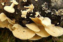
Panus conchatus, commonly known as the lilac oysterling, smooth panus, or conch panus, is an inedible species of mushroom that occurs throughout the Northern Hemisphere. Its fruitbodies are characterized by a smooth, lilac- or tan-colored cap, and decurrent gills. The fungus is saprophytic and fruits on the decomposing wood of a wide variety of deciduous and coniferous trees. Despite being a gilled species, phylogenetic analysis has shown it is closely related to the pored species found in the family Polyporaceae.

<i>Armillaria gallica</i> Species of fungus in the family Physalacriaceae

Armillaria gallica is a species of honey mushroom in the family Physalacriaceae of the order Agaricales. The species is a common and ecologically important wood-decay fungus that can live as a saprobe, or as an opportunistic parasite in weakened tree hosts to cause root or butt rot. It is found in temperate regions of Asia, North America, and Europe. The species forms fruit bodies singly or in groups in soil or rotting wood. The fungus has been inadvertently introduced to South Africa. Armillaria gallica has had a confusing taxonomy, due in part to historical difficulties encountered in distinguishing between similar Armillaria species. The fungus received international attention in the early 1990s when an individual colony living in a Michigan forest was reported to cover an area of 15 hectares, weigh at least 9.5 tonnes, and be 1,500 years old. This individual is popularly known as the "humongous fungus", and is a tourist attraction and inspiration for an annual mushroom-themed festival in Crystal Falls. Recent studies have revised the fungus's age to 2,500 years and its size to about 400 tonnes, four times the original estimate.

<i>Mycena haematopus</i> Fungus species in the family Mycenaceae widespread and common in Europe and North America

Mycena haematopus, commonly known as the bleeding fairy helmet, the burgundydrop bonnet, or the bleeding Mycena, is a species of fungus in the family Mycenaceae, of the order Agaricales. It is widespread and common in Europe and North America, and has also been collected in Japan and Venezuela. It is saprotrophic—meaning that it obtains nutrients by consuming decomposing organic matter—and the fruit bodies appear in small groups or clusters on the decaying logs, trunks, and stumps of deciduous trees, particularly beech. The fungus, first described scientifically in 1799, is classified in the section Lactipedes of the genus Mycena, along with other species that produce a milky or colored latex.

<i>Amanita abrupta</i> Species of fungus

Amanita abrupta, commonly known as the American abrupt-bulbed amanita or the American abrupt-bulbed lepidella, is a possibly toxic species of fungus in the family Amanitaceae. Named for the characteristic shape of its fruit bodies, this white Amanita has a slender stem, a cap covered with conical white warts, and an "abruptly enlarged" swollen base. This terrestrial species grows in mixed woods in eastern North America and eastern Asia, where it is thought to exist in a mycorrhizal relationship with a variety of both coniferous and deciduous tree species.

<i>Mycena polygramma</i> Species of fungus

Mycena polygramma, commonly known as the grooved bonnet, is a species of mushroom in the family Mycenaceae. The inedible fruit bodies are small, pale gray-brown mushrooms with broadly conical caps, pinkish gills. They are found in small troops on stumps and branches of deciduous and occasionally coniferous trees. The mushroom is found in Asia, Europe, and North America, where it is typically found on twigs or buried wood, carrying out its role in the forest ecosystem by decomposing organic matter, recycling nutrients, and forming humus in the soil. M. polygramma contains two uncommon hydroxy fatty acids and is also a bioluminescent fungus whose intensity of light emission follows a diurnal pattern.

<i>Mycena sanguinolenta</i> Species of fungus

Mycena sanguinolenta, commonly known as the bleeding bonnet, the smaller bleeding Mycena, or the terrestrial bleeding Mycena, is a species of mushroom in the family Mycenaceae. It is a common and widely distributed species, and has been found in North America, Europe, Australia, and Asia. The fungus produces reddish-brown to reddish-purple fruit bodies with conic to bell-shaped caps up to 1.5 cm (0.6 in) wide held by slender stipes up to 6 cm (2.4 in) high. When fresh, the fruit bodies will "bleed" a dark reddish-purple sap. The similar Mycena haematopus is larger, and grows on decaying wood, usually in clumps. M. sanguinolenta contains alkaloid pigments that are unique to the species, may produce an antifungal compound, and is bioluminescent. The edibility of the mushroom has not been determined.

<i>Dendrocollybia</i> Genus of fungi in the family Tricholomataceae

Dendrocollybia is a fungal genus in the family Tricholomataceae of the order Agaricales. It is a monotypic genus, containing the single species Dendrocollybia racemosa, commonly known as the branched collybia or the branched shanklet. The somewhat rare species is found in the Northern Hemisphere, including the Pacific Northwest region of western North America, and Europe, where it is included in several Regional Red Lists. It usually grows on the decaying fruit bodies of other agarics—such as Lactarius and Russula—although the host mushrooms may be decayed to the point of being difficult to recognize.

<i>Panellus</i> Genus of fungi

Panellus is a genus of more than 50 mushroom species of fungi in the family Mycenaceae as defined molecularly. Prior to molecular analyses the generic name had been used for any white-spored pleurotoid with amyloid spores. Unrelated but similar species are now classified in Sarcomyxa and Scytinotus. In older guides and other literature the type species had been placed in either Pleurotus or Panus and the poroid species had been classified in the synonymous genus Dictyopanus or in broadly defined genera like Polyporus (Polyporaceae) or the more closely allied Favolaschia (Mycenaceae). The closest molecular allies are Resinomycena and Cruentomycena.

<i>Hygrophorus agathosmus</i> Species of fungus

Hygrophorus agathosmus, commonly known as the gray almond waxy cap or the almond woodwax, is a species of fungus in the family Hygrophoraceae. It was first described by Elias Magnus Fries in 1815; Fries gave it its current name in 1838. A widespread species, it is distributed in the United States, Europe, Africa, and India, and is found growing under spruce and pine in mixed forests. The fruit bodies are characterized by a light grayish cap that measures up to 8 cm (3.1 in) in diameter, waxy gills, a dry stem, and the distinct odor of bitter almonds. An edible but bland-tasting mushroom, extracts of the fruit bodies have been shown in laboratory tests to have antimicrobial activity against various bacteria that are pathogenic to humans.

<i>Lactarius alnicola</i> Species of fungus

Lactarius alnicola, commonly known as the golden milkcap, is a species of fungus in the family Russulaceae. The fruit bodies produced by the fungus are characterized by a sticky, vanilla-colored cap up to 20 cm (7.9 in) wide with a mixture of yellow tones arranged in faint concentric bands. The stem is up to 5 cm (2.0 in) long and has yellow-brown spots. When it is cut or injured, the mushroom oozes a white latex, which has an intensely peppery taste. The acrid taste of the fruit bodies renders them unpalatable. The fungus is found in the western United States and Mexico, where it grows in mycorrhizal associations with various coniferous trees species, such as spruce, pine and fir, and deciduous species such as oak and alder. It has also been collected in India. Two varieties have been named: var. pitkinensis, known from Colorado, and var. pungens, from Michigan.

<i>Mycena stylobates</i> Species of fungus

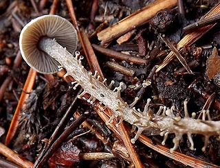
Mycena stylobates, commonly known as the bulbous bonnet, is a species of inedible mushroom in the family Mycenaceae. Found in North America and Europe, it produces small whitish to gray fruit bodies with bell-shaped caps that are up to 15 mm (0.6 in) in diameter. The distinguishing characteristic of the mushroom is the fragile stipe, which is seated on a flat disk marked with distinct grooves, and fringed with a row of bristles. The mushrooms grow in small troops on leaves and other debris of deciduous and coniferous trees. The mushroom's spores are white in deposit, smooth, and ellipsoid-shaped with dimensions of 6–10 by 3.5–4.5 μm. In the development of the fruit body, the preliminary stipe and cap structures appear at the same time within the primordium, and hyphae originating from the stipe form a cover over the developing structures. The mycelia of the mushroom is believed to have bioluminescent properties.

<i>Palaeoagaracites</i> Extinct genus of fungi

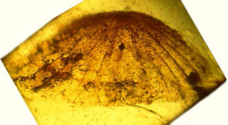
Palaeoagaracites is an extinct monotypic genus of gilled fungus in the order Agaricales. It contains the single species Palaeoagaracites antiquus.

<i>Mycena maculata</i> Species of fungus in the family Mycenaceae

Mycena maculata, commonly known as the reddish-spotted Mycena, is a species of fungus in the family Mycenaceae. The fruit bodies, or mushrooms, have conic to bell-shaped to convex caps that are initially dark brown but fade to brownish-gray when young, reaching diameters of up to 4 cm. They are typically wrinkled or somewhat grooved, and have reddish-brown spots in age, or after being cut or bruised. The whitish to pale gray gills also become spotted reddish-brown as they mature. The stem, up to 8 cm (3 in) long and covered with whitish hairs at its base, can also develop reddish stains. The mycelium of M. maculata has bioluminescent properties. The saprobic fungus is found in Europe and North America, where it grows in groups or clusters on the rotting wood of both hardwoods and conifers. The edibility of the fungus is unknown. Although the species is known for, and named after its propensity to stain reddish, occasionally these stains do not appear, making it virtually indistinguishable from M. galericulata.

<i>Collybia tuberosa</i> Species of fungus

Collybia tuberosa, commonly known as the lentil shanklet or the appleseed coincap, is an inedible species of fungus in the family Tricholomataceae, and the type species of the genus Collybia. Like the two other members of its genus, it lives on the decomposing remains of other fleshy mushrooms. The fungus produces small whitish fruit bodies with caps up to 1 cm (0.4 in) wide held by thin stems up to 5 cm (2.0 in) long. On the underside of the cap are closely spaced white gills that are broadly attached to the stem. At the base of the stem, embedded in the substrate is a small reddish-brown sclerotium that somewhat resembles an apple seed. The appearance of the sclerotium distinguishes it from the other two species of Collybia, which are otherwise very similar in overall appearance. C. tuberosa is found in Europe, North America, and Japan, growing in dense clusters on species of Lactarius and Russula, boletes, hydnums, and polypores.

<i>Auriscalpium vulgare</i> Inedible European fungi

Auriscalpium vulgare, commonly known as the pinecone mushroom, the cone tooth, or the ear-pick fungus, is a species of fungus in the family Auriscalpiaceae of the order Russulales. It was first described in 1753 by Carl Linnaeus, who included it as a member of the tooth fungi genus Hydnum, but British mycologist Samuel Frederick Gray recognized its uniqueness and in 1821 transferred it to the genus Auriscalpium that he created to contain it. The fungus is widely distributed in Europe, Central America, North America, and temperate Asia. Although common, its small size and nondescript colors lead it to be easily overlooked in the pine woods where it grows. A. vulgare is not generally considered edible because of its tough texture, but some historical literature says it used to be consumed in France and Italy.

<i>Mycena chlorophos</i> Species of agaric fungus in the family Mycenaceae

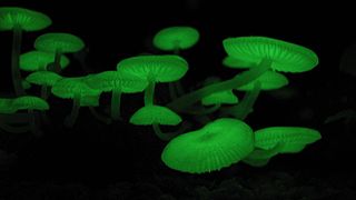
Mycena chlorophos is a species of agaric fungus in the family Mycenaceae. First described in 1860, the fungus is found in subtropical Asia, including India, Japan, Taiwan, Polynesia, Indonesia, and Sri Lanka, in Australia, and Brazil. Fruit bodies (mushrooms) have pale brownish-grey sticky caps up to 30 mm (1.2 in) in diameter atop stems 6–30 mm (0.2–1.2 in) long and up to a millimeter thick. The mushrooms are bioluminescent and emit a pale green light. Fruiting occurs in forests on fallen woody debris such as dead twigs, branches, and logs. The fungus can be made to grow and fruit in laboratory conditions, and the growth conditions affecting bioluminescence have been investigated.

<i>Filoboletus manipularis</i> Species of fungus

Filoboletus manipularis is a species of agaric fungus in the family Mycenaceae. Found in Australasia, Malaysia, and the Pacific islands, the mycelium and fruit bodies of the fungus grow in forests and can be bioluminescent. The fruiting bodies also display a variety of morphologies that have no current genetic attributions. References to Filoboletus manipularis can be found in Japanese folklore and Indonesian food culture.

References

  1. 1 2 3 Jin J, Petersen RH (2001). "Phylogenetic relationships of Panellus (Agaricales) and related species based on morphology and ribosomal large subunit DNA sequences". Mycotaxon. 79: 7–21.
  2. Bulliard JBF. (1783). Herbier de la France. Vol. 3 (in French). pp. 97–144. Archived from the original on 2010-02-12.
  3. Fries EM. (1821). Systema Mycologicum I (in Latin). Lundin, Sweden: ex officina Berlingiana. p. 188. Archived from the original on 2010-02-12.
  4. Fries EM. (1838). Epicrisis Systematis Mycologici (in Latin). Uppsala, Sweden: Typographia Academica. p. 399.
  5. Gmelin JF (1792). Systema Naturae per regna tria naturae: secundum classes, ordines, genera, species, cum characteribus, differentiis, synonymis, locis (in Latin). Vol. 2 (13th ed.). Leipzig, Germany: Impensis Georg. Emanuel. Beer. p. 1411.
  6. Kuntze O. (1898). Revisio Generum Plantarum (in Latin). Vol. 3. Leipzig: Arthur Felix. p. 506.
  7. Wallroth CFW. (1833). Flora Cryptogamica Germaniae (in Latin). Vol. 2. Nuremberg, Germany: J.L. Schrag. p. 742.
  8. Gray SF. (1821). A Natural Arrangement of British Plants. London, UK: Baldwin, Cradock, and Joy. p. 616.
  9. Kummer P. (1871). Der Führer in die Pilzkunde (in German). Zerbst, Germany: E. Luppe Staude. p. 105.
  10. Schröter J. (1885). Kryptogamen-Flora von Schlesien (in German). Vol. 3–1(1). Breslau, Poland: J.U. Kern's Verlag. p. 554.
  11. "Panellus stipticus – Species synonymy". Index Fungorum. CAB International. Archived from the original on 2012-10-04. Retrieved 2009-12-29.
  12. Karsten P. (1879). "Rysslands, Finlands och den Skandinaviska Halföns Hattsvampar. Förra Delen: Skifsvampar". Bidrag till Kännedom of Finlands Natur Folk (in Swedish). 32: 96–7.
  13. 1 2 Kuo M. (April 2007). "Panellus stipticus". MushroomExpert.Com. Retrieved 2010-01-01.
  14. 1 2 Singer R. (1986). The Agaricales in Modern Taxonomy (4th ed.). Königstein im Taunus, Germany: Koeltz Scientific Books. pp. 349–50. ISBN   978-3-87429-254-2.
  15. 1 2 3 4 5 6 7 Bursdall HH Jr, Miller OK Jr (1975). "A reevaluation of Panellus and Dictyopanus (Agaricales)". Nova Hedwigia. 51: 79–91.
  16. Kirk PM, Cannon PF, David JC, Stalpers JA (2001). Ainsworth & Bisby's Dictionary of the Fungi (9th ed.). Oxon, UK: CABI Bioscience. p. 373. ISBN   978-0-85199-377-5.
  17. "Panellus stipticus – Names Record". Index Fungorum . CAB International. Retrieved 2009-12-29.
  18. Kirk PM, Cannon PF, Minter DW, Stalpers JA (2008). Dictionary of the Fungi (10th ed.). Wallingford, UK: CABI. ISBN   978-0-85199-826-8.
  19. Moncalvo JM, Vilgalys R, Redhead SA, Johnson JE, James TY, Catherine Aime M, Hofstetter V, Verduin SJ, Larsson E, Baroni TJ, Greg Thorn R, Jacobsson S, Clémençon H, Miller OK (2002). "One hundred and seventeen clades of euagarics". Molecular Phylogenetics and Evolution. 23 (3): 357–400. doi:10.1016/S1055-7903(02)00027-1. PMID   12099793.
  20. 1 2 3 4 McKnight VB, McKnight KH (1987). A Field Guide to Mushrooms, North America. Boston, Massachusetts: Houghton Mifflin. p. 180. ISBN   978-0-395-91090-0.
  21. 1 2 Sundberg W, Bessette A (1987). Mushrooms: A Quick Reference Guide to Mushrooms of North America (Macmillan Field Guides). New York, New York: Collier Books. p. 58. ISBN   978-0-02-063690-8.
  22. Russell B. (2006). Field Guide to Wild Mushrooms of Pennsylvania and the Mid-Atlantic. Pennsylvania State University Press. p. 172. ISBN   978-0-271-02891-0.
  23. 1 2 3 4 5 Dickinson C, Lucas J (1982). VNR Color Dictionary of Mushrooms. Czechoslovakia: Van Nostrand Reinhold. p. 125. ISBN   978-0-442-21998-7.
  24. Bessette A, Bessette AR, Fischer DW (1997). Mushrooms of Northeastern North America. Syracuse, New York: Syracuse University Press. p. 219. ISBN   978-0-8156-0388-7.
  25. 1 2 3 Roody WC. (2003). Mushrooms of West Virginia and the Central Appalachians. Lexington, Kentucky: University Press of Kentucky. p. 23. ISBN   978-0-8131-9039-6.
  26. "styptic". Oxford English Dictionary online. Oxford University Press. Retrieved 2010-01-27.
  27. Forster EJ. (1888). "Agarics of the United States: genus Panus". The Journal of Mycology. 4 (2–3): 21–6. doi:10.2307/3752832. JSTOR   3752832.
  28. 1 2 Miller Jr., Orson K.; Miller, Hope H. (2006). North American Mushrooms: A Field Guide to Edible and Inedible Fungi. Guilford, CN: FalconGuide. p. 139. ISBN   978-0-7627-3109-1.
  29. Bas C, Kuyper Th W, Noordeloos ME, Van Crevel R, Vellinga EC (1995). Flora Agaricina Neerlandica. Vol. 3. CRC Press. pp. 168–9. ISBN   978-90-5410-616-6.
  30. Jordan M. (2004). The Encyclopedia of Fungi of Britain and Europe. London, UK: Frances Lincoln. p. 174. ISBN   978-0-7112-2378-3.
  31. 1 2 Zhishu B, Zheng G, Taihui L (1993). The Macrofungus Flora of China's Guangdong Province (Chinese University Press). New York, New York: Columbia University Press. p. 330. ISBN   978-962-201-556-2.
  32. 1 2 Reid DA. (1955). "New or interesting Records of Australasian Basidiomycetes". Kew Bulletin. 10 (4): 631–48. doi:10.2307/4113780. JSTOR   4113780.
  33. Orr DB, Orr RT (1979). Mushrooms of Western North America. Berkeley, California: University of California Press. p. 254. ISBN   978-0-520-03656-7.
  34. 1 2 3 4 5 6 Petersen RH, Bermudes D (1992). "Panellus stypticus: geographically separated interbreeding populations". Mycologia. 84 (2): 209–13. doi:10.2307/3760252. hdl: 10211.2/2129 . JSTOR   3760252.
  35. Hobbs CJ. (1995). Medicinal Mushrooms: An Exploration of Tradition, Healing & Culture. Portland, Oregon: Culinary Arts Ltd. p. 196. ISBN   978-1-884360-01-5.
  36. 1 2 3 4 5 Johnson MEM. (1919). "On the biology of Panus stypticus". Transactions of the British Mycological Society. 6: 348–52. doi:10.1016/s0007-1536(17)80054-1.
  37. Fuhrer BA. (1985). A Field Companion to Australian Fungi. Fitzroy, Victoria, Australia: Five Mile Press. p. 68. ISBN   978-0-86788-063-2.
  38. Segedin BP. (1987). "An annotated checklist of agarics and boleti recorded from New Zealand". New Zealand Journal of Botany. 25 (2): 185–215. doi:10.1080/0028825x.1987.10410067.
  39. Dennis RWG. (1990). "Fungi of the Hebrides: Supplement". Kew Bulletin. 45 (2): 287–301. doi:10.2307/4115687. JSTOR   4115687.
  40. 1 2 Jin J, Hughes KW, Petersen RH (2001). "Biogeographical patterns in Panellus stypticus". Mycologia. 93 (2): 309–316. doi:10.2307/3761652. JSTOR   3761652.
  41. Miller OK Jr. (1970). "The genus Panellus in North America". Michigan Botanist. 9: 17–30.
  42. 1 2 Buller AHR. (1924). "The bioluminescence of Panus stipticus". Researches on Fungi. Vol. III. London, UK: Longmans, Green and Company. pp. 357–431.
  43. 1 2 3 Macrae R. (1942). "Interfertility studies and inheritance of luminescence in Panus stypticus" (abstract). Canadian Journal of Research, Section C: Botanical Sciences. 20 (8): 411–34. doi:10.1139/cjr42c-037.
  44. 1 2 3 4 Lingle ML, Porter D, O'Kane DJ (1992). "Preliminary analysis of genetic complementation of bioluminescence in Panellus stypticus isolated from pine and hardwood". Mycologia. 84 (1): 94–104. doi:10.2307/3760407. JSTOR   3760407.
  45. 1 2 Dennis RWG. (1986). Fungi of the Hebrides. Kew, UK: Royal Botanic Gardens. p. 68. ISBN   978-0-947643-02-7.
  46. Banares Baudet A, Beltran Tejera E (1982). "Additions to the Canary Islands Spain 2. Gomera Island Garajonay national park". Collectanea Botanica (Barcelona) (in Spanish). 13 (2): 423–40. ISSN   0010-0730.
  47. Arora D. (1986). Mushrooms Demystified: a Comprehensive Guide to the Fleshy Fungi. Berkeley, California: Ten Speed Press. p. 138. ISBN   978-0-89815-169-5.
  48. Parker-Rhodes AF. (1953). "The Basidiomycetes of Thetford Chase. I. Correlation with age of plantation". New Phytologist. 52 (1): 65. doi: 10.1111/j.1469-8137.1953.tb05206.x .
  49. Atwood EL. (1941). "White-tailed deer food of the United States". The Journal of Wildlife Management. 5 (3): 314–32. doi:10.2307/3795797. JSTOR   3795797.
  50. Macrae R. (1937). "Interfertility phenomena of the American and European forms of Panus stipticus (Bull.) Fries". Nature . 139 (3520): 674. Bibcode:1937Natur.139..674M. doi:10.1038/139674b0. S2CID   4064765.
  51. 1 2 3 Petersen RH, Bermudes D (1992). "Intercontinental compatibility in Panellus stypticus with a note on bioluminescence". Persoonia. 14: 457–63.
  52. Petersen RH, Hughes KW (2003). "Phylogeographic examples of Asian biodiversity in mushrooms and their relatives" (PDF). Fungal Diversity. 13: 95–109.
  53. Oliveira AG, Stevani CV (2009). "The enzymatic nature of fungal bioluminescence". Photochemical & Photobiological Sciences. 8 (10): 1416–21. doi: 10.1039/b908982a . PMID   19789811.
  54. 1 2 3 Shimomura O. (1992). "The role of superoxide dismutase in regulating the light emission of luminescent fungi". Journal of Experimental Botany. 43 (256): 1519–25. doi:10.1093/jxb/43.11.1519.
  55. 1 2 Deheyn DD, Latz MI (2007). "Bioluminescence characteristics of a tropical terrestrial fungus (Basidiomycetes)". Luminescence . 22 (5): 462–7. doi: 10.1002/bio.985 . PMID   17610297.
  56. Ellis JB. (1886). "Phosphorescent Fungi". Journal of Mycology. 2 (6): 70–71. doi:10.2307/3752395. JSTOR   3752395.
  57. O'Kane DJ, Lingle WL, Porter D, Wampler JE (1990). "Spectral analysis of bioluminescence of Panellus stypticus". Mycologia. 82 (5): 607–16. doi:10.2307/3760051. JSTOR   3760051.
  58. 1 2 3 O'Kane DJ, Lingle WL, Porter D, Wampler JE (1990). "Localization of bioluminescent tissue during basidiocarp development in Panellus stypticus". Mycologia. 82 (5): 595–606. doi:10.2307/3760050. JSTOR   3760050.
  59. Weitz HJ, Campbell CD, Killham K (2002). "Development of a novel, bioluminescence-based, fungal bioassay for toxicity testing". Environmental Microbiology. 4 (7): 422–9. doi:10.1046/j.1462-2920.2002.00315.x. PMID   12123478.
  60. Desjardin DE, Oliveira AG, Stevani CV (2008). "Fungi bioluminescence revisited". Photochemical & Photobiological Sciences. 7 (2): 170–82. doi:10.1039/b713328f. PMID   18264584.
  61. 1 2 Bermudes D, Gerlach VL, Nealson KH (1990). "Effects of culture conditions on mycelial growth and luminescence in Panellus stypticus". Mycologia. 82 (3): 295–305. doi:10.2307/3759900. hdl: 10211.2/2144 . JSTOR   3759900.
  62. Airth RL, Foerster GE (1960). "Some aspects of fungal bioluminescence". Journal of Cellular and Comparative Physiology. 56 (3): 173–182. doi:10.1002/jcp.1030560307. PMID   13681827.
  63. Berliner MD, Brand PB (1962). "Effects of monochromatic ultraviolet radiation on luminescence in Panus stypticus". Mycologia. 54 (4): 415–21. doi:10.2307/3756571. JSTOR   3756571.
  64. Berliner MD. (1961). "Diurnal periodicity of luminescence in three basidiomycetes". Science. 134 (3481): 740. Bibcode:1961Sci...134..740B. doi:10.1126/science.134.3481.740. PMID   17795289. S2CID   21001720.
  65. Weitz HJ, Ballard AL, Campbell CD, Killham K (2001). "The effect of culture conditions on the mycelial growth and luminescence of naturally bioluminescent fungi". FEMS Microbiology Letters. 202 (2): 165–70. doi: 10.1111/j.1574-6968.2001.tb10798.x . PMID   11520609.
  66. Ewart AJ. (1906). "Note on the phosphorescence of Agaricus (Pleurotus) candescens". Victorian Naturalist. 13: 174.
  67. Lloyd JE. (1974). "Bioluminescent communication between fungi and insects". Florida Entomologist. 57 (1): 90. doi:10.2307/3493838. JSTOR   3493838.
  68. Sivinski J. (1981). "Arthropods attracted to luminous fungi". Psyche: A Journal of Entomology. 88 (3–4): 383–90. doi: 10.1155/1981/79890 .
  69. Alexopoulos CJ, Mims CW, Blackwell M (1996). Introductory Mycology. John Wiley and Sons. ISBN   978-0-471-52229-4.
  70. Shimomura O, Satoh S, Kishi Y (1993). "Structure and nonenzymatic light-emission of two luciferin precursors isolated from the luminous mushroom Panellus stipticus". Journal of Bioluminescence and Chemiluminescence. 8 (4): 201–5. doi: 10.1002/bio.1170080403 . PMID   8372704.
  71. Nakamura H, Kishi Y, Shimomura O (1988). "Panal – a possible precursor of fungal luciferin". Tetrahedron. 44 (6): 1597–1602. doi:10.1016/S0040-4020(01)86719-3.
  72. Shimomura O. (1989). "Chemi-luminescence of panal (a sesquiterpene) isolated from the luminous fungus Panellus stipticus". Photochemistry and Photobiology. 49 (3): 355–60. ISSN   0031-8655.
  73. Hanson JA. (2008). Chemistry of Fungi. Cambridge, UK: Royal Society of Chemistry. p. 92. ISBN   978-0-85404-136-7.
  74. Shimomura O. (1991). "Superoxide-triggered chemiluminescence of the extract of luminous mushroom Panellus stipticus after treatment with methylamine". Journal of Experimental Botany. 42 (237): 555–60. doi:10.1093/jxb/42.4.555.
  75. Aggelis G, Ehaliotis C, Nerud F, Stoychev I, Lyberatos G, Zervakis GI (2002). "Evaluation of white-rot fungi for detoxification and decolorization of effluents from the green olive debittering process". Applied Microbiology and Biotechnology. 59 (2–3): 353–60. doi:10.1007/s00253-002-1005-9. PMID   12111170. S2CID   10671228.
  76. Sato A, Watanabe T, Watanabe Y, Harazono K, Fukatsu T (2006). "Screening for basidiomycetous fungi capable of degrading 2,7-dichlorodibenzo-p-dioxin". FEMS Microbiology Letters. 213 (2): 213–7. doi: 10.1111/j.1574-6968.2002.tb11308.x . PMID   12167540.