
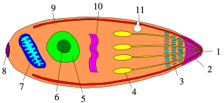
The Apicomplexa are a large phylum of parasitic alveolates. Most of them possess a unique form of organelle that comprises a type of plastid called an apicoplast, and an apical complex structure. The organelle is an adaptation that the apicomplexan applies in penetration of a host cell.
Toxoplasmosis is a parasitic disease caused by Toxoplasma gondii, an apicomplexan. Infections with toxoplasmosis usually cause no obvious symptoms in adults. Occasionally, people may have a few weeks or months of mild, flu-like illness such as muscle aches and tender lymph nodes. In a small number of people, eye problems may develop. In those with a weak immune system, severe symptoms such as seizures and poor coordination may occur. If a woman becomes infected during pregnancy, a condition known as congenital toxoplasmosis may affect the child.
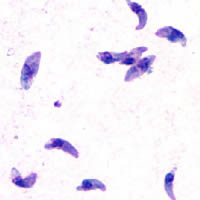
Toxoplasma gondii is an obligate intracellular parasitic protozoan eukaryote that causes the infectious disease toxoplasmosis. Found worldwide, T. gondii is capable of infecting virtually all warm-blooded animals, but felids, such as domestic cats, are the only known definitive hosts in which the parasite may undergo sexual reproduction.

Plasmodium is a genus of unicellular eukaryotes that are obligate parasites of vertebrates and insects. The life cycles of Plasmodium species involve development in a blood-feeding insect host which then injects parasites into a vertebrate host during a blood meal. Parasites grow within a vertebrate body tissue before entering the bloodstream to infect red blood cells. The ensuing destruction of host red blood cells can result in disease, called malaria. During this infection, some parasites are picked up by a blood-feeding insect, continuing the life cycle.

Plasmodium falciparum is a unicellular protozoan parasite of humans, and the deadliest species of Plasmodium that causes malaria in humans. The parasite is transmitted through the bite of a female Anopheles mosquito and causes the disease's most dangerous form, falciparum malaria. It is responsible for around 50% of all malaria cases. P. falciparum is therefore regarded as the deadliest parasite in humans. It is also associated with the development of blood cancer and is classified as Group 2A carcinogen.

Plasmodium malariae is a parasitic protozoan that causes malaria in humans. It is one of several species of Plasmodium parasites that infect other organisms as pathogens, also including Plasmodium falciparum and Plasmodium vivax, responsible for most malarial infection. Found worldwide, it causes a so-called "benign malaria", not nearly as dangerous as that produced by P. falciparum or P. vivax. The signs include fevers that recur at approximately three-day intervals – a quartan fever or quartan malaria – longer than the two-day (tertian) intervals of the other malarial parasites.

Micronemes are secretory organelles, possessed by parasitic apicomplexans. Micronemes are located on the apical third of the protozoan body. They are surrounded by a typical unit membrane. On electron microscopy they have an electron-dense matrix due to the high protein content. They are specialized secretory organelles important for host-cell invasion and gliding motility.
A rhoptry is a specialized secretory organelle. They are club-shaped organelles connected by thin necks to the extreme apical pole of the parasite. These organelles, like micronemes, are characteristic of the motile stages of Apicomplexa protozoans. They can vary in number and shape and contain numerous enzymes that are released during the penetration process. The proteins they contain are important in the interaction between the host and the parasite, including the formation of the parasitophorous vacuole.

Plasmodium knowlesi is a parasite that causes malaria in humans and other primates. It is found throughout Southeast Asia, and is the most common cause of human malaria in Malaysia. Like other Plasmodium species, P. knowlesi has a life cycle that requires infection of both a mosquito and a warm-blooded host. While the natural warm-blooded hosts of P. knowlesi are likely various Old World monkeys, humans can be infected by P. knowlesi if they are fed upon by infected mosquitoes. P. knowlesi is a eukaryote in the phylum Apicomplexa, genus Plasmodium, and subgenus Plasmodium. It is most closely related to the human parasite Plasmodium vivax as well as other Plasmodium species that infect non-human primates.
An apicoplast is a derived non-photosynthetic plastid found in most Apicomplexa, including Toxoplasma gondii, Plasmodium falciparum and other Plasmodium spp., but not in others such as Cryptosporidium. It originated from algae through secondary endosymbiosis. The apicoplast is surrounded by four membranes within the outermost part of the endomembrane system. The apicoplast hosts important metabolic pathways like fatty acid synthesis, isoprenoid precursor synthesis and parts of the heme biosynthetic pathway

Protozoan infections are parasitic diseases caused by organisms formerly classified in the Kingdom Protozoa. They are usually contracted by either an insect vector or by contact with an infected substance or surface and include organisms that are now classified in the supergroups Excavata, Amoebozoa, SAR, and Archaeplastida.
Acidocalcisomes are rounded electron-dense acidic organelles, rich in calcium and polyphosphate and between 100 nm and 200 nm in diameter.

Toll-like receptor 11 (TLR11) is a protein that in mice and rats is encoded by the gene TLR11, whereas in humans it is represented by a pseudogene. TLR11 belongs to the toll-like receptor (TLR) family and the interleukin-1 receptor/toll-like receptor superfamily. In mice, TLR11 has been shown to recognise flagellin and/or profilin present on certain microbes, it helps propagate a host immune response. TLR11 plays a fundamental role in both the innate and adaptive immune responses, through the activation of Tumor necrosis factor-alpha, the Interleukin 12 (IL-12) response, and Interferon-gamma (IFN-gamma) secretion. TLR11 mounts an immune response to multiple microbes, including Toxoplasma gondii, Salmonella species, and uropathogenic E. coli, and likely many other species due to the highly conserved nature of flagellin and profilin.
Apicomplexans, a group of intracellular parasites, have life cycle stages evolved to allow them to survive the wide variety of environments they are exposed to during their complex life cycle. Each stage in the life cycle of an apicomplexan organism is typified by a cellular variety with a distinct morphology and biochemistry.
Acroeimeria is a genus of parasites that contains those species which initially develop immediately beneath the brush-border of the intestinal epithelium, but the meronts and gamonts of which are early on extruded to form a layer on the surface of the gut mucosa. Morphologically they are similar to the Eimeria to which they are closely related. The genus was described in 1989 by Paperna and Landsberg.

Immunity Related Guanosine Triphosphatases or IRGs are proteins activated as part of an early immune response. IRGs have been described in various mammals but are most well characterized in mice. IRG activation in most cases is induced by an immune response and leads to clearance of certain pathogens.

In molecular biology, Duffy binding proteins are found in plasmodia. Plasmodium vivax and Plasmodium knowlesi merozoites invade Homo sapiens erythrocytes that express Duffy blood group surface determinants. The Duffy receptor family is localised in micronemes, an organelle found in all organisms of the phylum Apicomplexa.
Plasmodium falciparum erythrocyte membrane protein 1 (PfEMP1) is a family of proteins present on the membrane surface of red blood cells that are infected by the malarial parasite Plasmodium falciparum. PfEMP1 is synthesized during the parasite's blood stage inside the RBC, during which the clinical symptoms of falciparum malaria are manifested. Acting as both an antigen and adhesion protein, it is thought to play a key role in the high level of virulence associated with P. falciparum. It was discovered in 1984 when it was reported that infected RBCs had unusually large-sized cell membrane proteins, and these proteins had antibody-binding (antigenic) properties. An elusive protein, its chemical structure and molecular properties were revealed only after a decade, in 1995. It is now established that there is not one but a large family of PfEMP1 proteins, genetically regulated (encoded) by a group of about 60 genes called var. Each P. falciparum is able to switch on and off specific var genes to produce a functionally different protein, thereby evading the host's immune system. RBCs carrying PfEMP1 on their surface stick to endothelial cells, which facilitates further binding with uninfected RBCs, ultimately helping the parasite to both spread to other RBCs as well as bringing about the fatal symptoms of P. falciparum malaria.
Maurer's clefts are membranous structures seen in the red blood cell during infection with Plasmodium falciparum. The function and contents of Maurer's clefts are not completely known; however, they appear to play a role in trafficking of Plasmodium falciparum erythrocyte membrane protein 1 (PfEMP1) and other adhesins to the red blood cell surface.

A symbiosome is a specialised compartment in a host cell that houses an endosymbiont in a symbiotic relationship.
