Brown algae

Last updated

Brown algae
Temporal range: Late Jurassic to present 150–0  Ma [1] [2]
Kelp-forest-Monterey.jpg
Giant kelp ( Macrocystis pyrifera )
Scientific classification OOjs UI icon edit-ltr.svg
Domain: Eukaryota
Clade: Diaphoretickes
Clade: SAR
Clade: Stramenopiles
Phylum: Gyrista
Subphylum: Ochrophytina
Infraphylum: Chrysista
Superclass: Fucistia
Class: Phaeophyceae
Kjellman, 1891 [3]
Orders

See classification

Synonyms

Brown algae (SG: alga) are a large group of multicellular algae comprising the class Phaeophyceae. They include many seaweeds located in colder waters of the Northern Hemisphere. Brown algae are the major seaweeds of the temperate and polar regions. Many brown algae, such as members of the order Fucales, commonly grow along rocky seashores. Most brown algae live in marine environments, where they play an important role both as food and as a potential habitat. For instance, Macrocystis , a kelp of the order Laminariales, may reach 60 m (200 ft) in length and forms prominent underwater kelp forests that contain a high level of biodiversity. [4] Another example is Sargassum , which creates unique floating mats of seaweed in the tropical waters of the Sargasso Sea that serve as the habitats for many species. Some members of the class, such as kelps, are used by humans as food.

Contents

Between 1,500 and 2,000 species of brown algae are known worldwide. [5] Some species, such as Ascophyllum nodosum , have become subjects of extensive research in their own right due to their commercial importance. They also have environmental significance through carbon fixation. [4]

Brown algae belong to the Stramenopiles, a clade of eukaryotic organisms that are distinguished from green plants by having chloroplasts surrounded by four membranes, suggesting that they were acquired secondarily from a symbiotic relationship between a basal eukaryote and a red or green alga. Most brown algae contain the pigment fucoxanthin, which is responsible for the distinctive greenish-brown color that gives them their name. Brown algae are unique among Stramenopiles in developing into multicellular forms with differentiated tissues, but they reproduce by means of flagellated spores and gametes that closely resemble cells of single-celled Stramenopiles. Genetic studies show their closest relatives to be the yellow-green algae.

Morphology

Brown algae exist in a wide range of sizes and forms. The smallest members of the group grow as tiny, feathery tufts of threadlike cells no more than a few centimeters (a few inches) long. [6] Some species have a stage in their life cycle that consists of only a few cells, making the entire alga microscopic. Other groups of brown algae grow to much larger sizes. The rockweeds and leathery kelps are often the most conspicuous algae in their habitats. [7] Kelps can range in size from the 60-centimeter-tall (2 ft) sea palm Postelsia to the giant kelp Macrocystis pyrifera , which grows to over 50 m (150 ft) long [8] [9] and is the largest of all the algae. In form, the brown algae range from small crusts or cushions [10] to leafy free-floating mats formed by species of Sargassum . They may consist of delicate felt-like strands of cells, as in Ectocarpus , or of 30-centimeter-long (1 ft) flattened branches resembling a fan, as in Padina .

Regardless of size or form, two visible features set the Phaeophyceae apart from all other algae. First, members of the group possess a characteristic color that ranges from an olive green to various shades of brown. The particular shade depends upon the amount of fucoxanthin present in the alga. [11] Second, all brown algae are multicellular. There are no known species that exist as single cells or as colonies of cells, [11] and the brown algae are the only major group of seaweeds that does not include such forms. However, this may be the result of classification rather than a consequence of evolution, as all the groups hypothesized to be the closest relatives of the browns include single-celled or colonial forms.[ citation needed ] They can change color depending on salinity, ranging from reddish to brown.

Visible structures

Two specimens of Laminaria hyperborea, each showing the rootlike holdfast at lower left, a divided blade at upper right, and a stemlike stipe connecting the blade to the holdfast. Laminaria hyperborea.jpg
Two specimens of Laminaria hyperborea , each showing the rootlike holdfast at lower left, a divided blade at upper right, and a stemlike stipe connecting the blade to the holdfast.

Whatever their form, the body of all brown algae is termed a thallus , indicating that it lacks the complex xylem and phloem of vascular plants. This does not mean that brown algae completely lack specialized structures. But, because some botanists define "true" stems, leaves, and roots by the presence of these tissues, their absence in the brown algae means that the stem-like and leaf-like structures found in some groups of brown algae must be described using different terminology. [12] Although not all brown algae are structurally complex, those that are typically possess one or more characteristic parts.

A holdfast is a rootlike structure present at the base of the alga. Like a root system in plants, a holdfast serves to anchor the alga in place on the substrate where it grows, and thus prevents the alga from being carried away by the current. Unlike a root system, the holdfast generally does not serve as the primary organ for water uptake, nor does it take in nutrients from the substrate. The overall physical appearance of the holdfast differs among various brown algae and among various substrates. It may be heavily branched, or it may be cup-like in appearance. A single alga typically has just one holdfast, although some species have more than one stipe growing from their holdfast.

A stipe is a stalk or stemlike structure present in an alga. It may grow as a short structure near the base of the alga (as in Laminaria ), or it may develop into a large, complex structure running throughout the algal body (as in Sargassum or Macrocystis ). In the most structurally differentiated brown algae (such as Fucus ), the tissues within the stipe are divided into three distinct layers or regions. These regions include a central pith, a surrounding cortex, and an outer epidermis, each of which has an analog in the stem of a vascular plant. In some brown algae, the pith region includes a core of elongated cells that resemble the phloem of vascular plants both in structure and function. In others (such as Nereocystis ), the center of the stipe is hollow and filled with gas that serves to keep that part of the alga buoyant. The stipe may be relatively flexible and elastic in species like Macrocystis pyrifera that grow in strong currents, or may be more rigid in species like Postelsia palmaeformis that are exposed to the atmosphere at low tide.

Many algae have a flattened portion that may resemble a leaf, and this is termed a blade, lamina , or frond. The name blade is most often applied to a single undivided structure, while frond may be applied to all or most of an algal body that is flattened, but this distinction is not universally applied. The name lamina refers to that portion of a structurally differentiated alga that is flattened. It may be a single or a divided structure, and may be spread over a substantial portion of the alga. In rockweeds, for example, the lamina is a broad wing of tissue that runs continuously along both sides of a branched midrib. The midrib and lamina together constitute almost all of a rockweed, so that the lamina is spread throughout the alga rather than existing as a localized portion of it.

Species like Fucus vesiculosus produce numerous gas-filled pneumatocysts (air bladders) to increase buoyancy. Bladder Wrack (Fucus vesiculosus) - geograph.org.uk - 224125.jpg
Species like Fucus vesiculosus produce numerous gas-filled pneumatocysts (air bladders) to increase buoyancy.

In some brown algae, there is a single lamina or blade, while in others there may be many separate blades. Even in those species that initially produce a single blade, the structure may tear with rough currents or as part of maturation to form additional blades. These blades may be attached directly to the stipe, to a holdfast with no stipe present, or there may be an air bladder between the stipe and blade. The surface of the lamina or blade may be smooth or wrinkled; its tissues may be thin and flexible or thick and leathery. In species like Egregia menziesii , this characteristic may change depending upon the turbulence of the waters in which it grows. [6] In other species, the surface of the blade is coated with slime to discourage the attachment of epiphytes or to deter herbivores. Blades are also often the parts of the alga that bear the reproductive structures.

Gas-filled floats called pneumatocysts provide buoyancy in many kelps and members of the Fucales. These bladder-like structures occur in or near the lamina, so that it is held nearer the water surface and thus receives more light for photosynthesis. Pneumatocysts are most often spherical or ellipsoidal, but can vary in shape among different species. Species such as Nereocystis luetkeana and Pelagophycus porra bear a single large pneumatocyst between the top of the stipe and the base of the blades. In contrast, the giant kelp Macrocystis pyrifera bears many blades along its stipe, with a pneumatocyst at the base of each blade where it attaches to the main stipe. Species of Sargassum also bear many blades and pneumatocysts, but both kinds of structures are attached separately to the stipe by short stalks. In species of Fucus , the pneumatocysts develop within the lamina itself, either as discrete spherical bladders or as elongated gas-filled regions that take the outline of the lamina in which they develop.

Growth

Growth in Dictyota dichotoma occurs at each frond tip, where new cells are produced. Capo Gallo Dicotoma.jpg
Growth in Dictyota dichotoma occurs at each frond tip, where new cells are produced.

The brown algae include the largest and fastest growing of seaweeds. [6] Fronds of Macrocystis may grow as much as 50 cm (20 in) per day, and the stipes can grow 6 cm (2.4 in) in a single day. [13]

Growth in most brown algae occurs at the tips of structures as a result of divisions in a single apical cell or in a row of such cells. They are single cellular organisms. [7] As this apical cell divides, the new cells that it produces develop into all the tissues of the alga. Branchings and other lateral structures appear when the apical cell divides to produce two new apical cells. However, a few groups (such as Ectocarpus ) grow by a diffuse, unlocalized production of new cells that can occur anywhere on the thallus. [11]

Tissue organization

The simplest browns are filamentous—that is, their cells are elongate and have septa cutting across their width. They branch by getting wider at their tip, and then dividing the widening. [14]

These filaments may be haplostichous or polystichous, multiaxial or monoaxial forming or not a pseudoparenchyma. [15] [16] Besides fronds, there are the large in size parenchyma tic kelps with three-dimensional development and growth and different tissues (meristoderm, cortex and medulla) which could be consider the trees of the sea. [17] [18] There are also the Fucales and Dictyotales smaller than kelps but still parenchymatic with the same kind of distinct tissues.

The cell wall consists of two layers; the inner layer bears the strength, and consists of cellulose; the outer wall layer is mainly algin, and is gummy when wet but becomes hard and brittle when it dries out. [15] Specifically, the brown algal cell wall consists of several components with alginates and sulphated fucan being its main ingredients, up to 40% each of them. [19] Cellulose, a major component from most plant cell walls, is present in a very small percentage, up to 8%. Cellulose and alginate biosynthesis pathways seem to have been acquired from other organisms through endosymbiotic and horizontal gene transfer respectively, while the sulphated polysaccharides are of ancestral origin. [20] Specifically, the cellulose synthases seem to come from the red alga endosymbiont of the photosynthetic stramenopiles ancestor, and the ancestor of brown algae acquired the key enzymes for alginates biosynthesis from an actinobacterium. The presence and fine control of alginate structure in combination with the cellulose which existed before it, gave potentially the brown algae the ability to develop complex structurally multicellular organisms like the kelps. [21]

Evolutionary history

Genetic and ultrastructural evidence place the Phaeophyceae among the heterokonts (Stramenopiles), [22] a large assemblage of organisms that includes both photosynthetic members with plastids (such as the diatoms) as well as non-photosynthetic groups (such as the slime nets and water molds). Although some heterokont relatives of the brown algae lack plastids in their cells, scientists believe this is a result of evolutionary loss of that organelle in those groups rather than independent acquisition by the several photosynthetic members. [23] Thus, all heterokonts are believed to descend from a single heterotrophic ancestor that became photosynthetic when it acquired plastids through endosymbiosis of another unicellular eukaryote. [24]

The closest relatives of the brown algae include unicellular and filamentous species, but no unicellular species of brown algae are known. However, most scientists assume that the Phaeophyceae evolved from unicellular ancestors. [25] DNA sequence comparison also suggests that the brown algae evolved from the filamentous Phaeothamniophyceae, [26] Xanthophyceae, [27] or the Chrysophyceae [28] between 150 [1] and 200 million years ago. [2] In many ways, the evolution of the brown algae parallels that of the green algae and red algae, [29] as all three groups possess complex multicellular species with an alternation of generations. Analysis of 5S rRNA sequences reveals much smaller evolutionary distances among genera of the brown algae than among genera of red or green algae, [2] [30] which suggests that the brown algae have diversified much more recently than the other two groups.

Fossils

The occurrence of Phaeophyceae as fossils is rare due to their generally soft-bodied nature, [31] and scientists continue to debate the identification of some finds. [32] Part of the problem with identification lies in the convergent evolution of morphologies between many brown and red algae. [33] Most fossils of soft-tissue algae preserve only a flattened outline, without the microscopic features that permit the major groups of multicellular algae to be reliably distinguished. Among the brown algae, only species of the genus Padina deposit significant quantities of minerals in or around their cell walls. [34] Other algal groups, such as the red algae and green algae, have a number of calcareous members. Because of this, they are more likely to leave evidence in the fossil record than the soft bodies of most brown algae and more often can be precisely classified. [35]

Fossils comparable in morphology to brown algae are known from strata as old as the Upper Ordovician, [36] but the taxonomic affinity of these impression fossils is far from certain. [37] Claims that earlier Ediacaran fossils are brown algae [38] have since been dismissed. [26] While many carbonaceous fossils have been described from the Precambrian, they are typically preserved as flattened outlines or fragments measuring only millimeters long. [39] Because these fossils lack features diagnostic for identification at even the highest level, they are assigned to fossil form taxa according to their shape and other gross morphological features. [40] A number of Devonian fossils termed fucoids, from their resemblance in outline to species in the genus Fucus , have proven to be inorganic rather than true fossils. [31] The Devonian megafossil Prototaxites , which consists of masses of filaments grouped into trunk-like axes, has been considered a possible brown alga. [11] However, modern research favors reinterpretation of this fossil as a terrestrial fungus or fungal-like organism. [41] Likewise, the fossil Protosalvinia was once considered a possible brown alga, but is now thought to be an early land plant. [42]

A number of Paleozoic fossils have been tentatively classified with the brown algae, although most have also been compared to known red algae species. Phascolophyllaphycus possesses numerous elongate, inflated blades attached to a stipe. It is the most abundant of algal fossils found in a collection made from Carboniferous strata in Illinois. [43] Each hollow blade bears up to eight pneumatocysts at its base, and the stipes appear to have been hollow and inflated as well. This combination of characteristics is similar to certain modern genera in the order Laminariales (kelps). Several fossils of Drydenia and a single specimen of Hungerfordia from the Upper Devonian of New York have also been compared to both brown and red algae. [33] Fossils of Drydenia consist of an elliptical blade attached to a branching filamentous holdfast, not unlike some species of Laminaria , Porphyra , or Gigartina . The single known specimen of Hungerfordia branches dichotomously into lobes and resembles genera like Chondrus and Fucus [33] or Dictyota . [44]

The earliest known fossils that can be assigned reliably to the Phaeophyceae come from Miocene diatomite deposits of the Monterey Formation in California. [24] Several soft-bodied brown macroalgae, such as Julescraneia , have been found. [45]

Classification

Phylogeny

Based on the work of Silberfeld, Rousseau & de Reviers 2014. [46]

Discosporangiales

Choristocarpaceae

Discosporangiaceae

Ishigeales

Ishigeaceae

Petrodermataceae

Dictyotophycidae

Onslowiales

Dictyotales

Syringodermatales

Sphacelariales

Lithodermataceae

Phaeostrophiaceae

Stypocaulaceae

Cladostephaceae

Sphacelariaceae

Fucophycidae

Bachelotiaceae

Desmarestiales

Sporochnales

Ascoseirales

Ralfsiales

Tilopteridales

Cutleriaceae

Tilopteridaceae

Phyllariaceae

Nemodermatales

Fucales

Sargassaceae

Durvillaeaceae

Himanthaliaceae

Fucaceae

Scytothamnales

Asteronemataceae

Splachnidiaceae

Laminariales

Phaeosiphoniellaceae

Akkesiphycaceae

Pseudochordaceae

Chordaceae

Alariaceae

Agaraceae

Laminariaceae

Asterocladales

Ectocarpales

Adenocystaceae

Scytosiphonaceae

Petrospongiaceae

Ectocarpaceae

Acinetosporaceae

Chordariaceae

Taxonomy

This is a list of the orders in the class Phaeophyceae: [46] [47]

The life cycle of a representative species Laminaria. Most Brown Algae follow this form of sexual reproduction. Laminaria Life Cycle.png
The life cycle of a representative species Laminaria. Most Brown Algae follow this form of sexual reproduction.
A closeup of a Fucus's conceptacle, showing the gametes coming together to form a fertilized zygote. Fucus conceptacle XS3.jpg
A closeup of a Fucus's conceptacle, showing the gametes coming together to form a fertilized zygote.

Life cycle

Most brown algae, with the exception of the Fucales, perform sexual reproduction through sporic meiosis. [48] Between generations, the algae go through separate sporophyte (diploid) and gametophyte (haploid) phases. The sporophyte stage is often the more visible of the two, though some species of brown algae have similar diploid and haploid phases. Free floating forms of brown algae often do not undergo sexual reproduction until they attach themselves to substrate. The haploid generation consists of male and female gametophytes. [49] The fertilization of egg cells varies between species of brown algae, and may be isogamous, oogamous, or anisogamous. Fertilization may take place in the water with eggs and motile sperm, or within the oogonium itself.

Certain species of brown algae can also perform asexual reproduction through the production of motile diploid zoospores. These zoospores form in plurilocular sporangium, and can mature into the sporophyte phase immediately.

In a representative species Laminaria , there is a conspicuous diploid generation and smaller haploid generations. Meiosis takes place within several unilocular sporangium along the algae's blade, each one forming either haploid male or female zoospores. The spores are then released from the sporangia and grow to form male and female gametophytes. The female gametophyte produces an egg in the oogonium, and the male gametophyte releases motile sperm that fertilize the egg. The fertilized zygote then grows into the mature diploid sporophyte.

In the order Fucales, sexual reproduction is oogamous, and the mature diploid is the only form for each generation. Gametes are formed in specialized conceptacles that occur scattered on both surfaces of the receptacle, the outer portion of the blades of the parent plant. Egg cells and motile sperm are released from separate sacs within the conceptacles of the parent algae, combining in the water to complete fertilization. The fertilized zygote settles onto a surface and then differentiates into a leafy thallus and a finger-like holdfast. Light regulates differentiation of the zygote into blade and holdfast.(by samay the great )

Saccharina latissima on a beach. Saccharina latissima NOAA.jpg
Saccharina latissima on a beach.

Ecology

Brown algae have adapted to a wide variety of marine ecological niches including the tidal splash zone, rock pools, the whole intertidal zone and relatively deep near shore waters. They are an important constituent of some brackish water ecosystems, and have colonized freshwater on a minimum of six known occasions. [50] A large number of Phaeophyceae are intertidal or upper littoral, [26] and they are predominantly cool and cold water organisms that benefit from nutrients in up welling cold water currents and inflows from land; Sargassum being a prominent exception to this generalisation.

Brown algae growing in brackish waters are almost solely asexual. [26]

Chemistry

Algal groupδ13C range [51]
HCO3-using red algae 22.5 to 9.6
CO2-using red algae34.5 to 29.9
Brown algae 20.8 to 10.5
Green algae 20.3 to 8.8

Brown algae have a δ13C value in the range of 30.0 to 10.5, in contrast with red algae and greens. This reflects their different metabolic pathways. [52]

They have cellulose walls with alginic acid and also contain the polysaccharide fucoidan in the amorphous sections of their cell walls. A few species (of Padina ) calcify with aragonite needles. [26]

In addition to alginates, fucoidan and cellulose, the carbohydrate composition of brown algae consists of mannitol, laminarin and glucan. [53]

The photosynthetic system of brown algae is made of a P700 complex containing chlorophyll a. Their plastids also contain chlorophyll c and carotenoids (the most widespread of those being fucoxanthin). [54]

Brown algae produce a specific type of tannin called phlorotannins in higher amounts than red algae do.

Importance and uses

Brown algae include a number of edible seaweeds. All brown algae contain alginic acid (alginate) in their cell walls, which is extracted commercially and used as an industrial thickening agent in food and for other uses. [55] One of these products is used in lithium-ion batteries. [56] Alginic acid is used as a stable component of a battery anode. This polysaccharide is a major component of brown algae, and is not found in land plants.

Alginic acid can also be used in aquaculture. For example, alginic acid enhances the immune system of rainbow trout. Younger fish are more likely to survive when given a diet with alginic acid. [57]

Brown algae including kelp beds also fix a significant portion of the earth's carbon dioxide yearly through photosynthesis. [58] Additionally, they can store a great amount of carbon dioxide which can help us in the fight against climate change. [59] Sargachromanol G, an extract of Sargassum siliquastrum, has been shown to have anti-inflammatory effects. [60]

Edible brown algae

See also

Related Research Articles

<span class="mw-page-title-main">Kelp</span> Large brown seaweeds in the order Laminariales

Kelps are large brown algae or seaweeds that make up the order Laminariales. There are about 30 different genera. Despite its appearance, kelp is not a plant but a stramenopile, a group containing many protists.

<i>Nereocystis</i> Genus of kelp

Nereocystis is a monotypic genus of subtidal kelp containing the species Nereocystis luetkeana. Some English names include edible kelp, bull kelp, bullwhip kelp, ribbon kelp, bladder wrack, and variations of these names. Due to the English name, bull kelp can be confused with southern bull kelps, which are found in the Southern Hemisphere. Nereocystis luetkeana forms thick beds on subtidal rocks, and is an important part of kelp forests.

<i>Laminaria</i> Genus of algae

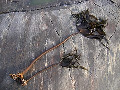
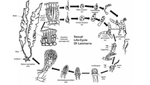
Laminaria is a genus of brown seaweed in the order Laminariales (kelp), comprising 31 species native to the north Atlantic and northern Pacific Oceans. This economically important genus is characterized by long, leathery laminae and relatively large size. Some species are called Devil's apron, due to their shape, or sea colander, due to the perforations present on the lamina. Others are referred to as tangle. Laminaria form a habitat for many fish and invertebrates.

<i>Macrocystis</i> Genus of large brown algae

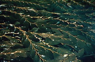
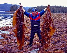
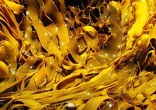
Macrocystis is a monospecific genus of kelp with all species now synonymous with Macrocystis pyrifera. It is commonly known as giant kelp or bladder kelp. This genus contains the largest of all the Phaeophyceae or brown algae. Macrocystis has pneumatocysts at the base of its blades. Sporophytes are perennial and the individual may live for up to three years; stipes/fronds within a whole individual undergo senescence, where each frond may persist for approximately 100 days. The genus is found widely in subtropical, temperate, and sub-Antarctic oceans of the Southern Hemisphere and in the northeast Pacific from Baja California to Sitka, Alaska. Macrocystis is often a major component of temperate kelp forests.

<span class="mw-page-title-main">Fucales</span> Order of brown algae

The Fucales (fucoids) are an order in the brown algae. The list of families in the Fucales, as well as additional taxonomic information on algae, is publicly accessible at Algaebase.

<span class="mw-page-title-main">Sporochnaceae</span> Family of algae

Sporochnacaeae is the only family in the order Sporochnales in the brown algae. Member of this family are thread-like algae growing by means of an intercalary row of dome shaped cells at the base of the hairs.

<span class="mw-page-title-main">Ectocarpales</span> Order of algae

Ectocarpales is a very large order in the brown algae. The order includes families with pseudoparenchymatous (Splachnidiaceae) or true parenchymatous (Scytosiphonaceae) tissue. Pseudoparenchymatous refers to a filamentous alga with cells packed very close together to give an appearance of parenchymatous tissue, the latter being composed of cells which can truly divide in three dimensions, unusual among the algae. Filamentous algae are composed of cells that divide along a single plane, allowing only elongation to form filaments of one or more rows of cells. Algae that can divide in two planes can form sheet-like thalli or bodies. Cells that can divide in a third plane potentially allow for the organism to develop a more complex body plan, and diversification of body plans into an erect thallus of some sort and a holdfast for attaching the upright portion to the substrate.

<i>Postelsia</i> Species of kelp

Postelsia palmaeformis, also known as the sea palm or palm seaweed, is a species of kelp and classified within brown algae. It is the only known species in the genus Postelsia. The sea palm is found along the western coast of North America, on rocky shores with constant waves. It is one of the few algae that can survive and remain erect out of the water; in fact, it spends most of its life cycle exposed to the air. It is an annual, and edible, though harvesting of the alga is discouraged due to the species' sensitivity to overharvesting.

<span class="mw-page-title-main">Sargassaceae</span> Family of seaweeds

Sargassaceae is a family of brown algae in the order Fucales.

<i>Eisenia arborea</i> Species of kelp

Eisenia arborea, or the southern sea palm, is a dominant species of kelp that is found on the western Pacific coast of North America, from Vancouver Island, Canada south to Mexico's Isla Magdalena and Baja California, as well as in Japan. They are commonly found from the midtidal areas stretching to the subtidal areas. It is an edible seaweed, a source of nutrients for grazing marine invertebrates and a source of alginic acid, a food thickener. Some of the algas have a hollow stipe above its holdfast with two branches terminating in multiple blades. Eisenia arborea is studied in order to predict environmental stress in oceans intertidal zones. Hollow stipes where present when the Eisenia arborea did not receive essential nutrients for its thalli development. Eisenia arborea with hollow stripes are believed to be evolved algae in order to increase their survival in harsh living conditions. They play a huge role in determining environmental stress.

<i>Durvillaea antarctica</i> Species of seaweed

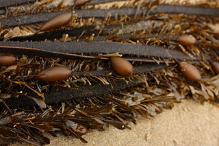
Durvillaea antarctica, also known as cochayuyo and rimurapa, is a large, robust species of southern bull kelp found on the coasts of Chile, southern New Zealand, and Macquarie Island. D. antarctica, an alga, does not have air bladders, but floats due to a unique honeycomb structure within the alga's blades, which also helps the kelp avoid being damaged by the strong waves.

<i>Egregia</i> Species of alga

Egregia menziesii is a species of kelp known commonly as feather boa kelp. It is the only species in the monotypic genus Egregia. It is native to the coastline of western North America from Alaska to Baja California, where it is a common kelp of the intertidal zone.

<span class="mw-page-title-main">Choristocarpaceae</span> Family of algae

Choristocarpaceae is a family in the order Discosporangiales of the brown algae. The family contains a single genus, Choristocarpus. The species is mostly located in the cold waters of the Northern hemisphere. A type of seaweed, Choristocarpaceae attaches itself to rocky substrate in places that are near continental shelves and the shore. Due to the species having morphological similarity, they were classified in closer relation with D. mesarthrocarpum. But due to many other differing characteristics Choristocarpaceae were put into their own family with a single genus and a single species of brown algae.

Pterygophora californica is a large species of kelp, commonly known as stalked kelp. It is the only species in its genus Pterygophora. It grows in shallow water on the Pacific coast of North America where it forms part of a biodiverse community in a "kelp forest". It is sometimes also referred to as woody-stemmed kelp, walking kelp, or winged kelp.

<i>Himanthalia</i> Genus of seaweeds

Himanthalia is a genus of brown algae. It is the only genus in the family Himanthaliaceae in the order Fucales. It includes two species: Himanthalia durvillei and Himanthalia elongata.

Sunamphitoe femorata is a species of amphipod crustacean in the family Ampithoidae. It is a herbivore and constructs a tubular nest-like home on a blade of the sporophyte of the giant kelp Macrocystis pyrifera. This home is made by rolling the sides of the blade together and securing them with silk. As the kelp blade grows, the nest is advanced down the blade towards the base, approximately keeping pace with the algal growth.

Pleurophycus gardneri is a species of brown alga. It is a deciduous kelp, primarily found in lower, rocky inter-tidal and shallow, rocky sub-tidal locations and is one of the most abundant kelps found within the Pleurophycus Zone. It is not commonly present deeper in the ocean than 30m and is considered a stipitate kelp. P. gardneri forms aggregates of densities up to 10m−2. These kelp beds reside below giant kelp forests, and were therefore often overlooked by researchers for many years. This kelp has a range from Central California to British Columbia, Canada, with a lifespan of only 3 – 6 years.

Fucophycidae is a subclass of Phaeophyceae which contains the most complex and evolved orders of Chromista algae. The members of this subclass have stalks with several morphological forms and distinct structures, characterized by an intercalary growth and a basic heteromorphic, sometimes secondarily iso- or sub-isomorphic life cycle.

<i>Padina</i> (alga) Genus of brown algae

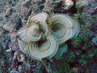
Padina is a genus of brown macroalgae in the family Dictyotaceae.

References

  1. 1 2 Medlin, L. K.; et al. (1997). "Phylogenetic relationships of the 'golden algae' (Haptophytes, heterokont chromophytes) and their plastids". Origins of Algae and their Plastids (PDF). Plant Systematics and Evolution. Vol. 11. pp. 187–219. doi:10.1007/978-3-7091-6542-3_11. hdl:10013/epic.12690. ISBN   978-3-211-83035-2. Archived (PDF) from the original on 5 October 2013. Retrieved 27 August 2012.
  2. 1 2 3 Lim, B.-L.; Kawai, H.; Hori, H.; Osawa, S. (1986). "Molecular evolution of 5S ribosomal RNA from red and brown algae". Japanese Journal of Genetics . 61 (2): 169–176. doi: 10.1266/jjg.61.169 . Archived from the original on 10 January 2015. Retrieved 27 August 2012.
  3. Kjellman, F. R. (1891). "Phaeophyceae (Fucoideae)". In Engler, A.; Prantl, K. (eds.). Die natürlichen Pflanzenfamilien. Vol. 1. Leipzig: Wilhelm Engelmann. pp. 176–192.
  4. 1 2 Cock, J. Mark; Peters, Akira F.; Coelho, Susana M. (9 August 2011). "Brown algae". Current Biology. 21 (15): R573–R575. doi: 10.1016/j.cub.2011.05.006 . PMID   21820616.
  5. Hoek, Christiaan; den Hoeck, Hoeck Van; Mann, David; Jahns, H.M. (1995). Algae: an introduction to phycology. Cambridge University Press. p. 166. ISBN   9780521316873. OCLC   443576944. Archived from the original on 16 October 2023. Retrieved 26 April 2018.
  6. 1 2 3 Connor, J.; Baxter, C. (1989). Kelp Forests. Monterey Bay Aquarium. ISBN   978-1-878244-01-7.
  7. 1 2 Dittmer, H. J. (1964). Phylogeny and Form in the Plant Kingdom . Princeton, NJ: D. Van Nostrand Company. pp.  115–137. ISBN   978-0-88275-167-2.
  8. Abbott, I. A.; Hollenberg, G. J. (1976). Marine Algae of California. California: Stanford University Press. ISBN   978-0-8047-0867-8.
  9. Cribb, A. B. (1953). "Macrocystis pyrifera (L.) Ag. in Tasmanian waters". Australian Journal of Marine and Freshwater Research . 5 (1): 1–34. doi:10.1071/MF9540001.
  10. Jones, W. E. (1962). "A key to the genera of the British seaweeds" (PDF). Field Studies . 1 (4): 1–32.[ permanent dead link ]
  11. 1 2 3 4 Bold, H. C.; Alexopoulos, C. J.; Delevoryas, T. (1987). Morphology of Plants and Fungi (5th ed.). New York: Harper & Row Publishers. pp. 112–131, 174–186. ISBN   978-0-06-040839-8.
  12. Raven, P. H.; Evert, R. F.; Eichhorn, S. E. (2005). Biology of Plants (7th ed.). New York: W. H. Freeman and Company. pp.  316–321, 347. ISBN   978-0-7167-1007-3.
  13. Round, F. E. (1981). The Ecology of Algae. Cambridge: Cambridge University Press. p. 103. ISBN   978-0-521-26906-3.
  14. Wynne, M. J. (1981). "The Biology of seaweeds". In Lobban, C. S.; Wynne, M. J. (eds.). Phaeophyta: Morphology and Classification. Botanical Monographs. Vol. 17. University of California Press. p. 52. ISBN   978-0-520-04585-9. Archived from the original on 16 October 2023. Retrieved 11 May 2020.
  15. 1 2 Sharma, O. P (1986). Textbook of Algae. Tata McGraw-Hill. p. 298. ISBN   978-0-07-451928-8.
  16. Graham; Wilcox; Graham (2009). Algae, 2nd Edition. Pearson. ISBN   9780321603128.{{cite book}}: CS1 maint: multiple names: authors list (link)
  17. Fritsch, F. E. 1945. The Structure And Reproduction Of The Algae. Cambridge University Press, Cambridge.
  18. Charrier, Bénédicte; Le Bail, Aude; de Reviers, Bruno (August 2012). "Plant Proteus: brown algal morphological plasticity and underlying developmental mechanisms". Trends in Plant Science. 17 (8): 468–477. doi:10.1016/j.tplants.2012.03.003. ISSN   1360-1385. PMID   22513108. Archived from the original on 16 October 2023. Retrieved 13 November 2020.
  19. Charrier, Bénédicte; Rabillé, Hervé; Billoud, Bernard (2019). "Gazing at Cell Wall Expansion under a Golden Light" (PDF). Trends in Plant Science. 24 (2): 130–141. doi:10.1016/j.tplants.2018.10.013. PMID   30472067. S2CID   53725259. Archived (PDF) from the original on 8 January 2022. Retrieved 17 September 2022.
  20. Michel, Gurvan; Tonon, Thierry; Scornet, Delphine; Cock, J. Mark; Kloareg, Bernard (2010). "The cell wall polysaccharide metabolism of the brown alga Ectocarpus siliculosus. Insights into the evolution of extracellular matrix polysaccharides in Eukaryotes". New Phytologist. 188 (1): 82–97. doi: 10.1111/j.1469-8137.2010.03374.x . PMID   20618907.
  21. Deniaud-Bouët, Estelle; Kervarec, Nelly; Michel, Gurvan; Tonon, Thierry; Kloareg, Bernard; Hervé, Cécile (2014). "Chemical and enzymatic fractionation of cell walls from Fucales: Insights into the structure of the extracellular matrix of brown algae". Annals of Botany. 114 (6): 1203–1216. doi: 10.1093/aob/mcu096 . PMC   4195554 . PMID   24875633.
  22. Adl, S. M.; et al. (2005). "The new higher level classification of eukaryotes with emphasis on the taxonomy of protists" (PDF). Journal of Eukaryotic Microbiology . 52 (5): 399–451. doi: 10.1111/j.1550-7408.2005.00053.x . PMID   16248873. Archived (PDF) from the original on 29 May 2013. Retrieved 27 August 2012.
  23. Lane, C. E.; Archibald, J. M. (2008). "The eukaryotic tree of life: Endosymbiosis takes its TOL" (PDF). Trends in Ecology and Evolution . 23 (5): 268–275. CiteSeerX   10.1.1.555.2930 . doi:10.1016/j.tree.2008.02.004. PMID   18378040. Archived (PDF) from the original on 20 June 2013. Retrieved 27 August 2012.
  24. 1 2 van den Hoek, C.; Mann, D. G.; Jahns, H. M. (1995). Algae: An Introduction to Phycology. Cambridge: Cambridge University Press. pp. 165–218. ISBN   978-0-521-31687-3.
  25. Niklas, K. J. (1997). The Evolutionary Biology of Plants. Chicago: University of Chicago Press. p. 156. ISBN   978-0-226-58082-1.
  26. 1 2 3 4 5 Lee, R. E. (2008). Phycology (4th ed.). Cambridge University Press. ISBN   978-0-521-63883-8.
  27. Ariztia, E. V.; Andersen, R. A.; Sogin, M. L. (1991). "A new phylogeny of chromophyte algae using 16S-like rRNA sequences from Mallomonas papillosa (Synurophyceae) and Tribonema aequale (Xanthophyceae)". Journal of Phycology . 27 (3): 428–436. doi:10.1111/j.0022-3646.1991.00428.x. S2CID   84693030.
  28. Taylor, T. N.; Taylor, E. L. (1993). The Biology and Evolution of Fossil Plants. Englewood Cliffs, NJ: Prentice Hall. pp. 128–131. ISBN   978-0-13-651589-0.
  29. Dittmer, H. J. (1964). Phylogeny and Form in the Plant Kingdom . Princeton, NJ: D. Van Nostrand Company. pp.  115–137. ISBN   978-0-88275-167-2.
  30. Hori, H.; Osawa, S. (1987). "Origin and evolution of organisms as deduced from 5S ribosomal RNS sequences". Molecular Biology and Evolution . 4 (5): 445–472. doi: 10.1093/oxfordjournals.molbev.a040455 . PMID   2452957.
  31. 1 2 Arnold, C. A. (1947). An Introduction to Paleobotany. New York; London: McGraw-Hill. p.  48. ISBN   978-1-4067-1861-4.
  32. Coyer, J. A.; Smith, G. J.; Andersen, R. A. (2001). "Evolution of Macrocystis spp. (Phaeophyta) as determined by ITS1 and ITS2 sequences" (PDF). Journal of Phycology . 37 (4): 574–585. doi:10.1046/j.1529-8817.2001.037001574.x. S2CID   84074495. Archived from the original on 16 October 2023. Retrieved 27 August 2012.
  33. 1 2 3 Fry, W. L.; Banks, H. P. (1955). "Three new genera of algae from the Upper Devonian of New York". Journal of Paleontology . 29 (1): 37–44. JSTOR   1300127.
  34. Prescott, G. W. (1968). The Algae: A Review . Boston: Houghton Mifflin Company. pp.  207–231, 371–372. ISBN   978-3-87429-244-3.
  35. Simpson, G. G. (1953). Life of the Past: An Introduction to Paleontology . New Haven: Yale University Press. pp.  158–159.
  36. Fry, W. L. (1983). "An algal flora from the Upper Ordovician of the Lake Winnipeg region, Manitoba, Canada". Review of Palaeobotany and Palynology . 39 (3–4): 313–341. Bibcode:1983RPaPa..39..313F. doi:10.1016/0034-6667(83)90018-0.
  37. Speer, B. R.; Waggoner, B. M. (2000). "Phaeophyta: Fossil Record". Archived from the original on 9 June 2007. Retrieved 24 April 2007.
  38. Loeblich, A. R. (1974). "Protistan Phylogeny as Indicated by the Fossil Record". Taxon . 23 (2/3): 277–290. doi:10.2307/1218707. JSTOR   1218707.
  39. Hofmann, H. J. (1985). "Precambrian Carbonaceous Megafossils". In D. F. Toomey; M. H. Nitecki (eds.). Paleoalgology: Contemporary Research and Applications. Berlin: Springer-Verlag. pp. 20–33.
  40. Hofmann, H. J. (1994). "Proterozoic carbonaceous compressions ("metaphytes" and "worms")". In Bengtson, S. (ed.). Life on Earth. Nobel Symposium. Vol. 84. New York: Columbia University Press. pp. 342–357.
  41. Hueber, F. M. (2001). "Rotted wood-alga-fungus: the history and life of Prototaxites Dawson 1859". Review of Palaeobotany and Palynology . 116 (1): 123–158. Bibcode:2001RPaPa.116..123H. doi:10.1016/S0034-6667(01)00058-6.
  42. Taylor, W. A.; Taylor, T. N. (1987). "Spore wall ultrastructure of Protosalvinia" (PDF). American Journal of Botany . 74 (3): 437–443. doi:10.2307/2443819. JSTOR   2443819. Archived from the original (PDF) on 17 June 2010.
  43. Leary, R. L. (1986). "Three new genera of fossil noncalcareous algae from Valmeyeran (Mississippian) strata of Illinois". American Journal of Botany . 73 (3): 369–375. doi:10.2307/2444080. JSTOR   2444080.
  44. Bold, H. C.; Wynne, M. J. (1978). Introduction to the Algae (2nd ed.). Prentice-Hall. p.  27. ISBN   978-0-13-477786-3.
  45. Parker, B. C.; Dawson, E. Y. (1965). "Non-calcareous marine algae from California Miocene deposits". Nova Hedwigia . 10: 273–295, plates 76–96.
  46. 1 2 Silberfeld, Thomas; Rousseau, Florence; de Reviers, Bruno (2014). "An Updated Classification of Brown Algae (Ochrophyta, Phaeophyceae)". Cryptogamie, Algologie. 35 (2): 117–156. doi:10.7872/crya.v35.iss2.2014.117. S2CID   86227768.
  47. Guiry, M. D.; Guiry, G. M. (2009). "AlgaeBase". National University of Ireland. Archived from the original on 12 October 2009. Retrieved 31 December 2012.
  48. Bold, Harold Charles; Wynne, Michael James (1 January 1985). Introduction to the algae: structure and reproduction. Prentice-Hall. ISBN   9780134777467. Archived from the original on 16 October 2023. Retrieved 11 May 2020.
  49. Lesley., Lovett-Doust, Jon and (1 January 1990). Plant Reproductive Ecology : Patterns and Strategies. Oxford University Press. ISBN   9780198021926. OCLC   437173314.{{cite book}}: CS1 maint: multiple names: authors list (link)
  50. Dittami, SM; Heesch, S; Olsen, JL; Collén, J (2017). "Transitions between marine and freshwater environments provide new clues about the origins of multicellular plants and algae" (PDF). J Phycol. 53 (4): 731–745. doi:10.1111/jpy.12547. PMID   28509401. S2CID   43325392. Archived (PDF) from the original on 26 February 2020. Retrieved 11 May 2020.
  51. Maberly, S. C.; Raven, J. A.; Johnston, A. M. (1992). "Discrimination between 12C and 13C by marine plants". Oecologia . 91 (4): 481. doi:10.1007/BF00650320. JSTOR   4220100.
  52. Fletcher, B. J.; Beerling, D. J.; Chaloner, W. G. (2004). "Stable carbon isotopes and the metabolism of the terrestrial Devonian organism Spongiophyton". Geobiology . 2 (2): 107–119. Bibcode:2004Gbio....2..107F. doi:10.1111/j.1472-4677.2004.00026.x. S2CID   85079041.
  53. Li, Y.; Zheng, Y.; Zhang, Y.; Yang, Y.; Wang, P.; Imre, B.; Wong, A. C.; Hsieh, Y. S.; Wang, D. (2021). "Brown Algae Carbohydrates: Structures, Pharmaceutical Properties, and Research Challenges". Marine Drugs. 19 (11): 620. doi: 10.3390/md19110620 . PMC   8623139 . PMID   34822491.
  54. Berkaloff, Claire (1990). "Subunit organization of PSI particles from brown algae and diatoms: polypeptide and pigment analysis". Photosynthesis Research. 23 (2): 181–193. doi:10.1007/BF00035009. PMID   24421060. S2CID   7160955.
  55. "Alginic acid". www.fao.org. Archived from the original on 20 April 2017. Retrieved 20 April 2017.
  56. Kovalenko, Igor; Zdyrko, Bogdan; Magasinski, Alexandre; Hertzberg, Benjamin; Milicev, Zoran; Burtovyy, Ruslan; Luzinov, Igor; Yushin, Gleb (1 January 2011). "A Major Constituent of Brown Algae for Use in High-Capacity Li-Ion Batteries". Science. 334 (6052): 75–79. Bibcode:2011Sci...334...75K. doi: 10.1126/science.1209150 . JSTOR   23059304. PMID   21903777. S2CID   6523029.
  57. Gioacchini, Giorgia; Lombardo, Francesco; Avella, Matteo Alessandro; Olivotto, Ike; Carnevali, Oliana (1 April 2010). "Welfare improvement using alginic acid in rainbow trout (Oncorhynchus mykiss) juveniles". Chemistry and Ecology. 26 (2): 111–121. Bibcode:2010ChEco..26..111G. doi:10.1080/02757541003627738. hdl: 11566/39838 . ISSN   0275-7540. S2CID   86175912.
  58. Vásquez, Julio A.; Zuñiga, Sergio; Tala, Fadia; Piaget, Nicole; Rodríguez, Deni C.; Vega, J. M. Alonso (1 April 2014). "Economic valuation of kelp forests in northern Chile: values of goods and services of the ecosystem". Journal of Applied Phycology. 26 (2): 1081–1088. doi:10.1007/s10811-013-0173-6. ISSN   0921-8971. S2CID   14492051.
  59. Krause-Jensen, D.; Duarte, C. (21 February 2020). "Substantial role of macroalgae in marine carbon sequestration". Nature Geoscience. 9 (10): 737–742. doi:10.1038/ngeo2790.
  60. Yoon, Weon-Jong; Heo, Soo-Jin; Han, Sang-Chul; Lee, Hye-Ja; Kang, Gyeoung-Jin; Kang, Hee-Kyoung; Hyun, Jin-Won; Koh, Young-Sang; Yoo, Eun-Sook (1 August 2012). "Anti-inflammatory effect of sargachromanol G isolated from Sargassum siliquastrum in RAW 264.7 cells". Archives of Pharmacal Research. 35 (8): 1421–1430. doi:10.1007/s12272-012-0812-5. ISSN   0253-6269. PMID   22941485. S2CID   39748571.