Choanoflagellate

Last updated

Choanoflagellates
Temporal range: 100.5–0 Ma
Only possible fossils are known from Cretaceous (Cenomanian/Turonian), [1] molecular clock evidence for origin 1050-800 Ma [2]
1singlelate.jpg
Codosiga sp.
Scientific classification OOjs UI icon edit-ltr.svg
Domain: Eukaryota
Clade: Amorphea
Clade: Obazoa
(unranked): Opisthokonta
(unranked): Holozoa
(unranked): Filozoa
Clade: Choanozoa
Class: Choanoflagellata
Kent, 1880–1882 [3] [4]
Type species
Monosiga brevicollis [5]
Orders & families
Synonyms
  • Craspedmonadina Stein, 1878
  • Craspedomonadaceae Senn, 1900
  • Craspedophyceae Chadefaud, 1960
  • Craspédomonadophycidées Bourrelly, 1968
  • Craspedomonadophyceae Hibberd, 1976
  • Choanomonadea Krylov et al., 1980
  • Choanoflagellida Levine et al., 1980, Lee et al., 1985
  • Choanoflagellea Cavalier-Smith, 1997
  • Choanomonada Adl et al. 2005 [6]
  • Choanoflagellatea Cavalier-Smith, 1998 [7] [8]

The choanoflagellates are a group of free-living unicellular and colonial flagellate eukaryotes considered to be the closest living relatives of the animals. Choanoflagellates are collared flagellates, having a funnel shaped collar of interconnected microvilli at the base of a flagellum. Choanoflagellates are capable of both asexual and sexual reproduction. [9] They have a distinctive cell morphology characterized by an ovoid or spherical cell body 3–10  µm in diameter with a single apical flagellum surrounded by a collar of 30–40 microvilli (see figure). Movement of the flagellum creates water currents that can propel free-swimming choanoflagellates through the water column and trap bacteria and detritus against the collar of microvilli, where these foodstuffs are engulfed. This feeding provides a critical link within the global carbon cycle, linking trophic levels. In addition to their critical ecological roles, choanoflagellates are of particular interest to evolutionary biologists studying the origins of multicellularity in animals. As the closest living relatives of animals, choanoflagellates serve as a useful model for reconstructions of the last unicellular ancestor of animals.

Contents

Etymology

Choanoflagellate is a hybrid word from Greek χοάνη khoánē meaning "funnel" (due to the shape of the collar) and the Latin word flagellum (whence English flagellum ).[ citation needed ]

Appearance

Cell scheme Cronoflagelado2.svg
Cell scheme

Each choanoflagellate has a single flagellum, surrounded by a ring of actin-filled protrusions called microvilli, forming a cylindrical or conical collar (choanos in Greek). Movement of the flagellum draws water through the collar, and bacteria and detritus are captured by the microvilli and ingested. [10] Water currents generated by the flagellum also push free-swimming cells along, as in animal sperm. In contrast, most other flagellates are pulled by their flagella.[ citation needed ]

In addition to the single apical flagellum surrounded by actin-filled microvilli that characterizes choanoflagellates, the internal organization of organelles in the cytoplasm is constant. [11] A flagellar basal body sits at the base of the apical flagellum, and a second, non-flagellar basal body rests at a right angle to the flagellar base. The nucleus occupies an apical-to-central position in the cell, and food vacuoles are positioned in the basal region of the cytoplasm. [11] [12] Additionally, the cell body of many choanoflagellates is surrounded by a distinguishing extracellular matrix or periplast. These cell coverings vary greatly in structure and composition and are used by taxonomists for classification purposes. Many choanoflagellates build complex basket-shaped "houses", called lorica, from several silica strips cemented together. [11] The functional significance of the periplast is unknown, but in sessile organisms, it is thought to aid attachment to the substrate. In planktonic organisms, there is speculation that the periplast increases drag, thereby counteracting the force generated by the flagellum and increasing feeding efficiency. [13]

Choanoflagellates are either free-swimming in the water column or sessile, adhering to the substrate directly or through either the periplast or a thin pedicel. [14] Although choanoflagellates are thought to be strictly free-living and heterotrophic, a number of choanoflagellate relatives, such as members of Ichthyosporea or Mesomycetozoa, follow a parasitic or pathogenic lifestyle. [15] The life histories of choanoflagellates are poorly understood. Many species are thought to be solitary; however, coloniality seems to have arisen independently several times within the group, and colonial species still retain a solitary stage. [14]

Ecology

Drawing of a choanoflagellate colony by Metchnikoff, 1886 Choanoflagellates (Mechnikov).png
Drawing of a choanoflagellate colony by Metchnikoff, 1886

Over 125 extant species of choanoflagellates [10] are known, distributed globally in marine, brackish and freshwater environments from the Arctic to the tropics, occupying both pelagic and benthic zones. Although most sampling of choanoflagellates has occurred between 0 and 25 m (0 and 82 ft), they have been recovered from as deep as 300 m (980 ft) in open water [16] and 100 m (330 ft) under Antarctic ice sheets. [17] Many species are hypothesized to be cosmopolitan on a global scale [e.g., Diaphanoeca grandis has been reported from North America, Europe and Australia (OBIS)], while other species are reported to have restricted regional distributions. [18] Co-distributed choanoflagellate species can occupy quite different microenvironments, but in general, the factors that influence the distribution and dispersion of choanoflagellates remain to be elucidated.[ citation needed ]

A number of species, such as those in the genus Proterospongia , form simple colonies, [10] planktonic clumps that resemble a miniature cluster of grapes in which each cell in the colony is flagellated or clusters of cells on a single stalk. [11] [19] In October 2019, scientists found a new band behaviour of choanoflagellates: they apparently can coordinate to respond to light. [20]

The choanoflagellates feed on bacteria and link otherwise inaccessible forms of carbon to organisms higher in the trophic chain. [21] Even today, they are important in the carbon cycle and microbial food web. [10] There is some evidence that choanoflagellates feast on viruses as well. [22]

Life cycle

The calcium homeostasis of a modern sperm cell (B) looks very similar to that of an ancient choanoflagellate (A). Farnesol is very ancient in evolution, and its use goes back at least as far as the choanoflagellates which preceded the animals. Choanoflagellate and human spermatozoon.jpg
The calcium homeostasis of a modern sperm cell (B) looks very similar to that of an ancient choanoflagellate (A). Farnesol is very ancient in evolution, and its use goes back at least as far as the choanoflagellates which preceded the animals.

Choanoflagellates grow vegetatively, with multiple species undergoing longitudinal fission; [12] however, the reproductive life cycle of choanoflagellates remains to be elucidated. A paper released in August 2017 showed that environmental changes, including the presence of certain bacteria, trigger the swarming and subsequent sexual reproduction of choanoflagellates. [9] The ploidy level is unknown; [24] however, the discovery of both retrotransposons and key genes involved in meiosis [25] previously suggested that they used sexual reproduction as part of their life cycle. Some choanoflagellates can undergo encystment, which involves the retraction of the flagellum and collar and encasement in an electron dense fibrillar wall. On transfer to fresh media, excystment occurs; though it remains to be directly observed. [26]

Evidence for sexual reproduction has been reported in the choanoflagellate species Salpingoeca rosetta . [27] [28] Evidence has also been reported for the presence of conserved meiotic genes in the choanoflagellates Monosiga brevicollis and Monosiga ovata. [29]

Silicon biomineralization

The Acanthoecid choanoflagellates produce an extracellular basket structure known as a lorica. The lorica is composed of individual costal strips, made of a silica-protein biocomposite. Each costal strip is formed within the choanoflagellate cell and is then secreted to the cell surface. In nudiform choanoflagellates, lorica assembly takes place using a number of tentacles once sufficient costal strips have been produced to comprise a full lorica. In tectiform choanoflagellates, costal strips are accumulated in a set arrangement below the collar. During cell division, the new cell takes these costal strips as part of cytokinesis and assembles its own lorica using only these previously produced strips. [30]

Choanoflagellate biosilicification requires the concentration of silicic acid within the cell. This is carried out by silicon transporter (SiT) proteins. Analysis of choanoflagellate SiTs shows that they are similar to the SiT-type silicon transporters of diatoms and other silica-forming stramenopiles. The SiT gene family shows little or no homology to any other genes, even to genes in non-siliceous choanoflagellates or stramenopiles. This suggests that the SiT gene family evolved via a lateral gene transfer event between Acanthoecids and Stramenopiles. This is a remarkable case of horizontal gene transfer between two distantly related eukaryotic groups, and has provided clues to the biochemistry and silicon-protein interactions of the unique SiT gene family. [31]

Classification

Relationship to metazoans

Dujardin, a French biologist interested in protozoan evolution, recorded the morphological similarities of choanoflagellates and sponge choanocytes and proposed the possibility of a close relationship as early as 1841. [13] Over the past decade, this hypothesized relationship between choanoflagellates and animals has been upheld by independent analyses of multiple unlinked sequences: 18S rDNA, nuclear protein-coding genes, and mitochondrial genomes (Steenkamp, et al., 2006; Burger, et al., 2003; [15] Wainright, et al., 1993). Importantly, comparisons of mitochondrial genome sequences from a choanoflagellate and three sponges confirm the placement of choanoflagellates as an outgroup to Metazoa and negate the possibility that choanoflagellates evolved from metazoans (Lavrov, et al., 2005). Finally, a 2001 study of genes expressed in choanoflagellates have revealed that choanoflagellates synthesize homologues of metazoan cell signaling and adhesion genes. [32] Genome sequencing shows that, among living organisms, the choanoflagellates are most closely related to animals. [10] Because choanoflagellates and metazoans are closely related, comparisons between the two groups promise to provide insights into the biology of their last common ancestor and the earliest events in metazoan evolution. The choanocytes (also known as "collared cells") of sponges (considered among the most basal metazoa) have the same basic structure as choanoflagellates. Collared cells are found in other animal groups, such as ribbon worms, [33] suggesting this was the morphology of their last common ancestor. The last common ancestor of animals and choanoflagellates was unicellular, perhaps forming simple colonies; in contrast, the last common ancestor of all eumetazoan animals was a multicellular organism, with differentiated tissues, a definite "body plan", and embryonic development (including gastrulation). [10] The timing of the splitting of these lineages is difficult to constrain, but was probably in the late Precambrian, > 600  million years ago. [10]

External relationships of Choanoflagellatea. [34]

Opisthokonta

Phylogenetic relationships

The choanoflagellates were included in Chrysophyceae until Hibberd, 1975. [35] Recent molecular phylogenetic reconstruction of the internal relationships of choanoflagellates allows the polarization of character evolution within the clade. Large fragments of the nuclear SSU and LSU ribosomal RNA, alpha tubulin, and heat-shock protein 90 coding genes were used to resolve the internal relationships and character polarity within choanoflagellates. [19] Each of the four genes showed similar results independently and analysis of the combined data set (concatenated) along with sequences from other closely related species (animals and fungi) demonstrate that choanoflagellates are strongly supported as monophyletic and confirm their position as the closest known unicellular living relative of animals.

Previously, Choanoflagellida was divided into these three families based on the composition and structure of their periplast: Codonosigidae, Salpingoecidae and Acanthoecidae. Members of the family Codonosigidae appear to lack a periplast when examined by light microscopy, but may have a fine outer coat visible only by electron microscopy. The family Salpingoecidae consists of species whose cells are encased in a firm theca that is visible by both light and electron microscopy. The theca is a secreted covering predominately composed of cellulose or other polysaccharides. [36] These divisions are now known to be paraphyletic, with convergent evolution of these forms widespread. The third family of choanoflagellates, the Acanthoecidae, has been supported as a monophyletic group. This clade possess a synapomorphy of the cells being found within a basket-like lorica, providing the alternative name of "Loricate Choanoflagellates". The Acanthoecid lorica is composed of a series of siliceous costal strips arranged into a species-specific lorica pattern." [11] [13]

The choanoflagellate tree based on molecular phylogenetics divides into three well supported clades. [19] Clade 1 and Clade 2 each consist of a combination of species traditionally attributed to the Codonosigidae and Salpingoecidae, while Clade 3 comprises species from the group taxonomically classified as Acanthoecidae. [19] The mapping of character traits on to this phylogeny indicates that the last common ancestor of choanoflagellates was a marine organism with a differentiated life cycle with sedentary and motile stages. [19]

Taxonomy

Choanoflagellates; [8]

Genomes and transcriptomes

Monosiga brevicollis genome

The genome of Monosiga brevicollis , with 41.6 million base pairs, [10] is similar in size to filamentous fungi and other free-living unicellular eukaryotes, but far smaller than that of typical animals. [10] In 2010, a phylogenomic study revealed that several algal genes are present in the genome of Monosiga brevicollis. This could be due to the fact that, in early evolutionary history, choanoflagellates consumed algae as food through phagocytosis. [37] Carr et al. (2010) [29] screened the M. brevicollis genome for known eukaryotic meiosis genes. Of 19 known eukaryotic meiotic genes tested (including 8 that function in no other process than meiosis), 18 were identified in M. brevicollis. The presence of meiotic genes, including meiosis specific genes, indicates that meiosis, and by implication, sex is present within the choanoflagellates.

Salpingoeca rosetta genome

The genome of Salpingoeca rosetta is 55 megabases in size. [38] Homologs of cell adhesion, neuropeptide and glycosphingolipid metabolism genes are present in the genome. S. rosetta has a sexual life cycle and transitions between haploid and diploid stages. [28] In response to nutrient limitation, haploid cultures of S. rosetta become diploid. This ploidy shift coincides with mating during which small, flagellated cells fuse with larger flagellated cells. There is also evidence of historical mating and recombination in S. rosetta.

S. rosetta is induced to undergo sexual reproduction by the marine bacterium Vibrio fischeri . [27] A single V. fischeri protein, EroS fully recapitulates the aphrodisiac-like activity of live V. fisheri.

Other genomes

The single-cell amplified genomes of four uncultured marine choanoflagellates, tentatively called UC1UC4, were sequenced in 2019. The genomes of UC1 and UC4 are relatively complete. [39]

Transcriptomes

An EST dataset from Monosiga ovata was published in 2006. [40] The major finding of this transcriptome was the choanoflagellate Hoglet domain and shed light on the role of domain shuffling in the evolution of the Hedgehog signaling pathway. M. ovata has at least four eukaryotic meiotic genes. [29]

The transcriptiome of Stephanoeca diplocostata was published in 2013. This first transcriptome of a loricate choanoflagellate [31] led to the discovery of choanoflagellate silicon transporters. Subsequently, similar genes were identified in a second loricate species, Diaphanoeca grandis. Analysis of these genes found that the choanoflagellate SITs show homology to the SIT-type silicon transporters of diatoms and have evolved through horizontal gene transfer.

An additional 19 transcriptomes were published in 2018. A large number of gene families previously thought to be animal-only were found. [41]

Related Research Articles

<span class="mw-page-title-main">Flagellum</span> Cellular appendage functioning as locomotive or sensory organelle

A flagellum is a hairlike appendage that protrudes from certain plant and animal sperm cells, from fungal spores (zoospores), and from a wide range of microorganisms to provide motility. Many protists with flagella are known as flagellates.

<span class="mw-page-title-main">Choanocyte</span>

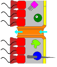
Choanocytes are cells that line the interior of asconoid, syconoid and leuconoid body types of sponges that contain a central flagellum, or cilium, surrounded by a collar of microvilli which are connected by a thin membrane.

<span class="mw-page-title-main">Opisthokont</span> Group of eukaryotes which includes animals and fungi, among other groups

The opisthokonts are a broad group of eukaryotes, including both the animal and fungus kingdoms. The opisthokonts, previously called the "Fungi/Metazoa group", are generally recognized as a clade. Opisthokonts together with Apusomonadida and Breviata comprise the larger clade Obazoa.

<span class="mw-page-title-main">Evolution of sexual reproduction</span> How sexually reproducing multicellular organisms could have evolved from a common ancestor species

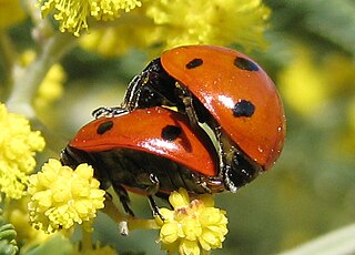
Sexual reproduction is an adaptive feature which is common to almost all multicellular organisms and various unicellular organisms. Currently, the adaptive advantage of sexual reproduction is widely regarded as a major unsolved problem in biology. As discussed below, one prominent theory is that sex evolved as an efficient mechanism for producing variation, and this had the advantage of enabling organisms to adapt to changing environments. Another prominent theory, also discussed below, is that a primary advantage of outcrossing sex is the masking of the expression of deleterious mutations. Additional theories concerning the adaptive advantage of sex are also discussed below. Sex does, however, come with a cost. In reproducing asexually, no time nor energy needs to be expended in choosing a mate and, if the environment has not changed, then there may be little reason for variation, as the organism may already be well-adapted. However, very few environments have not changed over the millions of years that reproduction has existed. Hence it is easy to imagine that being able to adapt to changing environment imparts a benefit. Sex also halves the amount of offspring a given population is able to produce. Sex, however, has evolved as the most prolific means of species branching into the tree of life. Diversification into the phylogenetic tree happens much more rapidly via sexual reproduction than it does by way of asexual reproduction.

<span class="mw-page-title-main">Amorphea</span> Members of the Unikonta, a taxonomic group proposed by Thomas Cavalier-Smith

Amorphea is a taxonomic supergroup that includes the basal Amoebozoa and Obazoa. That latter contains the Opisthokonta, which includes the Fungi, Animals and the Choanomonada, or Choanoflagellates. The taxonomic affinities of the members of this clade were originally described and proposed by Thomas Cavalier-Smith in 2002.

<i>Proterospongia</i> Genus of colony-forming single-celled organisms

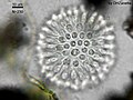
Proterospongia is a genus of single-celled aquatic organisms which form colonies. It belongs to the choanoflagellate class. As a colony-forming choanoflagellate, Proterospongia is of interest to scientists studying the mechanisms of intercellular signaling and adhesion present before animals appeared.

"Candidatus Midichloria" is a candidatus genus of Gram-negative, non-endospore-forming bacteria, with a bacillus shape around 0.45 µm in diameter and 1.2 µm in length. First described in 2004 with the temporary name IricES1, "Candidatus Midichloria" species are symbionts of several species of hard ticks. They live in the cells of the ovary of the females of this tick species. These bacteria have been observed in the mitochondria of the host cells, a trait that has never been described in any other symbiont of animals.

Metacaspases are members of the C14 class of cysteine proteases and thus related to caspases, orthocaspases and paracaspases. The metacaspases are arginine/lysine-specific, in contrast to caspases, which are aspartate-specific.

<span class="mw-page-title-main">Holozoa</span> Clade containing animals and some protists

Holozoa is a clade of organisms that includes animals and their closest single-celled relatives, but excludes fungi and all other organisms. Together they amount to more than 1.5 million species of purely heterotrophic organisms, including around 300 unicellular species. It consists of various subgroups, namely Metazoa and the protists Choanoflagellata, Filasterea, Pluriformea and Ichthyosporea. Along with fungi and some other groups, Holozoa is part of the Opisthokonta, a supergroup of eukaryotes. Choanofila was previously used as the name for a group similar in composition to Holozoa, but its usage is discouraged now because it excludes animals and is therefore paraphyletic.

<i>Guillardia</i> Genus of single-celled organisms

Guillardia is a genus of marine biflagellate cryptomonad algae with a plastid obtained through secondary endosymbiosis of a red alga.

<span class="mw-page-title-main">Filozoa</span> Monophyletic grouping within the Opisthokonta

The Filozoa are a monophyletic grouping within the Opisthokonta. They include animals and their nearest unicellular relatives.

<span class="mw-page-title-main">Eukaryote</span> Domain of life whose cells have nuclei

The eukaryotes constitute the domain of Eukarya or Eukaryota, organisms whose cells have a membrane-bound nucleus. All animals, plants, fungi, and many unicellular organisms are eukaryotes. They constitute a major group of life forms alongside the two groups of prokaryotes: the Bacteria and the Archaea. Eukaryotes represent a small minority of the number of organisms, but given their generally much larger size, their collective global biomass is much larger than that of prokaryotes.

<span class="mw-page-title-main">Precambrian body plans</span> Structure and development of early multicellular organisms

Until the late 1950s, the Precambrian was not believed to have hosted multicellular organisms. However, with radiometric dating techniques, it has been found that fossils initially found in the Ediacara Hills in Southern Australia date back to the late Precambrian. These fossils are body impressions of organisms shaped like disks, fronds and some with ribbon patterns that were most likely tentacles.

<span class="mw-page-title-main">Choanozoa</span> Clade of opisthokont eukaryotes consisting of the choanoflagellates and the animals

Choanozoa is a clade of opisthokont eukaryotes consisting of the choanoflagellates (Choanoflagellatea) and the animals. The sister-group relationship between the choanoflagellates and animals has important implications for the origin of the animals. The clade was identified in 2015 by Graham Budd and Sören Jensen, who used the name Apoikozoa. The 2018 revision of the classification first proposed by the International Society of Protistologists in 2012 recommends the use of the name Choanozoa.

<i>Salpingoeca rosetta</i> Species of eukaryote

Salpingoeca rosetta is a species of Choanoflagellates in the family Salpingoecidae. It is a rare marine eukaryote consisting of a number of cells embedded in a jelly-like matrix. This organism demonstrates a very primitive level of cell differentiation and specialization. This is seen with flagellated cells and their collar structures that move the cell colony through the water.
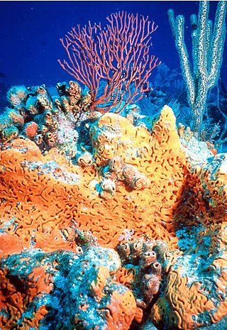
Similar low level cellular differentiation and specification can also be seen in sponges. They also have collar cells and amoeboid cells arranged in a gelatinous matrix.
Unlike S. rosetta, sponges also have other cell-types that can perform different functions. Also, the collar cells of sponges beat within canals in the sponge body, whereas Salpingoeca rosetta's collar cells reside on the inside and it lacks internal canals. Despite these minor differences, there is strong evidence that Proterospongia and Metazoa are highly related.

<span class="mw-page-title-main">Protist shell</span> Protective shell of a type of eukaryotic organism

Many protists have protective shells or tests, usually made from silica (glass) or calcium carbonate (chalk). Protists are a diverse group of eukaryote organisms that are not plants, animals, or fungi. They are typically microscopic unicellular organisms that live in water or moist environments.

Diaphanoeca grandis is a species of choanoflagellate in the family Acanthoecidae which is the type species of the genus Diaphanoeca. It is a unicellular micro-heterotroph with a large protective lorica that is found beneath sea ice in a wide distribution. The lorica is composed of silica and possibly originates from diatoms via Horizontal gene transfer.

<i>Syssomonas</i> Genus of protists

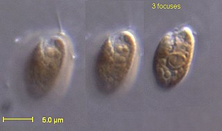
Syssomonas is a monotypic genus of unicellular flagellated protists containing the species Syssomonas multiformis. It is a member of Pluriformea inside the lineage of Holozoa, a clade containing animals and their closest protistan relatives. It lives in freshwater habitats. It has a complex life cycle that includes unicellular amoeboid and flagellated phases, as well as multicellular aggregates, depending on the growth medium and nutritional state.

Crinolina isefiordensis is a species of choanoflagellate in the family Acanthoecidae. It is the type species of the genus Crinolina and is named for the first location of its collection, the Ise Fjord in Denmark.

References

  1. Fonseca, Carolina; Mendonça Filho, João Graciano; Reolid, Matías; Duarte, Luís V.; de Oliveira, António Donizeti; Souza, Jaqueline Torres; Lézin, Carine (2023-01-23). "First putative occurrence in the fossil record of choanoflagellates, the sister group of Metazoa". Scientific Reports. 13 (1): 1242. Bibcode:2023NatSR..13.1242F. doi:10.1038/s41598-022-26972-8. ISSN   2045-2322. PMC   9870899 . PMID   36690681.
  2. Parfrey LW, Lahr DJ, Knoll AH, Katz LA (August 2011). "Estimating the timing of early eukaryotic diversification with multigene molecular clocks". Proceedings of the National Academy of Sciences of the United States of America. 108 (33): 13624–9. Bibcode:2011PNAS..10813624P. doi: 10.1073/pnas.1110633108 . PMC   3158185 . PMID   21810989.
  3. Saville-Kent, W. (1880). A manual of Infusoria. London, vol. 1, p. 324, .
  4. Adl SM, Bass D, Lane CE, Lukeš J, Schoch CL, Smirnov A, Agatha S, Berney C, Brown MW, Burki F, Cárdenas P, Čepička I, Chistyakova L, del Campo J, Dunthorn M, Edvardsen B, Eglit Y, Guillou L, Hampl V, Heiss AA, Hoppenrath M, James TY, Karnkowska A, Karpov S, Kim E, Kolisko M, Kudryavtsev A, Lahr DJG, Lara E, Le Gall L, Lynn DH, Mann DG, Massana R, Mitchell EAD, Morrow C, Park JS, Pawlowski JW, Powell MJ, Richter DJ, Rueckert S, Shadwick L, Shimano S, Spiegel FW, Torruella G, Youssef N, Zlatogursky V, Zhang Q (2019). "Revisions to the Classification, Nomenclature, and Diversity of Eukaryotes". Journal of Eukaryotic Microbiology. 66 (1): 4–119. doi:10.1111/jeu.12691. PMC   6492006 . PMID   30257078.
  5. King N, Westbrook MJ, Young SL, Kuo A, Abedin M, Chapman J, et al. (February 2008). "The genome of the choanoflagellate Monosiga brevicollis and the origin of metazoans". Nature. 451 (7180): 783–8. Bibcode:2008Natur.451..783K. doi:10.1038/nature06617. PMC   2562698 . PMID   18273011.
  6. Nitsche F, Carr M, Arndt H, Leadbeater BS (2011). "Higher level taxonomy and molecular phylogenetics of the Choanoflagellatea". The Journal of Eukaryotic Microbiology. 58 (5): 452–62. doi:10.1111/j.1550-7408.2011.00572.x. PMID   21895836. S2CID   2076733.
  7. Cavalier-Smith T (1998). "Neomonada and the origin of animals and fungi.". In Coombs GH, Vickerman K, Sleigh MA, Warren A (eds.). Evolutionary relationships among protozoa. London: Kluwer. pp. 375–407.
  8. 1 2 Leadbeater BS (2015). The choanoflagellates: evolution, biology, and ecology. University of Birmingham. ISBN   978-0-521-88444-0.
  9. 1 2 "Bacterial protein acts as aphrodisiac for choanoflagellates".
  10. 1 2 3 4 5 6 7 8 9 King N, Westbrook MJ, Young SL, Kuo A, Abedin M, Chapman J, et al. (February 2008). "The genome of the choanoflagellate Monosiga brevicollis and the origin of metazoans". Nature. 451 (7180): 783–8. Bibcode:2008Natur.451..783K. doi:10.1038/nature06617. PMC   2562698 . PMID   18273011.
  11. 1 2 3 4 5 Leadbeater BS, Thomsen H (2000). "Order Choanoflagellida". An Illustrated Guide to the Protozoa, Second Edition. Lawrence: Society of Protozoologists. 451: 14–38.
  12. 1 2 Karpov S, Leadbeater BS (May 1998). "Cytoskeleton structure and composition in choanoflagellates". Journal of Eukaryotic Microbiology. 45 (3): 361–367. doi:10.1111/j.1550-7408.1998.tb04550.x. S2CID   86287656.
  13. 1 2 3 Leadbeater BS, Kelly M (2001). "Evolution of animals choanoflagellates and sponges". Water and Atmosphere Online. 9 (2): 9–11.
  14. 1 2 Leadbeater BS (February 1983). "Life-History and Ultrastructure of a New Marine Species of Proterospongia (Choanoflagellida)". J. Mar. Biol. Assoc. U. K. 63 (1): 135–160. doi:10.1017/S0025315400049857. S2CID   84666673.
  15. 1 2 Mendoza L, Taylor JW, Ajello L (2002). "The class mesomycetozoea: a heterogeneous group of microorganisms at the animal-fungal boundary". Annual Review of Microbiology. 56: 315–44. doi:10.1146/annurev.micro.56.012302.160950. PMID   12142489. S2CID   14764188.
  16. Thomsen H (1982). Planktonic choanoflagellates from Disko Bugt, West Greenland, with a survey of the marine nanoplankton of the area. Meddelelser om Gronland, Bioscience. Vol. 8. pp. 3–63. ISBN   978-87-635-1149-0.
  17. Buck KR, Garrison DL (June 1988). "Distribution and abundance of choanoflagellates (Acanthoecidae) across the ice-edge zone in the Weddell Sea, Antarctica". Mar. Biol. 98 (2): 263–269. doi:10.1007/BF00391204. S2CID   84931348.
  18. Thomsen H, Buck K, Chavez F (1991). "Choanoflagellates of the central California waters: Taxonomy, morphology and species assemblages". Ophelia . 33 (2): 131–164. doi:10.1080/00785326.1991.10429736.
  19. 1 2 3 4 5 Carr M, Leadbeater BS, Hassan R, Nelson M, Baldauf SL (October 2008). "Molecular phylogeny of choanoflagellates, the sister group to Metazoa". Proceedings of the National Academy of Sciences of the United States of America. 105 (43): 16641–6. Bibcode:2008PNAS..10516641C. doi: 10.1073/pnas.0801667105 . PMC   2575473 . PMID   18922774.
  20. "Newly Discovered Microorganisms Band Together, 'Flip Out'". HHMI.org. Retrieved 2019-10-29.
  21. Butterfield NJ (April 1, 1997). "Plankton ecology and the Proterozoic-Phanerozoic transition". Paleobiology . 23 (2): 247–262. Bibcode:1997Pbio...23..247B. doi:10.1017/S009483730001681X. S2CID   140642074.
  22. Wu, Katherine J. (September 24, 2020). "Nothing Eats Viruses, Right? Meet Some Hungry Protists: New genetic evidence builds the case that single-celled marine microbes might chow down on viruses". The New York Times. p. D3 (September 29, 2020 print ed.). Retrieved December 1, 2020.
  23. De Loof, Arnold; Schoofs, Liliane (2019). "Mode of Action of Farnesol, the "Noble Unknown" in Particular in Ca2+ Homeostasis, and its Juvenile Hormone-Esters in Evolutionary Retrospect". Frontiers in Neuroscience. 13: 141. doi: 10.3389/fnins.2019.00141 . PMC   6397838 . PMID   30858798.
  24. Claus Nielsen. Animal Evolution: Interrelationships of the Living Phyla. 3rd ed. Claus Nielsen. Oxford, UK: Oxford University Press, 2012, p. 14.
  25. Carr M, Leadbeater BS, Baldauf SL (2002). "Conserved meiotic genes point to sex in the choanoflagellates". The Journal of Eukaryotic Microbiology. 57 (1): 56–62. doi:10.1111/j.1550-7408.2009.00450.x. PMID   20015185. S2CID   205759832.
  26. Leadbeater BS, Karpov SA (September–October 2000). "Cyst formation in a freshwater strain of the choanoflagellate Desmarella moniliformis Kent". The Journal of Eukaryotic Microbiology. 47 (5): 433–9. doi:10.1111/j.1550-7408.2000.tb00071.x. PMID   11001139. S2CID   23357186.
  27. 1 2 Woznica A, Gerdt JP, Hulett RE, Clardy J, King N (September 2017). "Mating in the Closest Living Relatives of Animals Is Induced by a Bacterial Chondroitinase". Cell. 170 (6): 1175–1183.e11. doi:10.1016/j.cell.2017.08.005. PMC   5599222 . PMID   28867285.
  28. 1 2 Levin TC, King N (November 2013). "Evidence for sex and recombination in the choanoflagellate Salpingoeca rosetta". Current Biology. 23 (21): 2176–80. doi:10.1016/j.cub.2013.08.061. PMC   3909816 . PMID   24139741.
  29. 1 2 3 Carr M, Leadbeater BS, Baldauf SL (2010). "Conserved meiotic genes point to sex in the choanoflagellates". The Journal of Eukaryotic Microbiology. 57 (1): 56–62. doi:10.1111/j.1550-7408.2009.00450.x. PMID   20015185. S2CID   205759832.
  30. Leadbeater BS, Yu Q, Kent J, Stekel DJ (January 2009). "Three-dimensional images of choanoflagellate loricae". Proceedings. Biological Sciences. 276 (1654): 3–11. doi:10.1098/rspb.2008.0844. PMC   2581655 . PMID   18755674.
  31. 1 2 Marron AO, Alston MJ, Heavens D, Akam M, Caccamo M, Holland PW, Walker G (April 2013). "A family of diatom-like silicon transporters in the siliceous loricate choanoflagellates". Proceedings. Biological Sciences. 280 (1756): 20122543. doi:10.1098/rspb.2012.2543. PMC   3574361 . PMID   23407828.
  32. King N, Carroll SB (December 2001). "A receptor tyrosine kinase from choanoflagellates: molecular insights into early animal evolution". Proceedings of the National Academy of Sciences of the United States of America. 98 (26): 15032–7. Bibcode:2001PNAS...9815032K. doi: 10.1073/pnas.261477698 . PMC   64978 . PMID   11752452.
  33. Cantell CE, Franzén Å, Sensenbaugh T (October 1982). "Ultrastructure of multiciliated collar cells in the pilidium larva of Lineus bilineatus (Nemertini)". Zoomorphology . 101 (1): 1–15. doi:10.1007/BF00312027. S2CID   42242685.
  34. Torruella, Guifré; Mendoza, Alex de; Grau-Bové, Xavier; Antó, Meritxell; Chaplin, Mark A.; Campo, Javier del; Eme, Laura; Pérez-Cordón, Gregorio; Whipps, Christopher M. (2015). "Phylogenomics Reveals Convergent Evolution of Lifestyles in Close Relatives of Animals and Fungi". Current Biology. 25 (18): 2404–2410. doi: 10.1016/j.cub.2015.07.053 . PMID   26365255.
  35. Reviers, B. de. (2006). Biologia e Filogenia das Algas . Editora Artmed, Porto Alegre, p. 156.
  36. (Adl, et al., 2005)
  37. Sun G, Yang Z, Ishwar A, Huang J (December 2010). "Algal genes in the closest relatives of animals". Molecular Biology and Evolution. 27 (12): 2879–89. doi: 10.1093/molbev/msq175 . PMID   20627874.
  38. Fairclough SR, Chen Z, Kramer E, Zeng Q, Young S, Robertson HM, Begovic E, Richter DJ, Russ C, Westbrook MJ, Manning G, Lang BF, Haas B, Nusbaum C, King N (February 2013). "Premetazoan genome evolution and the regulation of cell differentiation in the choanoflagellate Salpingoeca rosetta". Genome Biology. 14 (2): R15. doi: 10.1186/gb-2013-14-2-r15 . PMC   4054682 . PMID   23419129.
  39. López-Escardó, D; Grau-Bové, X; Guillaumet-Adkins, A; Gut, M; Sieracki, ME; Ruiz-Trillo, I (25 November 2019). "Reconstruction of protein domain evolution using single-cell amplified genomes of uncultured choanoflagellates sheds light on the origin of animals". Philosophical Transactions of the Royal Society of London. Series B: Biological Sciences. 374 (1786): 20190088. doi: 10.1098/rstb.2019.0088 . PMC   6792448 . PMID   31587642.
  40. Snell EA, Brooke NM, Taylor WR, Casane D, Philippe H, Holland PW (February 2006). "An unusual choanoflagellate protein released by Hedgehog autocatalytic processing". Proceedings. Biological Sciences. 273 (1585): 401–7. doi:10.1098/rspb.2005.3263. PMC   1560198 . PMID   16615205.
  41. Richter, Daniel J; Fozouni, Parinaz; Eisen, Michael B; King, Nicole (31 May 2018). "Gene family innovation, conservation and loss on the animal stem lineage". eLife. 7: e34226. doi: 10.7554/eLife.34226 . PMC   6040629 . PMID   29848444.