Symbiodinium

Last updated

Symbiodinium
Symbiodinium.png
Scientific classification OOjs UI icon edit-ltr.svg
Domain: Eukaryota
Clade: Diaphoretickes
Clade: SAR
Clade: Alveolata
Phylum: Myzozoa
Superclass: Dinoflagellata
Class: Dinophyceae
Order: Suessiales
Family: Symbiodiniaceae
Genus: Symbiodinium
Freudenthal, 1962 [1]
Species

See text

Symbiodinium is a genus of dinoflagellates that encompasses the largest and most prevalent group of endosymbiotic dinoflagellates known. These unicellular microalgae commonly reside in the endoderm of tropical cnidarians such as corals, sea anemones, and jellyfish, where the products of their photosynthetic processing are exchanged in the host for inorganic molecules. They are also harbored by various species of demosponges, flatworms, mollusks such as the giant clams, foraminifera (soritids), and some ciliates. Generally, these dinoflagellates enter the host cell through phagocytosis, persist as intracellular symbionts, reproduce, and disperse to the environment. The exception is in most mollusks, where these symbionts are intercellular (between the cells). Cnidarians that are associated with Symbiodinium occur mostly in warm oligotrophic (nutrient-poor), marine environments where they are often the dominant constituents of benthic communities. These dinoflagellates are therefore among the most abundant eukaryotic microbes found in coral reef ecosystems.

Contents

Symbiodinium are colloquially called zooxanthellae, and animals symbiotic with algae in this genus are said to be "zooxanthellate". The term was loosely used to refer to any golden-brown endosymbionts, including diatoms and other dinoflagellates. Continued use of the term in the scientific literature is discouraged because of the confusion caused by overly generalizing taxonomically diverse symbiotic relationships. [2]

In 2018, the systematics of Symbiodiniaceae was revised, and the distinct clades have been reassigned into seven genera. [3] Following this revision, the name Symbiodinium is now sensu stricto a genus name for only species that were previously classified as Clade A. [3] The other clades were reclassified as distinct genera (see Molecular Systematics below).

Light and confocal images of Symbiodinium cells in hospite (living in a host cell) within scyphistomae of the jellyfish Cassiopea xamachana. This animal requires infection by these algae to complete its life cycle. The chloroplast imaged in 3-D is highly reticulated and distributed around the cell's periphery SymbiodiniumCell Lightandconfocal copy.png
Light and confocal images of Symbiodinium cells in hospite (living in a host cell) within scyphistomae of the jellyfish Cassiopea xamachana . This animal requires infection by these algae to complete its life cycle. The chloroplast imaged in 3-D is highly reticulated and distributed around the cell's periphery

Intracellular symbionts

Symbiodinium reach high cell densities through prolific mitotic division in the endodermal tissues of many shallow tropical and sub-tropical cnidarians. This is a SEM of a freeze-fractured internal mesentery from a reef coral polyp (Porites porites) that shows the distribution and density of symbiont cells. HostTissue section.png
Symbiodinium reach high cell densities through prolific mitotic division in the endodermal tissues of many shallow tropical and sub-tropical cnidarians. This is a SEM of a freeze-fractured internal mesentery from a reef coral polyp ( Porites porites ) that shows the distribution and density of symbiont cells.

Many Symbiodinium species are known primarily for their role as mutualistic endosymbionts. In hosts, they usually occur in high densities, ranging from hundreds of thousands to millions per square centimeter. [4] The successful culturing of swimming gymnodinioid cells from coral led to the discovery that "zooxanthellae" were actually dinoflagellates. [5] [6] Each Symbiodinium cell is coccoid in hospite (living in a host cell) and surrounded by a membrane that originates from the host cell plasmalemma during phagocytosis. This membrane probably undergoes some modification to its protein content, which functions to limit or prevent phago-lysosome fusion. [7] [8] [9] The vacuole structure containing the symbiont is therefore termed the symbiosome. A single symbiont cell occupies each symbiosome. It is unclear how this membrane expands to accommodate a dividing symbiont cell. Under normal conditions, symbiont and host cells exchange organic and inorganic molecules that enable the growth and proliferation of both partners.

Natural services and economic value

Symbiodinium is one of the most studied symbionts. Their mutualistic relationships with reef-building corals form the basis of a highly diverse and productive ecosystem. Coral reefs have economic benefits – valued at hundreds of billions of dollars each year – in the form of ornamental, subsistence and commercial fisheries, tourism and recreation, coastal protection from storms, a source of bioactive compounds for pharmaceutical development, and more. [10]

Coral bleaching

The study of Symbiodinium biology is driven largely by a desire to understand global coral reef decline. A chief mechanism for widespread reef degradation has been stress-induced coral bleaching caused by unusually high seawater temperature. Bleaching is the disassociation of the coral and the symbiont and/or loss of chlorophyll within the alga, resulting in a precipitous loss in the animal's pigmentation. Many Symbiodinium-cnidarian associations are affected by sustained elevation of sea surface temperatures, [11] but may also result from exposure to high irradiance levels (including UVR), [12] [13] extreme low temperatures, [14] low salinity [15] and other factors. [16] The bleached state is associated with decreased host calcification, [17] increased disease susceptibility [18] and, if prolonged, partial or total mortality. [19] The magnitude of mortality from a single bleaching event can be global in scale as it was in 2015. These episodes are predicted to become more common and severe as temperatures worldwide continue to rise. [20] The physiology of a resident Symbiodinium species often regulates the bleaching susceptibility of a coral. [21] [22] Therefore, a significant amount of research has focused on characterizing the physiological basis of thermal tolerance [23] [24] [25] [26] and in identifying the ecology and distribution of thermally tolerant symbiont species. [27] [28] [29]

The symbiosis Symbodinium-coral could provide higher resistance to multiple stress (desiccation, high UVR) to the coral holobiont through its mycosporine-like amino acids (MAAs). The concentration of MAAs increases with stress and ROS in Symbodinium. [30] These UV-absorbing MAAs may also support light-harvesting pigments during photosynthesis, be source of nitrogen storage and for reproduction. More than half of the Symbodinium taxa contain MAAs. [31] [32] [33]

Symbiodinium trenchi is a stress-tolerant species and is able to form mutualistic relationships with many species of coral. It is present in small numbers in coral globally and is common in the Andaman Sea, where the water is about 4 °C (7 °F) warmer than in other parts of the Indian Ocean. [34] [ self-published source? ] In the Caribbean Sea in late 2005, water temperature was elevated for several months and it was found that S. trenchi, a symbiont not normally abundant, took up residence in many corals in which it had not previously been observed. Those corals did not bleach. Two years later, it had largely been replaced as a symbiont by the species normally found in the Caribbean. [28]

S. thermophilum was recently found to make up the bulk of the algal population inside the corals of the Persian Gulf. It is also present in the Gulf of Oman and the Red Sea, at a much lower concentration. Coral that hosted this species was able to tolerate the 35 °C (95 °F) waters of the Persian Gulf, much hotter than the 31 °C (88 °F) of coral reefs globally. [35]

Molecular systematics

Genetic disparity between clades in the legacy genus Symbiodinium sensu lato compared to other dinoflagellates. Analysis of conserved mitochondrial sequences (CO1) and rDNA (SSU) suggest that a taxonomic revision of this group was required. See clades A--F, Polarella, Scrippsiella, Pfiesteria, Peridinium, Lingulodinium, Alexandrium, Karlodinium, Karenia, Gymnodinium, Gyrodinium, Akashiwo, and Prorocentrum. CO1 Phylogeny.png
Genetic disparity between clades in the legacy genus Symbiodinium sensu lato compared to other dinoflagellates. Analysis of conserved mitochondrial sequences (CO1) and rDNA (SSU) suggest that a taxonomic revision of this group was required. See clades A—F, Polarella , Scrippsiella , Pfiesteria , Peridinium , Lingulodinium , Alexandrium , Karlodinium , Karenia , Gymnodinium , Gyrodinium , Aka­shiwo , and Prorocentrum .

The advent of DNA sequence comparison initiated a rebirth in the ordering and naming of all organisms. The application of this methodology helped overturn the long-held belief that (traditional understood) Symbiodinium comprised a single genus, a process which began in earnest with the morphological, physiological, and biochemical comparisons of cultured isolates. Currently, genetic markers are exclusively used to describe ecological patterns and deduce evolutionary relationships among morphologically cryptic members of this group. Foremost in the molecular systematics of Symbiodinium is to resolve ecologically relevant units of diversity (i.e. species).

Phylogenetic disparity among "clades"

The earliest ribosomal gene sequence data indicated that Symbiodinium had lineages whose genetic divergence was similar to that seen in other dinoflagellates from different genera, families, and even orders. [36] This large phylogenetic disparity among clades A, B, C, etc. was confirmed by analyses of the sequences of the mitochondrial gene coding for cytochrome c oxidase subunit I (CO1) among Dinophyceae. [37] Most of these clade groupings comprise numerous reproductively isolated, genetically distinct lineages (see ‘Species diversity’), exhibiting different ecological and biogeographic distributions (see ‘Geographic distributions and patterns of ‘diversity’).

Recently (2018), these distinct clades within the family of Symbiodiniaceae have been reassigned, although not exclusively, into seven genera: Symbiodinium (understood sensu stricto , i. e. clade A), Breviolum (clade B), Cladocopium (clade C), Durusdinium (clade D), Effrenium (clade E), Fugacium (clade F), and Gerakladium (clade G). [3]

Species diversity

The investigation of Symbiodinium diversity, ecology, and evolution is enhanced by analysis of ribosomal and single copy nuclear, plastid, and mitochondrial DNA. The use of multiple markers, along with a hierarchical phylogenetic classification provides the genetic resolution necessary for investigating species diversity, biogeography, dispersal, natural selection, and adaptive radiations. MolecularMarkersSymbiodinium.png
The investigation of Symbiodinium diversity, ecology, and evolution is enhanced by analysis of ribosomal and single copy nuclear, plastid, and mitochondrial DNA. The use of multiple markers, along with a hierarchical phylogenetic classification provides the genetic resolution necessary for investigating species diversity, biogeography, dispersal, natural selection, and adaptive radiations.

The recognition of species diversity in this genus remained problematic for many decades due to the challenges of identifying morphological and biochemical traits useful for diagnosing species. [38] Presently, phylogenetic, ecological, and population genetic data can be more rapidly acquired to resolve Symbiodinium into separate entities that are consistent with Biological, Evolutionary, and Ecological Species Concepts. [39] [40] Most genetics-based measures of diversity have been estimated from the analysis of one genetic marker (e. g. LSU, ITS2, or cp23S [41] ), yet in recent studies these and other markers were analyzed in combination. The high concordance found among nuclear, mitochondrial and chloroplast DNA argues that a hierarchical phylogenetic scheme, combined with ecological and population genetic data, can unambiguously recognize and assign nomenclature to reproductively isolated lineages, i.e. species. [3] [39] [40]

The analysis of additional phylogenetic markers show that some Symbiodinium that were initially identified by slight differences in ITS sequences may comprise members of the same species [40] whereas, in other cases, two or more genetically divergent lineages can possess the same ancestral ITS sequence. [42] [43] When analysed in the context of the major species concepts, [44] the majority of ITS2 sequence data provide a reasonable proxy for species diversity. [39] [40] [45] Currently, ITS2 types number in the hundreds, but most communities of symbiotic cnidaria around the world still require comprehensive sampling. Furthermore, there appears to be a large number of unique species found in association with equally diverse species assemblages of soritid foraminifera, [46] as well as many other Symbiodinium that are exclusively free-living and found in varied, often benthic, habitats. [47] Given the potential species diversity of these ecologically cryptic Symbiodinium, the total species number may never be accurately assessed. [46]

Clone diversity and population genetics

Through the use of microsatellite markers, multilocus genotypes identifying a single clonal line of Symbiodinium can be resolved from samples of host tissue. It appears that most individual colonies harbor a single multilocus genotype (i.e. clone). [48] [49] Extensive sampling within colonies confirms that many colonies harbor a homogeneous (clonal) Symbiodinium population. Additional genotypes do occur in some colonies, yet rarely more than two or three are found. When present in the same colony, multiple clones often exhibit narrow zones of overlap. [49] Colonies adjacent to each other on a reef may harbor identical clones, but across the host population the clone diversity of a particular Symbiodinium species is potentially large and comprises recombinant genotypes that are the product of sexual recombination. A clone tends to remain dominant in a colony over many months and years, but may be occasionally displaced or replaced. The few studies examining clone dispersal find that most genotypes have limited geographic distributions, but that dispersal and gene flow is likely influenced by host life history and mode of symbiont acquisition (e. g. horizontal vs. vertical). [49] [48]

Species diversity, ecology, and biogeography

Geographic distributions and patterns of diversity

Global distribution and preliminary diversity estimate of common Symbiodinium species associated with cnidarians (rare "species" excluded). Internal transcribed spacer region 2 (ITS2) data (sensu LaJeunesse 2002) are used here as a proxy for species diversity. Species Diversity Biogeography.png
Global distribution and preliminary diversity estimate of common Symbiodinium species associated with cnidarians (rare “species” excluded). Internal transcribed spacer region 2 (ITS2) data (sensu LaJeunesse 2002) are used here as a proxy for species diversity.

Symbiodinium are perhaps the best group for studying micro-eukaryote physiology and ecology for several reasons. Firstly, available phylogenetic and population genetic markers allow for detailed examination of their genetic diversity over broad spatial and temporal scales. Also, large quantities of Symbiodinium cells are readily obtained through the collection of hosts that harbor them. Lastly, their association with animals provides an additional axis by which to compare and contrast ecological distributions.[ citation needed ]

The earliest genetic methods for assessing Symbiodinium diversity relied on low-resolution molecular markers that separated the genus into a few evolutionarily divergent lineages, referred to as "clades". Previous characterizations of geographic distribution and dominance have focused on the clade-level of genetic resolution, but more detailed assessments of diversity at the species level are needed. While members of a given clade may be ubiquitous, the species diversity within each clade is potentially large, with each species often having different ecological and geographic distributions related to their dispersal ability, host biogeography, and external environmental conditions. A small number of species occur in temperate environments where few symbiotic animals occur. As a result, these high latitude associations tend to be highly species specific.[ citation needed ]

The distributions of Symbiodinium species comprising different ecological guilds in a coral reef ecosystem. A. Host cnidarians can expel millions of symbiont cells (viable and necrotic) per day into the surrounding environment (a). In turn these animals pass large volumes of water through their gastrovascular system for respiration and cellular waste removal, a process that introduces numerous small particles including food and various other Symbiodinium spp. (indicated by differences in cell color) (b). B. The ecological niche, from the viewpoint of functional groups, differs among species of Symbiodinium. Several ecological guilds exist, including abundant host-specific and host-generalist species (1), low background and potentially opportunistic species (2), and non-symbiotic species closely associated with the coral's biome (3) and/or occupying unrelated habitats (4) Symbiodinium Ecology.png
The distributions of Symbiodinium species comprising different ecological guilds in a coral reef ecosystem. A. Host cnidarians can expel millions of symbiont cells (viable and necrotic) per day into the surrounding environment (a). In turn these animals pass large volumes of water through their gastrovascular system for respiration and cellular waste removal, a process that introduces numerous small particles including food and various other Symbiodinium spp. (indicated by differences in cell color) (b). B. The ecological niche, from the viewpoint of functional groups, differs among species of Symbiodinium. Several ecological guilds exist, including abundant host-specific and host-generalist species (1), low background and potentially opportunistic species (2), and non-symbiotic species closely associated with the coral's biome (3) and/or occupying unrelated habitats (4)

Species diversity assigned to different ecological guilds

The large diversity of Symbiodinium revealed by genetic analyses is distributed non-randomly and appears to comprise several guilds with distinct ecological habits. Of the many Symbiodinium characterized genetically, most are host-specific, mutualistic, and dominate their host. [50] Others may represent compatible symbionts that remain as low-abundance background populations because of competitive inferiority under the prevailing external environmental conditions (e.g. high light vs. low light). [51] Some may also comprise opportunistic species that may proliferate during periods of physiological stress and displace the normal resident symbiont and remain abundant in the host's tissues for months to years before being replaced by the original symbiont. [28] [52] [53] There are also those that rapidly infect and establish populations in host juveniles until being replaced by symbionts that normally associate with host adult colonies. [54] Finally, there appears to be another group of Symbiodinium that are incapable of establishing endosymbiosis yet exist in environments around the animal or associate closely with other substrates (i.e. macro-algal surfaces, sediment surface) [47] [55] Symbiodinium from functional groups 2, 3, and 4 are known to exist because they culture easily, however species with these life histories are difficult to study because of their low abundance in the environment.

Free-living and "non-symbiotic" populations

There are few examples of documented populations of free-living Symbiodinium. [47] Given that most host larvae must initially acquire their symbionts from the environment, viable Symbiodinium cells occur outside the host. The motile phase is probably important in the external environment and facilitates the rapid infection of host larvae. The use of aposymbiotic host polyps deployed as "capture vessels" and the application of molecular techniques has allowed for the detection of environmental sources of Symbiodinium. [53] [56] With these methods employed, investigators may resolve the distribution of different species on various benthic surfaces [55] and cell densities suspended in the water column. [57] The genetic identities of cells cultured from the environment are often dissimilar to those found in hosts. These likely do not form endosymbioses and are entirely free-living; they are different from "dispersing" symbiotic species. [50] Learning more about the "private lives" of these environmental populations and their ecological function will further our knowledge about the diversity, dispersal success, and evolution among members within this large genus.

Culturing

Certain Symbiodinium strains and/or species are more easily cultured and can persist in artificial or supplemented seawater media (e.g. ASP–8A, F/2) [58] for decades. The comparison of cultured isolates under identical conditions show clear differences in morphology, size, biochemistry, gene expression, swimming behavior, growth rates, etc. [59] [60] [61] This pioneering comparative approach initiated a slow paradigm shift in recognizing that the traditional genus sensu lato comprised more than a single real genus.

Culturing is a selective process, and many Symbiodinium isolates growing on artificial media are not typical of the species normally associated with a particular host. Indeed, most host–specific species have yet to be cultured. Samples for genetic analysis should be acquired from the source colony in order to match the resulting culture with the identity of the dominant and ecologically relevant symbiont originally harbored by the animal. [50] [62] [63]

Life cycle

Symbiodinium life cycle Symbiodinium Life Cycle.png
Symbiodinium life cycle

The life cycle of Symbiodinium was first described from cells growing in culture media. For isolates that are in log phase growth, division rates occur every 1–3 days, with Symbiodinium cells alternating between a spherical, or coccoid, morphology and a smaller flagellated motile mastigote stage. While several similar schemes are published that describe how each morphological state transitions to other, the most compelling life history reconstruction was deduced from light and electron microscopy and nuclear staining evidence. [64] During asexual propagation (sometimes referred to as mitotic or vegetative growth), cells undergo a diel cycle of karyokinesis (chromosome/nuclear division) in darkness. The mother cell then divides (cytokinesis) soon after exposure to light and releases two motile cells. The initiation and duration of motility varies among species. [64] Approaching or at the end of the photoperiod the mastigotes cease swimming, release their flagella, and undergo a rapid metamorphosis into the coccoid form. As cultures reach stationary growth phase, fewer and fewer motile cells are observed, indicating slower division rates.

Large tetrads are occasionally observed, particularly when cells in stationary growth phase are transferred to fresh media. However, it is unknown whether this stage is the product of two consecutive mitotic divisions, or perhaps a process that generates sexually competent motile cells (i.e. gametes), or is the end result of meiosis (i. e. as meiotic tetrads) following gamete fusion. There is no cytological evidence for sexual recombination, and meiosis has never been observed, but population genetic evidence supports the view that Symbiodinium periodically undergo events of sexual recombination. How, when, and where the sexual phase in their life history occurs remains unknown. [42] [65] [66]

Morphology

The life stages of dinoflagellates in the genus Symbiodinium. (A) Electron micrographs of a Symbiodinium mastigote (motile cell) with characteristic gymnodinioid morphology (S. natans) and (B) the coccoid cell in hospite. As free-living cells the mastigote allows for short-range dispersal and can exhibit chemotaxis toward sources of nitrogen. Once within the host, these symbionts rapidly proliferate in and in many cases dominate the cytoplasm of the host cell. Duel life stages.png
The life stages of dinoflagellates in the genus Symbiodinium. (A) Electron micrographs of a Symbiodinium mastigote (motile cell) with characteristic gymnodinioid morphology (S. natans) and (B) the coccoid cell in hospite. As free-living cells the mastigote allows for short-range dispersal and can exhibit chemotaxis toward sources of nitrogen. Once within the host, these symbionts rapidly proliferate in and in many cases dominate the cytoplasm of the host cell.
Fine scale features and organelles found among Symbiodinium species. (A) Close-up of divided cells of a tetrad from a culture of S. corculorum that is genetically similar to S. pilosum (both are type/subclade A2). This particular lineage is unusual among described Symbiodinium by possessing mucocysts, and chloroplasts with parallel and peripheral thylakoid arrangements. Flagella are seen in cross section in the space between daughter cells. (B) A thick cellulosic cell wall is observed for many cultured isolates. (C) TEM of a cross-section through a chloroplast lobe at the periphery of the cell with parallel and peripheral thylakoids grouped in sets of three. Cell Wall Mucocysts Chloroplast.png
Fine scale features and organelles found among Symbiodinium species. (A) Close-up of divided cells of a tetrad from a culture of S. corculorum that is genetically similar to S. pilosum (both are type/subclade A2). This particular lineage is unusual among described Symbiodinium by possessing mucocysts, and chloroplasts with parallel and peripheral thylakoid arrangements. Flagella are seen in cross section in the space between daughter cells. (B) A thick cellulosic cell wall is observed for many cultured isolates. (C) TEM of a cross-section through a chloroplast lobe at the periphery of the cell with parallel and peripheral thylakoids grouped in sets of three.
TEM of cross-section through a dividing cell (the doublet). The light micrograph inset depicts a doublet in culture. Flagella are present from the daughter mastigote cells (white arrowheads) prior to emerging from the mother cell wall. n = nucleus; acc = accumulation body; pyr = pyrenoid. (Robert K. Trench et al. 1981) Dividing Cell.png
TEM of cross-section through a dividing cell (the doublet). The light micrograph inset depicts a doublet in culture. Flagella are present from the daughter mastigote cells (white arrowheads) prior to emerging from the mother cell wall. n = nucleus; acc = accumulation body; pyr = pyrenoid. (Robert K. Trench et al. 1981)

The morphological description of the genus Symbiodinium is originally based on the type species (holotype) S. microadriaticum. [38] [67] Because these dinoflagellates possess two major stages in their life history (see above), namely the mastigote (motile) and coccoid (non-motile) stages, the morphology of both is described in order to provide a complete diagnosis of the organism.

Flagellated (mastigote) cell

The motile flagellated form is gymnodinioid and athecate ("nude"). [68] The relative dimensions of the epicone and hypocone differ among species. [38] The alveoli are most visible in the motile phase but lack fibrous cellulosic structures found in thecate ("armored") dinoflagellates. Between the points of origin of the two flagella is an extensible structure of unknown function called the peduncle. In other dinoflagellates, an analogous structure has been implicated in heterotrophic feeding and sexual recombination. In Symbiodinium, it has been suggested that the peduncle may be involved in substrate attachment, explaining why certain cells seem to spin in place. [67] Compared to other gymnodinioid genera, there is little or no displacement at the sulcus where the ends of the cingulum (or cigulum) groove converge.

The internal organelles of the mastigote are essentially the same as described in the coccoid cell (see below). The transition from mastigote to coccoid stage in Symbiodinium occurs rapidly, but details about cellular changes are unknown. Mucocysts (an ejectile organelle [69] ) located beneath the plasmalemma are found in S. pilosum and their function is unknown, but may be involved in heterotrophic feeding.

Coccoid cell

The coccoid cell of Symbiodinium is spherical and ranges in average diameter from 6 to 13 µm, depending on the species (Blank et al. 1989). This stage is often wrongly interpreted as a dinocyst; hence, in published literature, the alga in hospite is often referred to as a vegetative cyst. [67] The term cyst usually refers to a dormant, metabolically quiescent stage in the life history of other dinoflagellates, initiated by several factors, including nutrient availability, temperature, and day length. [70] Such cysts permit extended resistance to unfavourable environmental conditions. Instead, coccoid Symbiodinium cells are metabolically active, as they photosynthesize, undergo mitosis, and actively synthesize proteins and nucleic acids. While most dinoflagellates undergo mitosis as a mastigote, in Symbiodinium, mitosis occurs exclusively in the coccoid cell. [64]

Cell wall

The coccoid cell is surrounded by a cellulosic, usually smooth cell wall that contains large-molecular-weight proteins and glycoproteins. [38] [71] Cell walls grow thicker in culture than in hospite. [7] The cell membrane (plasmalemma) is located beneath the cell wall, yet little is known about its composition and function in terms of the regulation of trans-membrane transport of metabolites. During karyokinesis and cytokinesis, the cell wall remains intact until the mastigotes escape the mother cell. In culture, the discarded walls accumulate at the bottom of the culture vessel. It is not known what becomes of the walls from divided cells in hospite. [72] One species, S. pilosum, possesses tufts of hair-like projections from the cell wall; this is the only known surface characteristic used to diagnose a species in the genus.

Chloroplast

Most described species possess a single, peripheral, reticulated chloroplast bounded by three membranes. The volume of the cell occupied by the chloroplast varies among species. [38] The lamellae comprise three closely appressed (stacked) thylakoids, and are attached by two stalks to the pyrenoid [38] surrounded by a starch sheath. In three of the described species, the thylakoids are in parallel arrays, but in S. pilosum, there are also peripheral lamellae. There are no thylakoid membranes invading the pyrenoid, which is unlike other symbiotic dinoflagellates. [73] [74] The lipid components of thylakoids include the galactolipids as monogalactosyl-diglycerides (MGDG) [75] and digalactosyl-diglycerides( DGDG),; [76] the sulpholipid sulphoquinovosyl-diglyceride (SQDG), [77] phosphatidyl glycerol, [78] and phosphatidyl choline. Associated with these are various fatty acids. [79] The light-harvesting and reaction centre components in the thylakoid membrane include a water-soluble peridinin-chlorophyll a-protein complex (PCP or PerCP), [80] and a membrane-bound chlorophyll a-chlorophyll c2-peridinin-protein-complex (acpPC), [81] along with typical photosynthetic electron transport systems such as the photosystem II reaction centre and the chlorophyll-a-P700 reaction centre complex of photosystem I. [82] [83] Also associated with the thylakoids are the xanthophylls dinoxanthin, diadinoxanthin, diatoxanthin and the carotene, β-Carotene. The pyrenoid contains the nuclear-encoded enzyme type II Ribulose-1,5-bis-phosphate-carboxylase-oxygenase (RuBisCO), [84] which is responsible for the catalysis of inorganic carbon dioxide (CO2) into organic compounds.

All cultured isolates (i.e. strains) are capable of phenotypic adjustment in their capacity for light harvesting (i.e. photoacclimation), such as by altering the cellular Chl. a and peridinin quota, as well as the size and number of photosynthetic units. [85] However, the ability to acclimate is a reflection of genetic differences among species that are differently adapted (evolved) to a particular photic environment. [86] [87] For example, S. pilosum is characterized as a high light-adapted species, while others are low light-adapted (S. kawagutii) or adapted to a larger range in varying light fields (S. microadriaticum).

The reticulated chloroplast structures of (A) Cladocopium goreaui (formerly S. goreaui type C1), (B) Symbiodinium fitti (type A3), and (C) Effrenium voratum (formerly S. californium type E1) imaged in 3-D using confocal microscopy of chlorophyll autofluorescence. Chloroplast 3.png
The reticulated chloroplast structures of (A) Cladocopium goreaui (formerly S. goreaui type C1), (B) Symbiodinium fitti (type A3), and (C) Effrenium voratum (formerly S. californium type E1) imaged in 3-D using confocal microscopy of chlorophyll autofluorescence.

Nucleus

In general, the nucleus is centrally located and the nucleolus is often associated with the inner nuclear membrane. The chromosomes, as in other dinoflagellates, are seen as ‘permanently super-coiled’ DNA in transmission electron micrographs (TEM). [88] The described species of Symbiodinium possess distinct chromosome numbers (ranging from 26 to 97 [38] ), which remain constant throughout all phases of the nuclear cycle. However, during M-phase, the volume of each chromosome is halved, as is the volume of each of the two resulting nuclei. Thus, the ratio of chromosome volume to nuclear volume remains constant. These observations are consistent with the interpretation that the algae are haploid, a conclusion supported by molecular genetic data. [89] During S-phase of the nuclear cycle the chromosomes do uncoil to facilitate DNA synthesis, and the volumes of both the chromosomes and the nucleus return to those seen in the G22 phase. [88]

Other cytoplasmic organelles

There are several additional organelles found in the cytoplasm of Symbiodinium. The most obvious of these is the structure referred to as the "accumulation body". This is a membrane-bound vesicle (vacuole) with contents that are unrecognizable, but appear red or yellow under the light microscope. It may serve to accumulate cellular debris or act as an autophagic vacuole in which non-functional organelles are digested and their components recycled. During mitosis, only one daughter cell appears to acquire this structure. There are other vacuoles that may contain membranous inclusions, [90] while still others contain crystalline material variously interpreted as oxalate crystals or crystalline uric acid.

Species

Symbiodinium kawagutii Symbiodinium kawagutii.jpg
Symbiodinium kawagutii

The following species are recognised by the World Register of Marine Species: [1]

Related Research Articles

<span class="mw-page-title-main">Endosymbiont</span> Organism that lives within the body or cells of another organism

An endosymbiont or endobiont is any organism that lives within the body or cells of another organism most often, though not always, in a mutualistic relationship. (The term endosymbiosis is from the Greek: ἔνδον endon "within", σύν syn "together" and βίωσις biosis "living".) Examples are nitrogen-fixing bacteria, which live in the root nodules of legumes, single-cell algae inside reef-building corals and bacterial endosymbionts that provide essential nutrients to insects.

<span class="mw-page-title-main">Coral</span> Marine invertebrates of the class Anthozoa

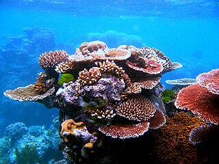
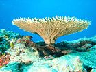
Corals are colonial marine invertebrates within the class Anthozoa of the phylum Cnidaria. They typically form compact colonies of many identical individual polyps. Coral species include the important reef builders that inhabit tropical oceans and secrete calcium carbonate to form a hard skeleton.

<span class="mw-page-title-main">Dinoflagellate</span> Unicellular algae with two flagella

The dinoflagellates are a monophyletic group of single-celled eukaryotes constituting the phylum Dinoflagellata and are usually considered protists. Dinoflagellates are mostly marine plankton, but they also are common in freshwater habitats. Their populations vary with sea surface temperature, salinity, and depth. Many dinoflagellates are photosynthetic, but a large fraction of these are in fact mixotrophic, combining photosynthesis with ingestion of prey.

<span class="mw-page-title-main">Coral bleaching</span> Phenomenon where coral expel algae tissue

Coral bleaching is the process when corals become white due to various stressors, such as changes in temperature, light, or nutrients. Bleaching occurs when coral polyps expel the zooxanthellae that live inside their tissue, causing the coral to turn white. The zooxanthellae are photosynthetic, and as the water temperature rises, they begin to produce reactive oxygen species. This is toxic to the coral, so the coral expels the zooxanthellae. Since the zooxanthellae produce the majority of coral colouration, the coral tissue becomes transparent, revealing the coral skeleton made of calcium carbonate. Most bleached corals appear bright white, but some are blue, yellow, or pink due to pigment proteins in the coral.

<span class="mw-page-title-main">Zooxanthellae</span> Dinoflagellates in symbiosis with coral, jellyfish and nudibranchs

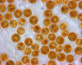
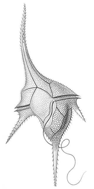
Zooxanthellae is a colloquial term for single-celled dinoflagellates that are able to live in symbiosis with diverse marine invertebrates including demosponges, corals, jellyfish, and nudibranchs. Most known zooxanthellae are in the genus Symbiodinium, but some are known from the genus Amphidinium, and other taxa, as yet unidentified, may have similar endosymbiont affinities. The true Zooxanthella K.brandt is a mutualist of the radiolarian Collozoum inerme and systematically placed in Peridiniales. Another group of unicellular eukaryotes that partake in similar endosymbiotic relationships in both marine and freshwater habitats are green algae zoochlorellae.

Symbiotic bacteria are bacteria living in symbiosis with another organism or each other. For example, rhizobia living in root nodules of legumes provide nitrogen fixing activity for these plants.

<i>Aiptasia</i> Genus of sea anemones

Aiptasia is a genus of a symbiotic cnidarian belonging to the class Anthozoa. Aiptasia is a widely distributed genus of temperate and tropical sea anemones of benthic lifestyle typically found living on mangrove roots and hard substrates. These anemones, as well as many other cnidarian species, often contain symbiotic dinoflagellate unicellular algae of the genus Symbiodinium living inside nutritive cells. The symbionts provide food mainly in the form of lipids and sugars produced from photosynthesis to the host while the hosts provides inorganic nutrients and a constant and protective environment to the algae. Species of Aiptasia are relatively weedy anemones able to withstand a relatively wide range of salinities and other water quality conditions. In the case of A. pallida and A. pulchella, their hardiness coupled with their ability to reproduce very quickly and out-compete other species in culture gives these anemones the status of pest from the perspective of coral reef aquarium hobbyists. These very characteristics make them easy to grow in the laboratory and thus they are extensively used as model organisms for scientific study. In this respect, Aiptasia have contributed a significant amount of knowledge regarding cnidarian biology, especially human understanding of cnidarian-algal symbioses, a biological phenomenon crucial to the survival of corals and coral reef ecosystems. The dependence of coral reefs on the health of the symbiosis is dramatically illustrated by the devastating effects experienced by corals due to the loss of algal symbionts in response to environmental stress, a phenomenon known as coral bleaching.

Cyanobionts are cyanobacteria that live in symbiosis with a wide range of organisms such as terrestrial or aquatic plants; as well as, algal and fungal species. They can reside within extracellular or intracellular structures of the host. In order for a cyanobacterium to successfully form a symbiotic relationship, it must be able to exchange signals with the host, overcome defense mounted by the host, be capable of hormogonia formation, chemotaxis, heterocyst formation, as well as possess adequate resilience to reside in host tissue which may present extreme conditions, such as low oxygen levels, and/or acidic mucilage. The most well-known plant-associated cyanobionts belong to the genus Nostoc. With the ability to differentiate into several cell types that have various functions, members of the genus Nostoc have the morphological plasticity, flexibility and adaptability to adjust to a wide range of environmental conditions, contributing to its high capacity to form symbiotic relationships with other organisms. Several cyanobionts involved with fungi and marine organisms also belong to the genera Richelia, Calothrix, Synechocystis, Aphanocapsa and Anabaena, as well as the species Oscillatoria spongeliae. Although there are many documented symbioses between cyanobacteria and marine organisms, little is known about the nature of many of these symbioses. The possibility of discovering more novel symbiotic relationships is apparent from preliminary microscopic observations.

Chromera velia, also known as a "chromerid", is a unicellular photosynthetic organism in the superphylum Alveolata. It is of interest in the study of apicomplexan parasites, specifically their evolution and accordingly, their unique vulnerabilities to drugs.

The resilience of coral reefs is the biological ability of coral reefs to recover from natural and anthropogenic disturbances such as storms and bleaching episodes. Resilience refers to the ability of biological or social systems to overcome pressures and stresses by maintaining key functions through resisting or adapting to change. Reef resistance measures how well coral reefs tolerate changes in ocean chemistry, sea level, and sea surface temperature. Reef resistance and resilience are important factors in coral reef recovery from the effects of ocean acidification. Natural reef resilience can be used as a recovery model for coral reefs and an opportunity for management in marine protected areas (MPAs).

The hologenome theory of evolution recasts the individual animal or plant as a community or a "holobiont" – the host plus all of its symbiotic microbes. Consequently, the collective genomes of the holobiont form a "hologenome". Holobionts and hologenomes are structural entities that replace misnomers in the context of host-microbiota symbioses such as superorganism, organ, and metagenome. Variation in the hologenome may encode phenotypic plasticity of the holobiont and can be subject to evolutionary changes caused by selection and drift, if portions of the hologenome are transmitted between generations with reasonable fidelity. One of the important outcomes of recasting the individual as a holobiont subject to evolutionary forces is that genetic variation in the hologenome can be brought about by changes in the host genome and also by changes in the microbiome, including new acquisitions of microbes, horizontal gene transfers, and changes in microbial abundance within hosts. Although there is a rich literature on binary host–microbe symbioses, the hologenome concept distinguishes itself by including the vast symbiotic complexity inherent in many multicellular hosts. For recent literature on holobionts and hologenomes published in an open access platform, see the following reference.

<i>Pocillopora damicornis</i> Species of coral

Pocillopora damicornis, commonly known as the cauliflower coral or lace coral, is a species of stony coral in the family Pocilloporidae. It is native to tropical and subtropical parts of the Indian and Pacific Oceans.

<i>Orbicella faveolata</i> Species of coral

Orbicella faveolata, commonly known as mountainous star coral, is a colonial stony coral in the family Merulinidae. Orbicella faveolata is native to the coral coast of the Caribbean Sea and the Gulf of Mexico and is listed as "endangered" by the International Union for Conservation of Nature. O. faveolata was formerly known as Montastraea faveolata.

Durusdinium trenchii is an endosymbiotic dinoflagellate, a unicellular alga which commonly resides in the tissues of tropical corals. It has a greater tolerance to fluctuations in water temperatures than do other species in the genus. It was named for the marine biologist R. K. Trench.

<span class="mw-page-title-main">Marine microbial symbiosis</span>

Microbial symbiosis in marine animals was not discovered until 1981. In the time following, symbiotic relationships between marine invertebrates and chemoautotrophic bacteria have been found in a variety of ecosystems, ranging from shallow coastal waters to deep-sea hydrothermal vents. Symbiosis is a way for marine organisms to find creative ways to survive in a very dynamic environment. They are different in relation to how dependent the organisms are on each other or how they are associated. It is also considered a selective force behind evolution in some scientific aspects. The symbiotic relationships of organisms has the ability to change behavior, morphology and metabolic pathways. With increased recognition and research, new terminology also arises, such as holobiont, which the relationship between a host and its symbionts as one grouping. Many scientists will look at the hologenome, which is the combined genetic information of the host and its symbionts. These terms are more commonly used to describe microbial symbionts.

<span class="mw-page-title-main">Marine microbiome</span>

All animals on Earth form associations with microorganisms, including protists, bacteria, archaea, fungi, and viruses. In the ocean, animal–microbial relationships were historically explored in single host–symbiont systems. However, new explorations into the diversity of marine microorganisms associating with diverse marine animal hosts is moving the field into studies that address interactions between the animal host and a more multi-member microbiome. The potential for microbiomes to influence the health, physiology, behavior, and ecology of marine animals could alter current understandings of how marine animals adapt to change, and especially the growing climate-related and anthropogenic-induced changes already impacting the ocean environment.

Robert Kent Trench was an American Biologist who was a professor at the University of California, Santa Barbara. His research considered corals and symbiotic algae, with a focus on the adaption of zooxanthellae. He was awarded the 1994 International Society of Endocytobiology Miescher-Ishida Prize.

<span class="mw-page-title-main">Mary Alice Coffroth</span> American marine biologist

Mary Alice Coffroth is an American marine biologist who is a professor at the State University of New York at Buffalo. She is known for her use of molecular tools to examined coral larval ecology, recruitment and cnidarian-dinoflagellate symbiosis.

<span class="mw-page-title-main">Symbiodiniaceae</span> Family of dinoflagellates (algae)

Symbiodiniaceae is a family of marine dinoflagellates notable for their symbiotic associations with reef-building corals, sea anemones, jellyfish, marine sponges, giant clams, acoel flatworms, and other marine invertebrates. Symbiotic Symbiodiniaceae are sometimes colloquially referred to as Zooxanthellae, though the latter term can be interpreted to include other families of symbiotic algae as well. While many Symbiodiniaceae species are endosymbionts, others are free living in the water column or sediment.

Durusdinium is a genus of dinoflagellate algae within the family Symbiodiniaceae. Durusdinium can be free living, or can form symbiotic associations with hard corals. Members of the genus have been documented in reef-building corals of the Indian and Pacific oceans, as well as the Caribbean. Prior to 2018, Durusdinium were classified as Symbiodinium Clade D.

References

  1. 1 2 Guiry, Michael D. (2014). "Symbiodinium Freudenthal, 1962". WoRMS. World Register of Marine Species . Retrieved 2015-01-29.
  2. Blank, Rudolf J.; Trench, Robert K. (May 1986). "Nomenclature of Endosymbiotic Dinoflagellates". Taxon. 35 (2): 286–94. doi:10.2307/1221270. JSTOR   1221270.
  3. 1 2 3 4 5 LaJeunesse, Todd C.; Parkinson, John E.; Gabrielson, Paul W.; Jeong, Hae Jin; Reimer, James D.; Voolstra, Christian R.; Santos, Scott R. (2018). "Systematic revision of Symbiodiniaceae highlights the antiquity and diversity of coral endosymbionts". Current Biology. 28 (16): P2570–2580. doi: 10.1016/j.cub.2018.07.008 . PMID   30100341.
  4. Stimson, J.; Sakai, K.; Sembali, H. (December 2002). "Interspecific comparison of the symbiotic relationship in corals with high and low rates of bleaching-induced mortality". Coral Reefs. 21 (4): 409–21. doi:10.1007/s00338-002-0264-3. S2CID   43084578.
  5. Kawaguti, Siro (1944). "On the physiology of reef corals VI. Study on the pigments". Palau Tropical Biological Station Studies. 2: 617–74.
  6. McLaughlin, John J. A.; Zahl, Paul A. (1959). "Axenic Zooxanthellae from Various Invertebrate Hosts". Annals of the New York Academy of Sciences. 77 (2): 55–72. Bibcode:1959NYASA..77...55M. doi:10.1111/j.1749-6632.1959.tb36892.x. S2CID   85877213.
  7. 1 2 Colley, Nansi J.; Trench, R. K. (1983). "Selectivity in Phagocytosis and Persistence of Symbiotic Algae by the Scyphistoma Stage of the Jellyfish Cassiopeia xamachana". Proceedings of the Royal Society of London. Series B. 219 (1214): 61–82. Bibcode:1983RSPSB.219...61C. doi:10.1098/rspb.1983.0059. JSTOR   35678. PMID   22470960. S2CID   22673808.
  8. Wakefield, Timothy S.; Kempf, Stephen C. (2001). "Development of host- and symbiont-specific monoclonal antibodies and confirmation of the origin of the symbiosome membrane in a cnidarian-dinoflagellate symbiosis". The Biological Bulletin. 200 (2): 127–43. doi:10.2307/1543306. JSTOR   1543306. PMID   11341574. S2CID   21833840.
  9. Peng, Shao-En; Wang, Yu-Bao; Wang, Li-Hsueh; Chen, Wan-Nan Uang; Lu, Chi-Yu; Fang, Lee-Shing; Chen, Chii-Shiarng (2010). "Proteomic analysis of symbiosome membranes in Cnidaria-dinoflagellate endosymbiosis". Proteomics. 10 (5): 1002–16. doi:10.1002/pmic.200900595. PMID   20049864. S2CID   27108503.
  10. Moberg, Fredrik; Folke, Carl (1999). "Ecological goods and services of coral reef ecosystems". Ecological Economics. 29 (2): 215–33. doi:10.1016/S0921-8009(99)00009-9.
  11. Jokiel, P. L.; Coles, S. L. (1990). "Response of Hawaiian and other Indo-Pacific reef corals to elevated temperature". Coral Reefs. 8 (4): 155–62. Bibcode:1990CorRe...8..155J. doi:10.1007/BF00265006. S2CID   5871917.
  12. Lesser, Michael P (1996). "Elevated temperatures and ultraviolet radiation cause oxidative stress and inhibit photosynthesis in symbiotic dinoflagellates". Limnology and Oceanography. 41 (2): 271–83. Bibcode:1996LimOc..41..271L. doi: 10.4319/lo.1996.41.2.0271 . S2CID   16428743.
  13. Fitt, William; Brown, Barbara; Warner, Mark; Dunne, Richard (2001). "Coral bleaching: Interpretation of thermal tolerance limits and thermal thresholds in tropical corals". Coral Reefs. 20 (1): 51–65. doi:10.1007/s003380100146. S2CID   19361052.
  14. Lajeunesse, Todd C.; Smith, Robin; Walther, Mariana; Pinzon, Jorge; Pettay, Daniel T.; McGinley, Michael; Aschaffenburg, Matthew; Medina-Rosas, Pedro; Cupul-Magana, Amilcar L.; Pérez, Andrés López; Reyes-Bonilla, Hector; Warner, Mark E. (2010). "Host-symbiont recombination versus natural selection in the response of coral-dinoflagellate symbioses to environmental disturbance". Proceedings of the Royal Society B: Biological Sciences. 277 (1696): 2925–34. doi:10.1098/rspb.2010.0385. JSTOR   27862400. PMC   2982020 . PMID   20444713.
  15. Goreau, Thomas F (1964). "Mass Expulsion of Zooxanthellae from Jamaican Reef Communities after Hurricane Flora". Science. 145 (3630): 383–6. Bibcode:1964Sci...145..383G. doi:10.1126/science.145.3630.383. PMID   17816975. S2CID   46191566.
  16. Brown, Barbara E. (2000). "The significance of pollution in eliciting the 'bleaching' response in symbiotic cnidarians". International Journal of Environment and Pollution. 13 (1–6): 392–415. doi:10.1504/IJEP.2000.002328.
  17. Colombo-Pallotta et al. 2010
  18. Brandt, Marilyn E.; McManus, John W. (2009). "Disease incidence is related to bleaching extent in reef-building corals". Ecology. 90 (10): 2859–67. doi:10.1890/08-0445.1. JSTOR   25592820. PMID   19886494. S2CID   3896954.
  19. Baker, Andrew C.; Glynn, Peter W.; Riegl, Bernhard (2008). "Climate change and coral reef bleaching: An ecological assessment of long-term impacts, recovery trends and future outlook". Estuarine. 80 (4): 435–71. Bibcode:2008ECSS...80..435B. doi:10.1016/j.ecss.2008.09.003.
  20. Hoegh-Guldberg, O.; Mumby, P. J.; Hooten, A. J.; Steneck, R. S.; Greenfield, P.; Gomez, E.; Harvell, C. D.; Sale, P. F.; Edwards, A. J.; Caldeira, K.; Knowlton, N.; Eakin, C. M.; Iglesias-Prieto, R.; Muthiga, N.; Bradbury, R. H.; Dubi, A.; Hatziolos, M. E. (2007). "Coral Reefs Under Rapid Climate Change and Ocean Acidification". Science. 318 (5857): 1737–42. Bibcode:2007Sci...318.1737H. CiteSeerX   10.1.1.702.1733 . doi:10.1126/science.1152509. PMID   18079392. S2CID   12607336.
  21. Berkelmans, R.; Van Oppen, M. J.H (2006). "The role of zooxanthellae in the thermal tolerance of corals: A 'nugget of hope' for coral reefs in an era of climate change". Proceedings of the Royal Society B: Biological Sciences. 273 (1599): 2305–12. doi:10.1098/rspb.2006.3567. PMC   1636081 . PMID   16928632.
  22. Sampayo, E. M.; Ridgway, T.; Bongaerts, P.; Hoegh-Guldberg, O. (2008). "Bleaching susceptibility and mortality of corals are determined by fine-scale differences in symbiont type". Proceedings of the National Academy of Sciences. 105 (30): 10444–9. Bibcode:2008PNAS..10510444S. doi: 10.1073/pnas.0708049105 . JSTOR   25463173. PMC   2492480 . PMID   18645181.
  23. Robison, Jennifer D.; Warner, Mark E. (2006). "Differential Impacts of Photoacclimation and Thermal Stress on the Photobiology of Four Different Phylotypes of Symbiodinium (Pyrrhophyta)". Journal of Phycology. 42 (3): 568–79. doi:10.1111/j.1529-8817.2006.00232.x. S2CID   84675726.
  24. Warner, Mark E.; Lajeunesse, Todd C.; Robison, Jennifer D.; Thur, Rebecca M. (2006). "The ecological distribution and comparative photobiology of symbiotic dinoflagellates from reef corals in Belize: Potential implications for coral bleaching". Limnology and Oceanography. 51 (4): 1887–97. Bibcode:2006LimOc..51.1887W. CiteSeerX   10.1.1.322.1206 . doi:10.4319/lo.2006.51.4.1887. S2CID   1240348.
  25. Ragni, Maria; Airs, Ruth L.; Hennige, Sebastian J.; Suggett, David J.; Warner, Mark E.; Geider, Richard J. (2010). "PSII photoinhibition and photorepair in Symbiodinium (Pyrrhophyta) differs between thermally tolerant and sensitive phylotypes". Marine Ecology Progress Series. 406: 57–70. Bibcode:2010MEPS..406...57R. doi: 10.3354/meps08571 .
  26. Takahashi, Shunichi; Whitney, Spencer; Itoh, Shigeru; Maruyama, Tadashi; Badger, Murray (2008). "Heat stress causes inhibition of the de novo synthesis of antenna proteins and photobleaching in cultured Symbiodinium". Proceedings of the National Academy of Sciences. 105 (11): 4203–8. Bibcode:2008PNAS..105.4203T. doi: 10.1073/pnas.0708554105 . JSTOR   25461395. PMC   2393757 . PMID   18322010.
  27. Lien, Yi-T.; Nakano, Y.; Plathong, S.; Fukami, H.; Wang, Jih-T.; Chen, C. A. (2007). "Occurrence of the putatively heat-tolerant Symbiodinium phylotype D in high-latitudinal outlying coral communities". Coral Reefs. 26 (1): 35–44. Bibcode:2007CorRe..26...35L. doi:10.1007/s00338-006-0185-7. S2CID   19100056.
  28. 1 2 3 Lajeunesse, Todd C.; Smith, Robin T.; Finney, Jennifer; Oxenford, Hazel (2009). "Outbreak and persistence of opportunistic symbiotic dinoflagellates during the 2005 Caribbean mass coral 'bleaching' event". Proceedings of the Royal Society B: Biological Sciences. 276 (1676): 4139–48. doi:10.1098/rspb.2009.1405. JSTOR   40506039. PMC   2821356 . PMID   19740874.
  29. Lajeunesse, Todd C.; Pettay, Daniel T.; Sampayo, Eugenia M.; Phongsuwan, Niphon; Brown, Barbara; Obura, David O.; Hoegh-Guldberg, Ove; Fitt, William K. (2010). "Long-standing environmental conditions, geographic isolation and host-symbiont specificity influence the relative ecological dominance and genetic diversification of coral endosymbionts in the genus Symbiodinium". Journal of Biogeography. 37 (5): 785–800. doi:10.1111/j.1365-2699.2010.02273.x. S2CID   30508571.
  30. Rosic, Nedeljka N. (November 2019). "Mycosporine-Like Amino Acids: Making the Foundation for Organic Personalised Sunscreens". Marine Drugs. 17 (11): 638. doi: 10.3390/md17110638 . PMC   6891770 . PMID   31726795.
  31. Shoguchi, Eiichi; Beedessee, Girish; Hisata, Kanako; Tada, Ipputa; Narisoko, Haruhi; Satoh, Noriyuki; Kawachi, Masanobu; Shinzato, Chuya (2021-02-03). Phadke, Sujal (ed.). "A New Dinoflagellate Genome Illuminates a Conserved Gene Cluster Involved in Sunscreen Biosynthesis". Genome Biology and Evolution. 13 (2): evaa235. doi:10.1093/gbe/evaa235. ISSN   1759-6653. PMC   7875005 . PMID   33146374.
  32. Shoguchi, Eiichi; Yoshioka, Yuki; Shinzato, Chuya; Arimoto, Asuka; Bhattacharya, Debashish; Satoh, Noriyuki (2020-03-01). McFadden, Geoff (ed.). "Correlation between Organelle Genetic Variation and RNA Editing in Dinoflagellates Associated with the Coral Acropora digitifera". Genome Biology and Evolution. 12 (3): 203–209. doi:10.1093/gbe/evaa042. ISSN   1759-6653. PMC   7144361 . PMID   32108224.
  33. Shoguchi, Eiichi; Beedessee, Girish; Tada, Ipputa; Hisata, Kanako; Kawashima, Takeshi; Takeuchi, Takeshi; Arakaki, Nana; Fujie, Manabu; Koyanagi, Ryo; Roy, Michael C.; Kawachi, Masanobu (2018-06-14). "Two divergent Symbiodinium genomes reveal conservation of a gene cluster for sunscreen biosynthesis and recently lost genes". BMC Genomics. 19 (1): 458. doi: 10.1186/s12864-018-4857-9 . ISSN   1471-2164. PMC   6001144 . PMID   29898658.
  34. David DeFranza (2010-02-17). "Andaman Sea Coral May Hold the Secret to Warm Water Reef Survival". Treehugger. Retrieved 2015-02-02.
  35. "A hot survivor". The Economist. April 9, 2016. ISSN   0013-0613 . Retrieved 2016-04-30.
  36. Rowan, Rob; Powers, Dennis A. (1992). "Ribosomal RNA Sequences and the Diversity of Symbiotic Dinoflagellates (Zooxanthellae)". Proceedings of the National Academy of Sciences of the United States of America. 89 (8): 3639–43. Bibcode:1992PNAS...89.3639R. doi: 10.1073/pnas.89.8.3639 . JSTOR   2359156. PMC   48924 . PMID   1565660.
  37. Stern, Rowena F.; Horak, Ales; Andrew, Rose L.; Coffroth, Mary-Alice; Andersen, Robert A.; Küpper, Frithjof C.; Jameson, Ian; Hoppenrath, Mona; Véron, Benoît; Kasai, Fumai; Brand, Jerry; James, Erick R.; Keeling, Patrick J. (2010). "Environmental Barcoding Reveals Massive Dinoflagellate Diversity in Marine Environments". PLOS ONE. 5 (11): e13991. Bibcode:2010PLoSO...513991S. doi: 10.1371/journal.pone.0013991 . PMC   2981561 . PMID   21085582.
  38. 1 2 3 4 5 6 7 Trench, Robert K.; Blank, Rudolf J. (1987). "Symbiodinium Microadriaticum Freudenthal, S. Goreauii Sp. Nov., S. Kawagutii Sp. Nov. And S. Pilosum Sp. Nov.: Gymnodinioid Dinoflagellate Symbionts of Marine Invertebrates". Journal of Phycology. 23 (3): 469–81. doi:10.1111/j.1529-8817.1987.tb02534.x. S2CID   83712799.
  39. 1 2 3 Sampayo, E. M.; Dove, S.; Lajeunesse, T. C. (2009). "Cohesive molecular genetic data delineate species diversity in the dinoflagellate genus Symbiodinium". Molecular Ecology. 18 (3): 500–19. doi:10.1111/j.1365-294X.2008.04037.x. PMID   19161470. S2CID   24285565.
  40. 1 2 3 4 Lajeunesse, Todd C.; Thornhill, Daniel J. (2011). "Improved Resolution of Reef-Coral Endosymbiont (Symbiodinium) Species Diversity, Ecology, and Evolution through psbA Non-Coding Region Genotyping". PLOS ONE. 6 (12): e29013. Bibcode:2011PLoSO...629013L. doi: 10.1371/journal.pone.0029013 . PMC   3247227 . PMID   22216157.
  41. Christian Renicke: Symbiodinium cp23S RFLP and sequence analysis, Pringle Lab, Grossman Lab, Jul 23, 2018
  42. 1 2 Santos, S. R.; Shearer, T. L.; Hannes, A. R.; Coffroth, M. A. (2004). "Fine-scale diversity and specificity in the most prevalent lineage of symbiotic dinoflagellates (Symbiodinium, Dinophyceae) of the Caribbean". Molecular Ecology. 13 (2): 459–69. doi:10.1046/j.1365-294X.2003.02058.x. PMID   14717900. S2CID   32227215.
  43. Finney, J. Christine; Pettay, Daniel Tye; Sampayo, Eugenia M.; Warner, Mark E.; Oxenford, Hazel A.; Lajeunesse, Todd C. (2010). "The Relative Significance of Host–Habitat, Depth, and Geography on the Ecology, Endemism, and Speciation of Coral Endosymbionts in the Genus Symbiodinium". Microbial Ecology. 60 (1): 250–63. doi:10.1007/s00248-010-9681-y. JSTOR   40802290. PMID   20502891. S2CID   5890376.
  44. De Queiroz, Kevin (2007). "Species Concepts and Species Delimitation". Systematic Biology. 56 (6): 879–86. doi: 10.1080/10635150701701083 . PMID   18027281.
  45. Thornhill, Daniel J.; Lajeunesse, Todd C.; Santos, Scott R. (2007). "Measuring rDNA diversity in eukaryotic microbial systems: How intragenomic variation, pseudogenes, and PCR artifacts confound biodiversity estimates". Molecular Ecology. 16 (24): 5326–40. doi:10.1111/j.1365-294X.2007.03576.x. PMID   17995924. S2CID   12791319.
  46. 1 2 Pochon, X.; Garcia-Cuetos, L.; Baker, A. C.; Castella, E.; Pawlowski, J. (2007). "One-year survey of a single Micronesian reef reveals extraordinarily rich diversity of Symbiodinium types in soritid foraminifera" (PDF). Coral Reefs. 26 (4): 867–82. Bibcode:2007CorRe..26..867P. doi:10.1007/s00338-007-0279-x. S2CID   24642825.
  47. 1 2 3 Reimer, James Davis; Shah, Md Mahfuzur Rahman; Sinniger, Frederic; Yanagi, Kensuke; Suda, Shoichiro (2010). "Preliminary analyses of cultured Symbiodinium isolated from sand in the oceanic Ogasawara Islands, Japan". Marine Biodiversity. 40 (4): 237–47. doi:10.1007/s12526-010-0044-1. S2CID   10186027.
  48. 1 2 Andras, Jason P.; Kirk, Nathan L.; Drew Harvell, C. (2011). "Range-wide population genetic structure of Symbiodinium associated with the Caribbean Sea fan coral, Gorgonia ventalina". Molecular Ecology. 20 (12): 2525–42. doi:10.1111/j.1365-294X.2011.05115.x. PMID   21545573. S2CID   25577087.
  49. 1 2 3 Pettay, Daniel T.; Wham, Drew C.; Pinzón, Jorge H.; Lajeunesse, Todd C. (2011). "Genotypic diversity and spatial-temporal distribution of Symbiodinium clones in an abundant reef coral". Molecular Ecology. 20 (24): 5197–212. doi:10.1111/j.1365-294X.2011.05357.x. PMC   5957298 . PMID   22082053.
  50. 1 2 3 LaJeunesse, T. (2002). "Diversity and community structure of symbiotic dinoflagellates from Caribbean coral reefs". Marine Biology. 141 (2): 387–400. doi:10.1007/s00227-002-0829-2. S2CID   84357806.
  51. Rowan, Rob; Knowlton, Nancy; Baker, Andrew; Jara, Javier (1997). "Landscape ecology of algal symbionts creates variation in episodes of coral bleaching". Nature. 388 (6639): 265–9. Bibcode:1997Natur.388..265R. doi: 10.1038/40843 . PMID   9230434.
  52. Toller, W. W.; Rowan, R; Knowlton, N (2001). "Repopulation of Zooxanthellae in the Caribbean corals Montastraea annularis and M. Faveolata following experimental and disease-associated bleaching". The Biological Bulletin. 201 (3): 360–73. doi:10.2307/1543614. JSTOR   1543614. PMID   11751248. S2CID   7765487.
  53. 1 2 Thornhill, Daniel J.; LaJeunesse, Todd C.; Kemp, Dustin W.; Fitt, William K.; Schmidt, Gregory W. (2005). "Multi-year, seasonal genotypic surveys of coral-algal symbioses reveal prevalent stability or post-bleaching reversion". Marine Biology. 148 (4): 711–22. doi:10.1007/s00227-005-0114-2. S2CID   36226084.
  54. Coffroth, Mary Alice; Santos, Scott R.; Goulet, Tamar L. (2001). "Early ontogenetic expression of specificity in a cnidarian-algal symbiosis" (PDF). Marine Ecology Progress Series. 222: 85–96. Bibcode:2001MEPS..222...85C. doi: 10.3354/meps222085 .
  55. 1 2 Porto, Isabel; Granados, Camila; Restrepo, Juan C.; Sánchez, Juan A. (2008). "Macroalgal-Associated Dinoflagellates Belonging to the Genus Symbiodinium in Caribbean Reefs". PLOS ONE. 3 (5): e2160. Bibcode:2008PLoSO...3.2160P. doi: 10.1371/journal.pone.0002160 . PMC   2364641 . PMID   18478069.
  56. Coffroth, Mary Alice; Lewis, Cynthia F.; Santos, Scott R.; Weaver, Jessica L. (2006). "Environmental populations of symbiotic dinoflagellates in the genus Symbiodinium can initiate symbioses with reef cnidarians". Current Biology. 16 (23): R985–7. doi: 10.1016/j.cub.2006.10.049 . PMID   17141602.
  57. Manning, Mackenzie M.; Gates, Ruth D. (2008). "Diversity in populations of free-living Symbiodinium from a Caribbean and Pacific reef". Limnology and Oceanography. 53 (5): 1853–61. Bibcode:2008LimOc..53.1853M. doi: 10.4319/lo.2008.53.5.1853 . S2CID   17895332.
  58. Culturing, Georgia State University, International Genetically Engineered Machine (iGEM)
  59. Schoenberg, D. A.; Trench, R. K. (1980). "Genetic Variation in Symbiodinium (=Gymnodinium) microadriaticum Freudenthal, and Specificity in its Symbiosis with Marine Invertebrates. I. Isoenzyme and Soluble Protein Patterns of Axenic Cultures of Symbiodinium microadriaticum". Proceedings of the Royal Society of London. Series B. 207 (1169): 405–27. Bibcode:1980RSPSB.207..405S. doi:10.1098/rspb.1980.0031. JSTOR   35362. S2CID   84190993.
  60. Schoenberg, D. A.; Trench, R. K. (1980). "Genetic Variation in Symbiodinium (=Gymnodinium) microadriaticum Freudenthal, and Specificity in its Symbiosis with Marine Invertebrates. II. Morphological Variation in Symbiodinium microadriaticum". Proceedings of the Royal Society B: Biological Sciences. 207 (1169): 429–44. Bibcode:1980RSPSB.207..429S. doi:10.1098/rspb.1980.0032. JSTOR   35363. S2CID   84395179.
  61. Schoenberg, D. A.; Trench, R. K. (1980). "Genetic Variation in Symbiodinium (=Gymnodinium) microadriaticum Freudenthal, and Specificity in its Symbiosis with Marine Invertebrates. III. Specificity and Infectivity of Symbiodinium microadriaticum". Proceedings of the Royal Society of London. Series B. 207 (1169): 445–60. Bibcode:1980RSPSB.207..445S. doi:10.1098/rspb.1980.0033. JSTOR   35364. S2CID   83896405.
  62. Santos, Scott R.; Taylor, Derek J.; Coffroth, Mary Alice (2001). "Genetic Comparisons of Freshly Isolated Versus Cultured Symbiotic Dinoflagellates: Implications for Extrapolating to the Intact Symbiosis". Journal of Phycology. 37 (5): 900–12. doi:10.1046/j.1529-8817.2001.00194.x. S2CID   2335127.
  63. Goulet, T.; Coffroth, M. (February 2003). "Genetic composition of zooxanthellae between and within colonies of the octocoral Plexaura kuna, based on small subunit rDNA and multilocus DNA fingerprinting". Marine Biology. 142 (2): 233–9. doi:10.1007/s00227-002-0936-0. S2CID   85919438.
  64. 1 2 3 Fitt, W. K.; Trench, R. K. (1983). "The Relation of Diel Patterns of Cell Division to Diel Patterns of Motility in the Symbiotic Dinoflagellate Symbiodinium Microadria Ticum Freudenthal in Culture". New Phytologist. 94 (3): 421–32. doi: 10.1111/j.1469-8137.1983.tb03456.x . JSTOR   2432757.
  65. Baillie, B. K.; Belda-Baillie, C. A.; Silvestre, V.; Sison, M.; Gomez, A. V.; Gomez, E. D.; Monje, V. (2000). "Genetic variation in Symbiodinium isolates from giant clams based on random-amplified-polymorphic DNA (RAPD) patterns". Marine Biology. 136 (5): 829–36. doi:10.1007/s002270000290. S2CID   84351451.
  66. Lajeunesse, Todd C. (2001). "Investigating the Biodiversity, Ecology, and Phylogeny of Endosymbiotic Dinoflagellates in the Genus Symbiodinium Using the Its Region: In Search of A 'species' Level Marker". Journal of Phycology. 37 (5): 866–80. doi:10.1046/j.1529-8817.2001.01031.x. S2CID   4208350.
  67. 1 2 3 Freudenthal, Hugo D. (1962). "Symbiodinium gen. Nov. And Symbiodinium microadriaticum sp. nov., a Zooxanthella: Taxonomy, Life Cycle, and Morphology". The Journal of Protozoology. 9 (1): 45–52. doi:10.1111/j.1550-7408.1962.tb02579.x.
  68. Taylor, FJR (1987). "Dinoflagellate morphology". In Taylor, F. J. R. (ed.). The Biology of Dinoflagellates. Botanical Monographs, vol. 21. Oxford: Blackwell Scientific Publications. pp.  24–91. ISBN   978-0-632-00915-2.
  69. Dodge, JD; Greuet, C (1987). "Dinoflagellate ultrastructure and complex organelles". In Taylor, F. J. R. (ed.). The Biology of Dinoflagellates. Botanical Monographs, vol. 21. Oxford: Blackwell Scientific Publications. pp.  92–142. ISBN   978-0-632-00915-2.
  70. Lee, Edward Lee (2008). Phycology (4th ed.). New York: Cambridge University Press. ISBN   978-1-139-46987-6.[ page needed ]
  71. Markell, DA; Trench, RK; Iglesias-Prieto, R (1992). "Macromolecules associated with the cell-walls of symbiotic dinoflagellates". Symbiosis. 12 (1): 19–31. INIST   5092729.
  72. Wakefield, Timothy S.; Farmer, Mark A.; Kempf, Stephen C. (August 2000). "Revised description of the fine structure of in situ 'zooxanthellae' genus Symbiodinium". The Biological Bulletin. 199 (1): 76–84. doi:10.2307/1542709. JSTOR   1542709. PMID   10975645.
  73. Trench, RK; Winsor, H (1987). "Symbiosis with dinoflagellates in two pelagic flatworms, Amphiscolops sp. and Haplodiscus sp". Symbiosis. 3 (1): 1–21. INIST   8265704.
  74. Banaszak, Anastazia T.; Iglestas-Prieto, Roberto; Trench, Robert K. (1993). "Scrippsiella velellae sp. nov. (Peridiniales) and Gloeokinium viscum sp. nov. (Phytodiniales), Dinoflagellate Symbionts of Two Hydrozoans (Cnidiaria)". Journal of Phycology. 29 (4): 517–28. doi:10.1111/j.1529-8817.1993.tb00153.x. S2CID   84895398.
  75. Monogalactosyl Diglyceride, biomol
  76. Digalactosyl diglyceride, Merck Sigma-Aldrich
  77. Sulfoquinovosyl diglyceride, Karel's Nutrition Blog
  78. Phosphatidylglycerol and Related Lipids, The LipidWeb
  79. Díaz-Almeyda, E.; Thomé, P. E.; El Hafidi, M.; Iglesias-Prieto, R. (2011). "Differential stability of photosynthetic membranes and fatty acid composition at elevated temperature in Symbiodinium". Coral Reefs. 30 (1): 217–25. Bibcode:2011CorRe..30..217D. doi:10.1007/s00338-010-0691-5. S2CID   20410939.
  80. Donatella Carbonera, Marilena Di Valentin, Riccardo Spezia, Alberto Mezzetti: The Unique Photophysical Properties of the Peridinin-Chlorophyll-a-Protein. In: Current Protein & Peptide Science, volume 15, issue 4, Jun 2014, doi:10.2174/1389203715666140327111139, PMC   4030626, PMID 24678668, PMC4030626 (Web Archive July 2021, 10th)
  81. Dariusz M. Niedzwiedzki, Jing Jiang, Cynthia S. Lo, Robert E. Blankenship: Spectroscopic properties of the Chlorophyll a–Chlorophyll c2–Peridinin-Protein-Complex (acpPC) from the coral symbiotic dinoflagellate Symbiodinium. In: Photosynthesis Research, volume 120, pp. 125–139, (May 2014)/ January 2013, 20th, doi:10.1007/s11120-013-9794-5
  82. Iglesias-Prieto, R.; Govind, N. S.; Trench, R. K. (1991). "Apoprotein Composition and Spectroscopic Characterization of the Water-Soluble Peridinin–Chlorophyll a–Proteins from Three Symbiotic Dinoflagellates". Proceedings: Biological Sciences. 246 (1317): 275–83. Bibcode:1991RSPSB.246..275I. doi:10.1098/rspb.1991.0155. JSTOR   76745. S2CID   84255943.
  83. Iglesias-Prieto, R.; Govind, N. S.; Trench, R. K. (1993). "Isolation and Characterization of Three Membrane-Bound Chlorophyll-Protein Complexes from Four Dinoflagellate Species". Philosophical Transactions of the Royal Society B: Biological Sciences. 340 (1294): 381–92. doi:10.1098/rstb.1993.0080. JSTOR   3030171.
  84. Rowan, Rob; Whitney, Spencer M.; Fowler, Amanda; Yellowlees, David (1996). "Rubisco in marine symbiotic dinoflagellates: Form II enzymes in eukaryotic oxygenic phototrophs encoded by a nuclear multigene family". The Plant Cell Online. 8 (3): 539–53. doi:10.1105/tpc.8.3.539. JSTOR   3870331. PMC   161119 . PMID   8721755.
  85. Hennige, S. J.; Suggett, D. J.; Warner, M. E.; McDougall, K. E.; Smith, D. J. (2009). "Photobiology of Symbiodinium revisited: Bio-physical and bio-optical signatures". Coral Reefs. 28 (1): 179–95. Bibcode:2009CorRe..28..179H. doi:10.1007/s00338-008-0444-x. S2CID   34202181.
  86. Iglesias-Prieto, Roberto; Trench, Robert K. (1994). "Acclimation and adaptation to irradiance in symbiotic dinoflagellates. I. Responses of the photosynthetic unit to changes in photon flux density". Marine Ecology Progress Series. 113 (1): 163–75. Bibcode:1994MEPS..113..163I. doi: 10.3354/meps113163 .
  87. Iglesias-Prieto, R.; Trench, R. K. (1997). "Acclimation and adaptation to irradiance in symbiotic dinoflagellates. II. Response of chlorophyll-protein complexes to different photon-flux densities". Marine Biology. 130 (1): 23–33. doi:10.1007/s002270050221. S2CID   84705656.
  88. 1 2 Blank, Rudolf J.; Trench, Robert K. (1985). "Speciation and Symbiotic Dinoflagellates". Science. 229 (4714): 656–8. Bibcode:1985Sci...229..656B. doi:10.1126/science.229.4714.656. PMID   17739379. S2CID   45228641.
  89. Santos, Scott R.; Coffroth, Mary Alice (February 2003). "Molecular genetic evidence that dinoflagellates belonging to the genus Symbiodinium freudenthal are haploid". The Biological Bulletin. 204 (1): 10–20. doi:10.2307/1543491. JSTOR   1543491. PMID   12588740. S2CID   7258554.
  90. Trench, R. K. (1974). "Nutritional potentials in Zoanthus sociathus (Coelenterata, Anthozoa)". Helgoländer Wissenschaftliche Meeresuntersuchungen. 26 (2): 174–216. Bibcode:1974HWM....26..174T. doi: 10.1007/BF01611382 .
  91. Drew C. Wham, Gang Ning, and Todd C. LaJeunesse (2017) [Symbiodinium glynnii sp. nov., a species of stress-tolerant symbiotic dinoflagellates from pocilloporid and montiporid corals in the Pacific Ocean]. Phycologia: 2017, Vol. 56, No. 4, pp. 396-409.
  92. Algal species helps corals survive in Earth's hottest reefs