
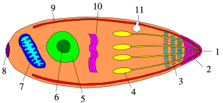
The Apicomplexa are organisms of a large phylum of mainly parasitic alveolates. Most possess a unique form of organelle structure that comprises a type of (non-photosynthetic) plastid called an apicoplast—with an apical complex membrane. The organelle's apical shape is an adaptation that the apicomplexan applies in penetrating a host cell.

Zygomycota, or zygote fungi, is a former division or phylum of the kingdom Fungi. The members are now part of two phyla: the Mucoromycota and Zoopagomycota. Approximately 1060 species are known. They are mostly terrestrial in habitat, living in soil or on decaying plant or animal material. Some are parasites of plants, insects, and small animals, while others form symbiotic relationships with plants. Zygomycete hyphae may be coenocytic, forming septa only where gametes are formed or to wall off dead hyphae. Zygomycota is no longer recognised as it was not believed to be truly monophyletic.

Plasmodium falciparum is a unicellular protozoan parasite of humans, and the deadliest species of Plasmodium that causes malaria in humans. The parasite is transmitted through the bite of a female Anopheles mosquito and causes the disease's most dangerous form, falciparum malaria. It is responsible for around 50% of all malaria cases. P. falciparum is therefore regarded as the deadliest parasite in humans. It is also associated with the development of blood cancer and is classified as a Group 2A (probable) carcinogen.

Coccidia (Coccidiasina) are a subclass of microscopic, spore-forming, single-celled obligate intracellular parasites belonging to the apicomplexan class Conoidasida. As obligate intracellular parasites, they must live and reproduce within an animal cell. Coccidian parasites infect the intestinal tracts of animals, and are the largest group of apicomplexan protozoa.

Plasmodium vivax is a protozoal parasite and a human pathogen. This parasite is the most frequent and widely distributed cause of recurring malaria. Although it is less virulent than Plasmodium falciparum, the deadliest of the five human malaria parasites, P. vivax malaria infections can lead to severe disease and death, often due to splenomegaly. P. vivax is carried by the female Anopheles mosquito; the males do not bite.

Eimeria tenella is a species of Eimeria that causes hemorrhagic cecal coccidiosis in young poultry. It is found worldwide.

Leucocytozoon is a genus of parasitic alveolates belonging to the phylum Apicomplexa.
Megaloschizonts are large schizonts that produce extremely high numbers of merozoites. They are found in various species of the Phylum Apicomplexa. The Apicomplexa phylum contains several parasitic protozoans. They have a very complex life cycle that includes several stages. Megaloschizonts and the smaller schizonts are the part of the life cycle that takes place inside the infected host organism and operates as an asexually reproductive cell. Megaloschizonts appear as grey-white nodules found in the smooth muscle of major organs, such as the heart, liver, lung or spleen.

Conoidasida is a class of parasitic alveolates in the phylum Apicomplexa. The class was defined in 1988 by Levine and contains two subclasses – the coccidia and the gregarines. All members of this class have a complete, hollow, truncated conoid. Gregarines tend to parasitize invertebrates with the mature gamonts being extracellular; the coccidia mostly infect vertebrates and have intracellular gamonts.
Plasmodium fieldi is a parasite of the genus Plasmodium sub genus Plasmodium found in Malaysia. This species is related to Plasmodium ovale and Plasmodium simiovale. As in all Plasmodium species, P. fieldi has both vertebrate and insect hosts. The vertebrate hosts for this parasite are primates.
Karyolysus is a genus of coccidia. With the exception of K. sonomae whose vertebrate host is the yellow-legged frog, species in this genus only infect lizards of the genus Lacerta.
Apicomplexans, a group of intracellular parasites, have life cycle stages that allow them to survive the wide variety of environments they are exposed to during their complex life cycle. Each stage in the life cycle of an apicomplexan organism is typified by a cellular variety with a distinct morphology and biochemistry.
Acroeimeria is a genus of parasites that contains those species which initially develop immediately beneath the brush-border of the intestinal epithelium, but the meronts and gamonts of which are early on extruded to form a layer on the surface of the gut mucosa. Morphologically they are similar to the Eimeria to which they are closely related. The genus was described in 1989 by Paperna and Landsberg.
Polychromophilus is a genus of obligate intracellular eukaryotic parasites that infect bats from every continent except Antarctica. They are transmitted by bat flies, which act as an insect vector as well as the parasite’s site of sporogeny. Polychromophilus follows a fairly typical Haemospororidian lifecycle, with gametocytes and gametes restricted to the bloodstream of the host and meronts infecting organs – most notably the lungs and the liver. The type species is Polychromophilus melanipherus, and was described by Dionisi in 1898.
The genus Schellackia comprises obligate unicellular eukaryotic parasites within the phylum Apicomplexa, and infects numerous species of lizards and amphibians worldwide. Schellackia is transmitted via insect vectors, primarily mites and mosquitoes, which take up the parasite in blood meals. These vectors then subsequently infect reptilian and amphibian which consume the infected insects. The parasites deform erythrocytes of the host into crescents, and can be visualized using a blood smear.
Chagasella is a genus of parasitic alveolates of the phylum Apicomplexa. Species in this genus infect insects of the order Hemiptera and of the family Termitoidae.
Ovivora is a genus in the phylum Apicomplexa.

Cystoisospora belli, previously known as Isospora belli, is a parasite that causes an intestinal disease known as cystoisosporiasis. This protozoan parasite is opportunistic in immune suppressed human hosts. It primarily exists in the epithelial cells of the small intestine, and develops in the cell cytoplasm. The distribution of this coccidian parasite is cosmopolitan, but is mainly found in tropical and subtropical areas of the world such as the Caribbean, Central and S. America, India, Africa, and S.E. Asia. In the U.S., it is usually associated with HIV infection and institutional living.
Schizocystidae is a genus of parasitic alveolates in the phylum Apicomplexa.
Coccidinium is a genus of parasitic syndinian dinoflagellates that infect the nucleus and cytoplasm of other marine dinoflagellates. Coccidinium, along with two other dinoflagellate genera, Amoebophyra and Duboscquella, contain species that are the primary endoparasites of marine dinoflagellates. While numerous studies have been conducted on the genus Amoebophyra, specifically Amoebophyra ceratii, little is known about Coccidinium. These microscopic organisms have gone relatively unstudied after the initial observations of Édouard Chatton and Berthe Biecheler in 1934 and 1936.