| Balantidiasis | |
|---|---|
 | |
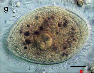
| Balantidium coli as seen in a wet mount of a stool specimen. The organism is surrounded by cilia. | |
| Specialty | Infectious disease |
Balantidiasis is a protozoan infection caused by infection with Balantidium coli . [1]
| Balantidiasis | |
|---|---|
| | |
| Balantidium coli as seen in a wet mount of a stool specimen. The organism is surrounded by cilia. | |
| Specialty | Infectious disease |
Balantidiasis is a protozoan infection caused by infection with Balantidium coli . [1]
Usually asymptomatic in immunocompetent individuals, but the symptoms of balantidiasis include:[ citation needed ]
The most common ones are intermittent diarrhea and constipation or inflammation of the colon combined with abdominal cramps and bloody stools.[ citation needed ]
Balantidium is the only ciliated protozoan known to infect humans. Balantidiasis is a zoonotic disease and is acquired by humans via the feco-oral route from the normal host, the pig, where it is asymptomatic. Fecally contaminated food and water are the common sources of infection in humans. [2]
Balantidium coli exists in either of two developmental stages: trophozoites and cysts. [3] In the trophozoite form, they can be oblong or spherical, and are typically 30 to 150 μm in length and 25 to 120 μm in width. [4] It is its size at this stage that allows Balantidium coli to be characterized as the largest protozoan parasite of humans. [3] Trophozoites possess both a macronucleus and a micronucleus, and both are usually visible. [3] The macronucleus is large and sausage-shaped while the micronucleus is less prominent. [4] At this stage, the organism is not infective but it can replicate by transverse binary fission. [3]
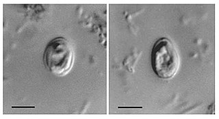
In its cyst stage, the parasite takes on a smaller, more spherical shape, with a diameter of around 40 to 60 μm. [4] Unlike the trophozoite, whose surface is covered only with cilia, the cyst form has a tough wall made of one or more layers. The cyst form also differs from the trophozoite form in being non-motile and does not undergo reproduction. The parasite must be ingested as a cyst in order to cause infection. [3]
The diagnosis of balantidiasis can be an intricate process, partly because the related symptoms may or may not all be present at once. However, the diagnosis of balantidiasis can be considered when a patient has diarrhea combined with a probable history of current exposure to pigs (as pigs are the primary reservoir), contact with infected persons, or anal sexual contact. [5] The diagnosis of balantidiasis can be made by microscopic examination of stools in search of trophozoites or cysts, [6] or colonoscopy or sigmoidoscopy to obtain a biopsy specimen from the large intestine, which may provide evidence for the presence of trophozoites.[ citation needed ]
Preventative measures require effective personal and community hygiene. Some specific safeguards include the following:[ citation needed ]
Balantidiasis can be treated with tetracycline, metronidazole or iodoquinol. [7]

The first study to generate balantidiasis in humans was undertaken by Cassagrandi and Barnagallo in 1896. [8] However, this experiment was not successful in creating an infection and it was unclear whether Balantidium coli was the actual parasite used. [8] The first case of balantidiasis in the Philippines, where it is the most common, was reported in 1904. [9] [4] Currently, Balantidium coli is distributed worldwide but less than 1% of the human population is infected. [10] [4] Pigs are a major reservoir of the parasite, and infection of humans occurs more frequently in areas where pigs commingle with people. [10] This includes places like the Philippines, as previously mentioned, but also includes countries such as Bolivia and Papua New Guinea. [10] [11] Pigs are not the sole species capable of hosting B. coli. For example, the parasite also has a high rate of occurrence in rats. [12] In a Japanese study that analyzed the fecal samples in 56 mammalian species, Balantidium coli was found to be present not just in all the wild boars tested (with wild boars and pigs being considered the same species), it was also found in five species of non human primate: Chimpanzee (Pan troglodytes), White-handed gibbon (Hylobates lar), Squirrel monkey (Saimiri sciurea), Sacred baboon (Comopithecus hamadryas), and Japanese macaque (Macaca fuscata). [13] In other studies, Balantidium coli has also been found in species from the order Carnivora. [13]

Trichinosis, also known as trichinellosis, is a parasitic disease caused by roundworms of the Trichinella type. During the initial infection, invasion of the intestines can result in diarrhea, abdominal pain, and vomiting. Migration of larvae to muscle, which occurs about a week after being infected, can cause swelling of the face, inflammation of the whites of the eyes, fever, muscle pains, and a rash. Minor infection may be without symptoms. Complications may include inflammation of heart muscle, central nervous system involvement, and inflammation of the lungs.
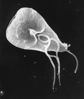
Giardia duodenalis, also known as Giardia intestinalis and Giardia lamblia, is a flagellated parasitic protozoan microorganism of the genus Giardia that colonizes the small intestine, causing a diarrheal condition known as giardiasis. The parasite attaches to the intestinal epithelium by an adhesive disc or sucker, and reproduces via binary fission. Giardiasis does not spread to other parts of the gastrointestinal tract, but remains confined to the lumen of the small intestine. The microorganism has an outer membrane that makes it possible to survive even when outside of its host, and which can render it tolerant to certain disinfectants. Giardia trophozoites are anaerobic, and absorb their nutrients from the intestinal lumen. If the organism is stained, its characteristic pattern resembles the familiar "smiley face" symbol.

Entamoeba histolytica is an anaerobic parasitic amoebozoan, part of the genus Entamoeba. Predominantly infecting humans and other primates causing amoebiasis, E. histolytica is estimated to infect about 35-50 million people worldwide. E. histolytica infection is estimated to kill more than 55,000 people each year. Previously, it was thought that 10% of the world population was infected, but these figures predate the recognition that at least 90% of these infections were due to a second species, E. dispar. Mammals such as dogs and cats can become infected transiently, but are not thought to contribute significantly to transmission.
Giardiasis is a parasitic disease caused by Giardia duodenalis. Infected individuals who experience symptoms may have diarrhoea, abdominal pain, and weight loss. Less common symptoms include vomiting and blood in the stool. Symptoms usually begin one to three weeks after exposure and, without treatment, may last two to six weeks or longer.
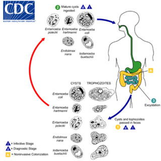
Entamoeba coli is a non-pathogenic species of Entamoeba that frequently exists as a commensal parasite in the human gastrointestinal tract. E. coli is important in medicine because it can be confused during microscopic examination of stained stool specimens with the pathogenic Entamoeba histolytica. This amoeba does not move much by the use of its pseudopod, and creates a "sur place (non-progressive) movement" inside the large intestine. Usually, the amoeba is immobile, and keeps its round shape. This amoeba, in its trophozoite stage, is only visible in fresh, unfixed stool specimens. Sometimes the Entamoeba coli have parasites as well. One is the fungus Sphaerita spp. This fungus lives in the cytoplasm of the E. coli. While this differentiation is typically done by visual examination of the parasitic cysts via light microscopy, new methods using molecular biology techniques have been developed. The scientific name of the amoeba, E. coli, is often mistaken for the bacterium, Escherichia coli. Unlike the bacterium, the amoeba is mostly harmless, and does not cause as many intestinal problems as some strains of the E. coli bacterium. To make the naming of these organisms less confusing, "alternate contractions" are used to name the species for the purpose making the naming easier; for example, using Esch. coli and Ent. coli for the bacterium and amoeba, instead of using E. coli for both.
Balantidium coli is a parasitic species of ciliate alveolates that causes the disease balantidiasis. It is the only member of the ciliate phylum known to be pathogenic to humans.
A trophozoite is the activated, feeding stage in the life cycle of certain protozoa such as malaria-causing Plasmodium falciparum and those of the Giardia group. The complementary form of the trophozoite state is the thick-walled cyst form. They are often different from the cyst stage, which is a protective, dormant form of the protozoa. Trophozoites are often found in the host's body fluids and tissues and in many cases, they are the form of the protozoan that causes disease in the host. In the protozoan, Entamoeba histolytica it invades the intestinal mucosa of its host, causing dysentery, which aid in the trophozoites traveling to the liver and leading to the production of hepatic abscesses.
Histomonas meleagridis is a species of parasitic protozoan that infects a wide range of birds including chickens, turkeys, peafowl, quail and pheasants, causing infectious enterohepatitis, or histomoniasis. H. meleagridis can infect many birds, but it is most deadly in turkeys. It inhabits the lumen of cecum and parenchyma of liver, where it causes extensive necrosis. It is transmitted by another cecal parasite, the nematode Heterakis gallinarum.
Cryptosporidium, sometimes called crypto, is an apicomplexan genus of alveolates which are parasites that can cause a respiratory and gastrointestinal illness (cryptosporidiosis) that primarily involves watery diarrhea, sometimes with a persistent cough.

Retortamonas is a genus of flagellated excavates. It is one of only two genera belonging to the family Retortamonadidae along with the genus Chilomastix. The genus parasitizes a large range of hosts including humans. Species within this genus are considered harmless commensals which reside in the intestine of their host. The wide host diversity is a useful factor given that species are distinguished based on their host rather than morphology. This is because all species share similar morphology, which would present challenges when trying to make classifications based on structural anatomy. Although Retortamonas currently includes over 25 known species, it is possible that some defined species are synonymous, given that such overlapping species have been discovered in the past. Further efforts into learning about this genus must be done such as cross-transmission testing as well as biochemical and genetic studies. One of the most well-known species within this genus is Retortamonas intestinalis, a human parasite that lives in the large intestine of humans.

Taeniasis is an infection within the intestines by adult tapeworms belonging to the genus Taenia. There are generally no or only mild symptoms. Symptoms may occasionally include weight loss or abdominal pain. Segments of tapeworm may be seen in the stool. Complications of pork tapeworm may include cysticercosis.

Babesia, also called Nuttallia, is an apicomplexan parasite that infects red blood cells and is transmitted by ticks. Originally discovered by the Romanian bacteriologist Victor Babeș in 1888, over 100 species of Babesia have since been identified.

Blastocystis is a genus of single-celled parasites belonging to the Stramenopiles that includes algae, diatoms, and water molds. There are several species, living in the gastrointestinal tracts of species as diverse as humans, farm animals, birds, rodents, reptiles, amphibians, fish, and cockroaches. Blastocystis has low host specificity, and many different species of Blastocystis can infect humans, and by current convention, any of these species would be identified as Blastocystis hominis.
Protozoan infections are parasitic diseases caused by organisms formerly classified in the kingdom Protozoa. These organisms are now classified in the supergroups Excavata, Amoebozoa, Harosa, and Archaeplastida. They are usually contracted by either an insect vector or by contact with an infected substance or surface.

Amoebiasis, or amoebic dysentery, is an infection of the intestines caused by a parasitic amoeba Entamoeba histolytica. Amoebiasis can be present with no, mild, or severe symptoms. Symptoms may include lethargy, loss of weight, colonic ulcerations, abdominal pain, diarrhea, or bloody diarrhea. Complications can include inflammation and ulceration of the colon with tissue death or perforation, which may result in peritonitis. Anemia may develop due to prolonged gastric bleeding.
Entamoeba polecki is an intestinal parasite of the genus Entamoeba. E. polecki is found primarily in pigs and monkeys and is largely considered non-pathogenic in humans, although there have been some reports regarding symptomatic infections of humans. Prevalence is concentrated in New Guinea, with distribution also recorded in areas of southeast Asia, France, and the United States.

Dientamoeba fragilis is a species of single-celled excavates found in the gastrointestinal tract of some humans, pigs and gorillas. It causes gastrointestinal upset in some people, but not in others. It is an important cause of travellers diarrhoea, chronic diarrhoea, fatigue and, in children, failure to thrive. Despite this, its role as a "commensal, pathobiont, or pathogen" is still debated. D. fragilis is one of the smaller parasites that are able to live in the human intestine. Dientamoeba fragilis cells are able to survive and move in fresh feces but are sensitive to aerobic environments. They dissociate when in contact or placed in saline, tap water or distilled water.

Cystoisospora belli, previously known as Isospora belli, is a parasite that causes an intestinal disease known as cystoisosporiasis. This protozoan parasite is opportunistic in immune suppressed human hosts. It primarily exists in the epithelial cells of the small intestine, and develops in the cell cytoplasm. The distribution of this coccidian parasite is cosmopolitan, but is mainly found in tropical and subtropical areas of the world such as the Caribbean, Central and S. America, India, Africa, and S.E. Asia. In the U.S., it is usually associated with HIV infection and institutional living.

Naegleria fowleri, also known as the brain-eating amoeba, is a species of the genus Naegleria. It belongs to the phylum Percolozoa and is technically classified as an amoeboflagellate excavate, rather than a true amoeba. This free-living microorganism primarily feeds on bacteria but can become pathogenic in humans, causing an extremely rare, sudden, severe, and usually fatal brain infection known as naegleriasis or primary amoebic meningoencephalitis (PAM).

Chilomastix is a genus of pyriform excavates within the family Retortamonadidae All species within this genus are flagellated, structured with three flagella pointing anteriorly and a fourth contained within the feeding groove. Chilomastix also lacks Golgi apparatus and mitochondria but does possess a single nucleus. The genus parasitizes a wide range of vertebrate hosts, but is known to be typically non-pathogenic, and is therefore classified as harmless. The life cycle of Chilomastix lacks an intermediate host or vector. Chilomastix has a resistant cyst stage responsible for transmission and a trophozoite stage, which is recognized as the feeding stage. Chilomastix mesnili is one of the more studied species in this genus due to the fact it is a human parasite. Therefore, much of the information on this genus is based on what is known about this one species.