
Trichuriasis, also known as whipworm infection, is an infection by the parasitic worm Trichuris trichiura (whipworm). If infection is only with a few worms, there are often no symptoms. In those who are infected with many worms, there may be abdominal pain, fatigue and diarrhea. The diarrhea sometimes contains blood. Infections in children may cause poor intellectual and physical development. Low red blood cell levels may occur due to loss of blood.

Cryptosporidiosis, sometimes informally called crypto, is a parasitic disease caused by Cryptosporidium, a genus of protozoan parasites in the phylum Apicomplexa. It affects the distal small intestine and can affect the respiratory tract in both immunocompetent and immunocompromised individuals, resulting in watery diarrhea with or without an unexplained cough. In immunosuppressed individuals, the symptoms are particularly severe and can be fatal. It is primarily spread through the fecal-oral route, often through contaminated water; recent evidence suggests that it can also be transmitted via fomites contaminated with respiratory secretions.
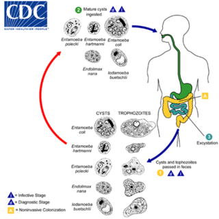
Entamoeba coli is a non-pathogenic species of Entamoeba that frequently exists as a commensal parasite in the human gastrointestinal tract. E. coli is important in medicine because it can be confused during microscopic examination of stained stool specimens with the pathogenic Entamoeba histolytica. This amoeba does not move much by the use of its pseudopod, and creates a "sur place (non-progressive) movement" inside the large intestine. Usually, the amoeba is immobile, and keeps its round shape. This amoeba, in its trophozoite stage, is only visible in fresh, unfixed stool specimens. Sometimes the Entamoeba coli have parasites as well. One is the fungus Sphaerita spp. This fungus lives in the cytoplasm of the E. coli. While this differentiation is typically done by visual examination of the parasitic cysts via light microscopy, new methods using molecular biology techniques have been developed. The scientific name of the amoeba, E. coli, is often mistaken for the bacterium, Escherichia coli. Unlike the bacterium, the amoeba is mostly harmless, and does not cause as many intestinal problems as some strains of the E. coli bacterium. To make the naming of these organisms less confusing, "alternate contractions" are used to name the species for the purpose making the naming easier; for example, using Esch. coli and Ent. coli for the bacterium and amoeba, instead of using E. coli for both.
Hymenolepiasis is infestation by one of two species of tapeworm: Hymenolepis nana or H. diminuta. Alternative names are dwarf tapeworm infection and rat tapeworm infection. The disease is a type of helminthiasis which is classified as a neglected tropical disease.

Coccidia (Coccidiasina) are a subclass of microscopic, spore-forming, single-celled obligate intracellular parasites belonging to the apicomplexan class Conoidasida. As obligate intracellular parasites, they must live and reproduce within an animal cell. Coccidian parasites infect the intestinal tracts of animals, and are the largest group of apicomplexan protozoa.

Eimeria tenella is a species of Eimeria that causes hemorrhagic cecal coccidiosis in young poultry. It is found worldwide.

Eimeria is a genus of apicomplexan parasites that includes various species capable of causing the disease coccidiosis in animals such as cattle, poultry and smaller ruminants including sheep and goats. Eimeria species are considered to be monoxenous because the life cycle is completed within a single host, and stenoxenous because they tend to be host specific, although a number of exceptions have been identified. Species of this genus infect a wide variety of hosts. Thirty-one species are known to occur in bats (Chiroptera), two in turtles, and 130 named species infect fish. Two species infect seals. Five species infect llamas and alpacas: E. alpacae, E. ivitaensis, E. lamae, E. macusaniensis, and E. punonensis. A number of species infect rodents, including E. couesii, E. kinsellai, E. palustris, E. ojastii and E. oryzomysi. Others infect poultry, rabbits and cattle. For full species list, see below.

Cryptosporidium parvum is one of several species that cause cryptosporidiosis, a parasitic disease of the mammalian intestinal tract.
Cryptosporidium, sometimes informally called crypto, is a genus of apicomplexan parasitic alveolates that can cause a respiratory and gastrointestinal illness (cryptosporidiosis) that primarily involves watery diarrhea, sometimes with a persistent cough.
Cyclosporiasis is a disease caused by infection with Cyclospora cayetanensis, a pathogenic protozoan transmitted by feces or feces-contaminated food and water. Outbreaks have been reported due to contaminated fruits and vegetables. It is not spread from person to person, but can be a hazard for travelers as a cause of diarrhea.
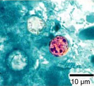
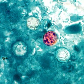
Cyclospora cayetanensis is a coccidian parasite that causes a diarrheal disease called cyclosporiasis in humans and possibly in other primates. Originally reported as a novel pathogen of probable coccidian nature in the 1980s and described in the early 1990s, it was virtually unknown in developed countries until awareness increased due to several outbreaks linked with fecally contaminated imported produce. C. cayetanensis has since emerged as an endemic cause of diarrheal disease in tropical countries and a cause of traveler's diarrhea and food-borne infections in developed nations. This species was placed in the genus Cyclospora because of the spherical shape of its sporocysts. The specific name refers to the Cayetano Heredia University in Lima, Peru, where early epidemiological and taxonomic work was done.

Sarcocystis is a genus of protozoan parasites, with many species infecting mammals, reptiles and birds. Its name is dervived from Greek sarx = flesh and kystis = bladder.
Karyolysus is a genus of coccidia. With the exception of K. sonomae whose vertebrate host is the yellow-legged frog, species in this genus only infect lizards of the genus Lacerta.

Heterophyes heterophyes was discovered by Theodor Maximaillian Bilharz in 1851. This parasite was found during an autopsy of an Egyptian mummy. H. heterophyes is found in the Middle East, West Europe and Africa. They use different species to complete their complex lifestyle. Humans and other mammals are the definitive host, first intermediate host are snails, and second intermediate are fish. Mammals that come in contact with the parasite are dogs, humans, and cats. Snails that are affected by this parasite are the Cerithideopsilla conica. Fish that come in contact with this parasite are Mugil cephalus, Tilapia milotica, Aphanius fasciatus, and Acanthgobius sp. Humans and mammals will come in contact with this parasite by the consumption of contaminated or raw fish. This parasite is one of the smallest endoparasite to infect humans. It can cause intestinal infection called heterophyiasis.
Apicomplexans, a group of intracellular parasites, have life cycle stages that allow them to survive the wide variety of environments they are exposed to during their complex life cycle. Each stage in the life cycle of an apicomplexan organism is typified by a cellular variety with a distinct morphology and biochemistry.

Hemolivia is a genus of the phylum Apicomplexia.

Cystoisospora is a genus of parasitic alveolates belonging to the phylum Apicomplexa.
Cystoisospora belli, previously known as Isospora belli, is a parasite that causes an intestinal disease known as cystoisosporiasis. This protozoan parasite is opportunistic in immune suppressed human hosts. It primarily exists in the epithelial cells of the small intestine, and develops in the cell cytoplasm. The distribution of this coccidian parasite is cosmopolitan, but is mainly found in tropical and subtropical areas of the world such as the Caribbean, Central and S. America, India, Africa, and S.E. Asia. In the U.S., it is usually associated with HIV infection and institutional living.
Cystoisospora canis, previously known as Isospora canis, is a microscopic, coccidian parasite that causes an intestinal tract infection in dogs. The intestinal tract infection is coccidiosis caused by a protozoa called coccidia.
Sarcocystis neurona is primarily a neural parasite of horses and its management is of concern in veterinarian medicine. The protozoan Sarcocystis neurona is a protozoan of single celled character and belongs to the family, Sarcocystidae, a group called coccidia. The protozoan, S. neurona, is a member of the genus Sarcocystis, and is most commonly associated with equine protozoal myeloencephalitis (EPM). The protozoan, S. neurona, can be easily cultivated and genetically manipulated, hence its common use as a model to study numerous aspects of cell biology.
