
Carrion's disease is an infectious disease produced by Bartonella bacilliformis infection.
Bartonellosis is an infectious disease produced by bacteria of the genus Bartonella. Bartonella species cause diseases such as Carrión's disease, trench fever, cat-scratch disease, bacillary angiomatosis, peliosis hepatis, chronic bacteremia, endocarditis, chronic lymphadenopathy, and neurological disorders.

Burkholderia pseudomallei is a Gram-negative, bipolar, aerobic, motile rod-shaped bacterium. It is a soil-dwelling bacterium endemic in tropical and subtropical regions worldwide, particularly in Thailand and northern Australia. It was reported in 2008 that there had been an expansion of the affected regions due to significant natural disasters, and it could be found in Southern China, Hong Kong, and countries in America. B. pseudomallei, amongst other pathogens, has been found in monkeys imported into the United States from Asia for laboratory use, posing a risk that the pathogen could be introduced into the country.

Protothecosis, otherwise known as Algaemia, is a disease found in dogs, cats, cattle, and humans caused by a type of green alga known as Prototheca that lacks chlorophyll and enters the human or animal bloodstream. It and its close relative Helicosporidium are unusual in that they are actually green algae that have become parasites. The two most common species are Prototheca wickerhamii and Prototheca zopfii. Both are known to cause disease in dogs, while most human cases are caused by P. wickerhami. Prototheca is found worldwide in sewage and soil. Infection is rare despite high exposure, and can be related to a defective immune system. In dogs, females and Collies are most commonly affected.

Pythiosis is a rare and deadly tropical disease caused by the oomycete Pythium insidiosum. Long regarded as being caused by a fungus, the causative agent was not discovered until 1987. It occurs most commonly in horses, dogs, and humans, with isolated cases in other large mammals. The disease is contracted after exposure to stagnant fresh water such as swamps, ponds, lakes, and rice paddies. P. insidiosum is different from other members of the genus in that human and horse hair, skin, and decaying animal and plant tissue are chemoattractants for its zoospores. Additionally, it is the only member in the genus known to infect mammals, while other members are pathogenic to plants and are responsible for some well-known plant diseases.
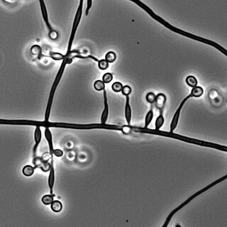
Pichia kudriavzevii is a budding yeast involved in chocolate production. P. kudriavzevii is an emerging fungal nosocomial pathogen primarily found in the immunocompromised and those with hematological malignancies. It has natural resistance to fluconazole, a standard antifungal agent. It is most often found in patients who have had prior fluconazole exposure, sparking debate and conflicting evidence as to whether fluconazole should be used prophylactically. Mortality due to P. kudriavzevii fungemia is much higher than the more common C. albicans. Other Candida species that also fit this profile are C. parapsilosis, C. glabrata, C. tropicalis, C. guillermondii and C. rugosa.

Sporothrix schenckii, a fungus that can be found worldwide in the environment, is named for medical student Benjamin Schenck, who in 1896 was the first to isolate it from a human specimen. The species is present in soil as well as in and on living and decomposing plant material such as peat moss. It can infect humans as well as animals and is the causative agent of sporotrichosis, commonly known as "rose handler's disease." The most common route of infection is the introduction of spores to the body through a cut or puncture wound in the skin. Infection commonly occurs in otherwise healthy individuals but is rarely life-threatening and can be treated with antifungals. In the environment it is found growing as filamentous hyphae. In host tissue it is found as a yeast. The transition between the hyphal and yeast forms is temperature dependent making S. schenckii a thermally dimorphic fungus.

Acrophialophora fusispora is a poorly studied ascomycete fungus found in soil, air and various plants. A. fusispora is morphologically similar to the genera Paecilomyces and Masonia, but differ in the presence of pigmented conidiophores, verticillate phialides, and frequent sympodial proliferation. Moreover, A. fusispora is distinguished by its pigmented spindle-shaped conidia, covered with spiral bands. The fungus is naturally found in soils of tropical to temperate regions. The fungus has been identified as a plant and animal pathogen, and has recently been recognized as an emerging opportunistic human pathogen. A. fusispora infection in human is rare and has few documented clinical cases, but due to the rarity of the fungus and potential misidentification, the infections may be underdiagnosed. Clinical cases of A. fusispora include cases of keratitis, pulmonary colonization and infection, and cerebral infections. The fungus also has two documented cases of infection in dogs.

Trichosporon is a genus of anamorphic fungi in the family Trichosporonaceae. All species of Trichosporon are yeasts with no known teleomorphs. Most are typically isolated from soil, but several species occur as a natural part of the skin microbiota of humans and other animals. Proliferation of Trichosporon yeasts in the hair can lead to an unpleasant but non-serious condition known as white piedra. Trichosporon species can also cause severe opportunistic infections (trichosporonosis) in immunocompromised individuals.

Pathogenic bacteria are bacteria that can cause disease. This article focuses on the bacteria that are pathogenic to humans. Most species of bacteria are harmless and are often beneficial but others can cause infectious diseases. The number of these pathogenic species in humans is estimated to be fewer than a hundred. By contrast, several thousand species are part of the gut flora present in the digestive tract.

Sabouraud agar or Sabouraud dextrose agar (SDA) is a type of agar growth medium containing peptones. It is used to cultivate dermatophytes and other types of fungi, and can also grow filamentous bacteria such as Nocardia. It has utility for research and clinical care.
Saksenaea vasiformis is an infectious fungus associated with cutaneous or subcutaneous lesions following trauma. It causes opportunistic infections as the entry of the fungus is through open spaces of cutaneous barrier ranging in severity from mild to severe or fatal. It lives in soils worldwide, but is considered as a rare human pathogen since only 38 cases were reported as of 2012. Saksenaea vasiformis usually fails to sporulate on the routine culture media, creating a challenge for early diagnosis, which is essential for a good prognosis. Infections are usually treated using a combination of amphotericin B and surgery. Saksenaea vasiformis is one of the few fungi known to cause necrotizing fasciitis or "flesh-eating disease".
Algaemia is a secondary term that refers to the emerging condition in which green algae enter the bloodstream. Members of the genus Prototheca are the most common algae that leads to algaemia. Prototheca and Chlorella, which is extremely rare, are the only two known algae genera capable of inflicting disease on mammals, including humans, through invasion of host tissue. The majority of cases are observed in dairy cattle as a cause of bovine mastitis as well as other domesticated animals. Cases of algaemia have been observed in dogs and cats as well. Few cases have been observed in humans. Human cases of algaemia or, protothecosis, are examined on a case-by-case basis due to the particularity of each case. Protothecosis infection is classified based on the symptoms: (i) cutaneous lesions, (ii) olecranon bursitis, and (iii) disseminated or systemic type infections.
Neisseria bacilliformis is a bacterium commonly found living as a commensal in the mucous membranes of mammals. However, depending on host immunocompetence, there have been documented cases of N. bacilliformis infections of the respiratory tract and oral cavity thus making it an opportunistic pathogen. It was originally isolated from patients being treated in a cancer center. Rarely, a more serious infection such as endocarditis can occur often as a result of a predisposing condition.
Staphylococcus pseudintermedius is a gram positive coccus bacteria of the genus Staphylococcus found worldwide. It is primarily a pathogen for domestic animals, but has been known to affect humans as well. S. pseudintermedius is an opportunistic pathogen that secretes immune modulating virulence factors, has many adhesion factors, and the potential to create biofilms, all of which help to determine the pathogenicity of the bacterium. Diagnoses of Staphylococcus pseudintermedius have traditionally been made using cytology, plating, and biochemical tests. More recently, molecular technologies like MALDI-TOF, DNA hybridization and PCR have become preferred over biochemical tests for their more rapid and accurate identifications. This includes the identification and diagnosis of antibiotic resistant strains.

Lichtheimia corymbifera is a thermophilic fungus in the phylum Zygomycota. It normally lives as a saprotrophic mold, but can also be an opportunistic pathogen known to cause pulmonary, CNS, rhinocerebral, or cutaneous infections in animals and humans with impaired immunity.
Emmonsia parva is a filamentous, saprotrophic fungus and one of three species within the genus Emmonsia. The fungus is most known for its causal association with the lung disease, adiaspiromycosis which occurs most commonly in small mammals but is also seen in humans. The disease was first described from rodents in Arizona, and the first human case was reported in France in 1964. Since then, the disease has been reported from Honduras, Brazil, the Czech Republic, Russia, the United States of America and Guatemala. Infections in general are quite rare, especially in humans.
Prototheca zopfii is an ubiquitous achlorophyllic green alga. It is a known cause of mastitis in cattle.
Metarhizium granulomatis is a fungus in the family Clavicipitaceae associated with systemic mycosis in veiled chameleons. The genus Metarhizium is known to infect arthropods, and collectively are referred to green-spored asexual pathogenic fungi. This species grows near the roots of plants and has been reported as an agent of disease in captive veiled chameleons. The etymology of the species epithet, "granulomatis" refers to the ability of the fungus to cause granulomatous disease in susceptible reptiles.
Sarocladium kiliense is a saprobic fungus that is occasionally encountered as a opportunistic pathogen of humans, particularly immunocompromised and individuals. The fungus is frequently found in soil and has been linked with skin and systemic infections. This species is also known to cause disease in the green alga, Cladophora glomerata as well as various fruit and vegetable crops grown in warmer climates.
