Babesiosis or piroplasmosis is a malaria-like parasitic disease caused by infection with a eukaryotic parasite in the order Piroplasmida, typically a Babesia or Theileria, in the phylumApicomplexa.[2] Human babesiosis transmission via tick bite is most common in the Northeastern and Midwestern United States and parts of Europe, and sporadic throughout the rest of the world. It occurs in warm weather.[3] People can get infected with Babesia parasites by the bite of an infected tick, by getting a blood transfusion from an infected donor of blood products, or by congenital transmission (an infected mother to her baby).[4] Ticks transmit the human strain of babesiosis, so it often presents with other tick-borne illnesses such as Lyme disease.[5] After trypanosomes, Babesia is thought to be the second-most common blood parasite of mammals. They can have major adverse effects on the health of domestic animals in areas without severe winters. In cattle, the disease is known as Texas cattle fever or redwater.[6]
Half of all children and a quarter of previously healthy adults with Babesia infection are asymptomatic. When people do develop symptoms, the most common are fever and hemolytic anemia, symptoms that are similar to those of malaria.[5] People with symptoms usually become ill 1 to 4 weeks after the bite, or 1 to 9 weeks after transfusion of contaminated blood products. A person infected with babesiosis gradually develops malaise and fatigue, followed by a fever. Hemolytic anemia, in which red blood cells are destroyed and removed from the blood, also develops. Chills, sweats, and thrombocytopenia are also common symptoms. Symptoms may last from several days to several months.[citation needed]
Less common symptoms and physical exam findings of mild-to-moderate babesiosis:[5]
In more severe cases, symptoms similar to malaria occur, with fevers up to 40.5°C (105°F), shaking chills, and severe anemia (hemolytic anemia). Organ failure may follow, including adult respiratory distress syndrome. Sepsis in people who have had a splenectomy can occur rapidly, consistent with overwhelming post-splenectomy infection. Severe cases are also more likely to occur in the very young, very old, and persons with immunodeficiency, such as HIV/AIDS patients.[citation needed]
A reported increase in human babesiosis diagnoses in the 2000s is thought to be caused by more widespread testing and higher numbers of people with immunodeficiencies coming in contact with ticks, the disease vector.[6] Little is known about the occurrence of Babesia species in malaria-endemic areas, where Babesia can easily be misdiagnosed as Plasmodium. Human patients with repeat babesiosis infection may exhibit premunity.[8]
B. divergens (cattle parasite seen mostly in Europe) and B.venatorum (roe deer parasite, formerly called EU1), most closely related to the large Babesia clade
Large Babesia (>3μm) mostly infects ungulates, but also includes K01 strain (an isolated case observed in South Korea, see isolated cases)
Pathophysiology
Babesia lifecycle
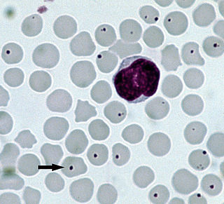
Babesia parasites reproduce in red blood cells, where they can be seen as cross-shaped inclusions (four merozoites asexually budding, but attached together forming a structure looking like a "Maltese cross")[9] and cause hemolytic anemia, quite similar to malaria.
Unlike the Plasmodium parasites that cause malaria, Babesia species lack an exoerythrocytic phase, so the liver is usually not affected.[citation needed]
In nonhuman animals, Babesia canis rossi, Babesia bigemina, and Babesia bovis cause particularly severe forms of the disease, including a severe haemolytic anaemia, with positive erythrocyte-in-saline-agglutination test indicating an immune-mediated component to the haemolysis. Common sequelae include haemoglobinuria "red-water", disseminated intravascular coagulation, and "cerebral babesiosis" caused by sludging of erythrocytes in cerebral capillaries.[citation needed]
In bovine species, the organism causes hemolytic anemia, so an infected animal shows pale mucous membranes initially. As the levels of bilirubin (a byproduct of red blood cell lysis) continue to increase, the visible mucous membranes become yellow in color (icterus) due to the failure of the liver to metabolize the excess bilirubin. Hemoglobinuria is seen due to excretion of red-blood-cell lysis byproducts via the kidneys. Fever of 40.5°C (105°F) develops due to release of inflammatory byproducts.[citation needed]
Diagnosis
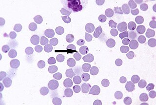
Only specialized laboratories can adequately diagnose Babesia infection in humans, so Babesia infections are considered highly under-reported. It develops in patients who live in or travel to an endemic area or receive a contaminated blood transfusion within the preceding 9 weeks, so this aspect of the medical history is vital.[10] Babesiosis may be suspected when a person with such an exposure history develops persistent fevers and hemolytic anemia. The definitive diagnostic test is the identification of parasites on a Giemsa-stainedthin-film blood smear.[10]
So-called "Maltese cross formations" on the blood film are diagnostic (pathognomonic) of babesiosis, since they are not seen in malaria, the primary differential diagnosis.[9] Careful examination of multiple smears may be necessary, since Babesia may infect less than 1% of circulating red blood cells, thus be easily overlooked.[11]
Serologic testing for antibodies against Babesia (both IgG and IgM) can detect low-level infection in cases with a high clinical suspicion, but negative blood film examinations. Serology is also useful for differentiating babesiosis from malaria in cases where people are at risk for both infections. Since detectable antibody responses require about a week after infection to develop, serologic testing may be falsely negative early in the disease course.[12]
A polymerase chain reaction (PCR) test has been developed for the detection of Babesia from the peripheral blood.[13] PCR may be at least as sensitive and specific as blood-film examination in diagnosing babesiosis, though it is also significantly more expensive.[14] Most often, PCR testing is used in conjunction with blood film examination and possibly serologic testing.[10]
In animals, babesiosis is suspected by observation of clinical signs (hemoglobinuria and anemia) in animals in endemic areas. Diagnosis is confirmed by observation of merozoites on thin film blood smear examined at maximum magnification under oil using Romonovski stains (methylene blue and eosin). This is a routine part of the veterinary examination of dogs and ruminants in regions where babesiosis is endemic.[citation needed]
Babesia canis and B.bigemina are "large Babesia species" that form paired merozoites in the erythrocytes, commonly described as resembling "two pears hanging together", rather than the "Maltese cross" of the "small Babesia species". Their merozoites are around twice the size of small ones.[citation needed]
Cerebral babesiosis is suspected in vivo when neurological signs (often severe) are seen in cattle that are positive for B.bovis on blood smear, but this has yet to be proven scientifically. Outspoken red discoloration of the grey matter post mortem further strengthens suspicion of cerebral babesiosis. Diagnosis is confirmed post mortem by observation of Babesia-infected erythrocytes sludged in the cerebral cortical capillaries in a brain smear.[citation needed]
Treatment
Treatment of asymptomatic carriers should be considered if parasites are still detected after 3 months. In mild-to-moderate babesiosis, the treatment of choice is a combination of atovaquone and azithromycin. This regimen is preferred to clindamycin and quinine because it has fewer side effects. The standard course is 7 to 10 days, but this is extended to at least 6 weeks in people with relapsing disease. Even mild cases are recommended to be treated to decrease the chance of inadvertently transmitting the infection by donating blood.[5] In severe babesiosis, the combination of clindamycin and quinine is preferred. In life-threatening cases, exchange transfusion is performed.[15] In this procedure, the infected red blood cells are removed and replaced with uninfected ones.[citation needed]
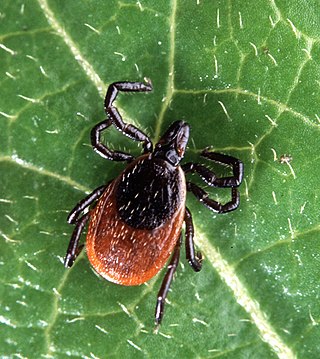
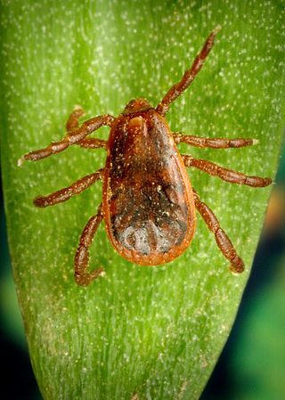
Babesiosis is a vector-borne illness usually transmitted by Ixodes scapularisticks. B.microti uses the same tick vector as Lyme disease, and may occur in conjunction with Lyme.[6] The organism can also be transmitted by blood transfusion.[18][19]Ticks of domestic animals, especially Rhipicephalus (Boophilus) microplus and R. (B.) decoloratus transmit several species of Babesia to livestock, causing considerable economic losses to farmers in tropical and subtropical regions.[citation needed]
In Europe, B.divergens is the primary cause of infectious babesiosis and is transmitted by I.ricinus.[5]
Babesiosis has emerged in Lower Hudson Valley, New York, since 2001.[27]
In Australia, one locally-acquired case of B.microti has been reported, which was fatal.[28] A subsequent investigation found no additional evidence of human Babesiosis in over 7000 patient samples, leading the authors to conclude that Babesiosis was rare in Australia.[29] A similar disease in cattle, commonly known as tick fever, is spread by Babesia bovis and B. bigemina in the introduced cattle tick Rhipicephalus microplus. This disease is found in eastern and northern Australia.[30]
Isolated cases
A table of isolated cases of babesiosis, which may be underestimated given how widely distributed the tick vectors are in temperate latitudes.[5]
The disease is named for the genus of the causative organism,[32] which was named after the RomanianbacteriologistVictor Babeș.[33] In 1888, Victor Babeș identified the microorganisms in red blood cells as the cause of febrile hemoglobinuria in cattle.[5] In 1893, Theobald Smith and Frederick Kilborne discovered that a tick was the vector for transmission in Texas cattle. The agent was B.bigemina. This was the first demonstration that an arthropod could act as a disease vector to transmit an infectious agent to a vertebrate host.[citation needed]
In 1957, the first human case was documented in a splenectomized Croatian herdsman.[5] The agent was B.divergens. In 1969, the first case was reported in an immunocompetent individual on Nantucket Island. The agent was B.microti, and the vector was the tick I. scapularis.[citation needed]Equine babesiosis (caused by the protozoan Theileria equi) is also known as piroplasmosis (from the Latinpiro, meaning pear + Greekplasma, a thing formed).[34]
Other animals
Veterinary treatment of babesiosis does not normally use antibiotics. In livestock and animals, diminazen (Berenil), imidocarb, or trypan blue would be the drugs of choice for treatment of B.canis rossi (dogs in Africa), B.bovis, and B.bigemina (cattle in Southern Africa). In acute cases in cattle, blood transfusion may be carried out. A vaccine is effective against B.canis canis (dogs in the Mediterranean region), but is ineffective against B.c.rossi. B.imitans causes a mild form of the disease that frequently resolves without treatment (dogs in Southeast Asia).[citation needed]
Related Research Articles
Arbovirus is an informal name for any virus that is transmitted by arthropod vectors. The term arbovirus is a portmanteau word. Tibovirus is sometimes used to more specifically describe viruses transmitted by ticks, a superorder within the arthropods. Arboviruses can affect both animals and plants. In humans, symptoms of arbovirus infection generally occur 3–15 days after exposure to the virus and last three or four days. The most common clinical features of infection are fever, headache, and malaise, but encephalitis and viral hemorrhagic fever may also occur.
Plasmodium knowlesi is a parasite that causes malaria in humans and other primates. It is found throughout Southeast Asia, and is the most common cause of human malaria in Malaysia. Like other Plasmodium species, P. knowlesi has a life cycle that requires infection of both a mosquito and a warm-blooded host. While the natural warm-blooded hosts of P. knowlesi are likely various Old World monkeys, humans can be infected by P. knowlesi if they are fed upon by infected mosquitoes. P. knowlesi is a eukaryote in the phylum Apicomplexa, genus Plasmodium, and subgenus Plasmodium. It is most closely related to the human parasite Plasmodium vivax as well as other Plasmodium species that infect non-human primates.
Babesia, also called Nuttallia, is an apicomplexan parasite that infects red blood cells and is transmitted by ticks. Originally discovered by the Romanian bacteriologist Victor Babeș in 1888, over 100 species of Babesia have since been identified.
Anaplasmosis is a tick-borne disease affecting ruminants, dogs, and horses, and is caused by Anaplasma bacteria. Anaplasmosis is an infectious but not contagious disease. Anaplasmosis can be transmitted through mechanical and biological vector processes. Anaplasmosis can also be referred to as "yellow bag" or "yellow fever" because the infected animal can develop a jaundiced look. Other signs of infection include weight loss, diarrhea, paleness of the skin, aggressive behavior, and high fever.
Babesia divergens is an intraerythrocytic parasite, transmitted by the tick Ixodes ricinus. It is the most common cause of human babesiosis. It is the main agent of bovine babesiosis, or "redwater fever", in Europe. Young cattle are less susceptible. The current emphasis in Europe on sustainable agriculture and extensification is likely to lead to an increase in vector tick populations with increased risk of infection. B. divergens is also prevalent in cottontail rabbits on Nantucket Island, MA, USA.
Ixodes scapularis is commonly known as the deer tick or black-legged tick, and in some parts of the US as the bear tick. It was also named Ixodes dammini until it was shown to be the same species in 1993. It is a hard-bodied tick found in the eastern and northern Midwest of the United States as well as in southeastern Canada. It is a vector for several diseases of animals, including humans and is known as the deer tick owing to its habit of parasitizing the white-tailed deer. It is also known to parasitize mice, lizards, migratory birds, etc. especially while the tick is in the larval or nymphal stage.
Avian malaria is a parasitic disease of birds, caused by parasite species belonging to the genera Plasmodium and Hemoproteus. The disease is transmitted by a dipteran vector including mosquitoes in the case of Plasmodium parasites and biting midges for Hemoproteus. The range of symptoms and effects of the parasite on its bird hosts is very wide, from asymptomatic cases to drastic population declines due to the disease, as is the case of the Hawaiian honeycreepers. The diversity of parasites is large, as it is estimated that there are approximately as many parasites as there are species of hosts. As research on human malaria parasites became difficult, Dr. Ross studied avian malaria parasites. Co-speciation and host switching events have contributed to the broad range of hosts that these parasites can infect, causing avian malaria to be a widespread global disease, found everywhere except Antarctica.
Babesia microti is a parasitic blood-borne piroplasm transmitted by deer ticks. B. microti is responsible for the disease babesiosis, a malaria-like disease which also causes fever and hemolysis.
Protozoan infections are parasitic diseases caused by organisms formerly classified in the kingdom Protozoa. These organisms are now classified in the supergroups Excavata, Amoebozoa, Harosa, and Archaeplastida. They are usually contracted by either an insect vector or by contact with an infected substance or surface.
A transfusion transmitted infection (TTI) is a virus, parasite, or other potential pathogen that can be transmitted in donated blood through a transfusion to a recipient. The term is usually limited to known pathogens, but also sometimes includes agents such as simian foamy virus which are not known to cause disease.
Human granulocytic anaplasmosis (HGA) is a tick-borne, infectious disease caused by Anaplasma phagocytophilum, an obligate intracellular bacterium that is typically transmitted to humans by ticks of the Ixodes ricinus species complex, including Ixodes scapularis and Ixodes pacificus in North America. These ticks also transmit Lyme disease and other tick-borne diseases.
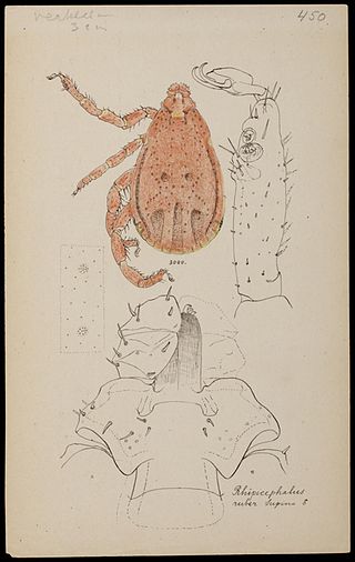
Rhipicephalus sanguineus, commonly called the brown dog tick, kennel tick, or pantropical dog tick, is a species of tick found worldwide, but more commonly in warmer climates. This species is unusual among ticks in that its entire lifecycle can be completed indoors. The brown dog tick is easily recognized by its reddish-brown color, elongated body shape, and hexagonal basis capituli. Adults are 2.28 to 3.18 mm in length and 1.11 to 1.68 mm in width. They do not have ornamentation on their backs.
Hematozoa is a subclass of blood parasites of the Apicomplexa clade. Well known examples include the Plasmodium spp. which cause malaria in humans and Theileria which causes theileriosis in cattle. A large number of species are known to infect birds and are transmitted by insect vectors. The pattern in which Haematozoa infect a host cell depends on the genera of the blood parasite. Plasmodium and Leucozytozoon displace the nucleus of the host cell so that the parasite can take control of the cell where as Hemoproteus completely envelops the nucleus in a host cell.
Babesia bovis is an Apicomplexan single-celled parasite of cattle which occasionally infects humans. The disease it and other members of the genus Babesia cause is a hemolytic anemia known as babesiosis and colloquially called Texas cattle fever, redwater or piroplasmosis. It is transmitted by bites from infected larval ticks of the order Ixodida. It was eradicated from the United States by 1943, but is still present in Mexico and much of the world's tropics. The chief vector of Babesia species is the southern cattle fever tick Rhipicephalus microplus.
Theileria parva is a species of parasites, named in honour of Arnold Theiler, that causes East Coast fever (theileriosis) in cattle, a costly disease in Africa. The main vector for T. parva is the tick Rhipicephalus appendiculatus. Theiler found that East Coast fever was not the same as redwater, but caused by a different protozoan.
Ticks of domestic animals directly cause poor health and loss of production to their hosts. Ticks also transmit numerous kinds of viruses, bacteria, and protozoa between domestic animals. These microbes cause diseases which can be severely debilitating or fatal to domestic animals, and may also affect humans. Ticks are especially important to domestic animals in tropical and subtropical countries, where the warm climate enables many species to flourish. Also, the large populations of wild animals in warm countries provide a reservoir of ticks and infective microbes that spread to domestic animals. Farmers of livestock animals use many methods to control ticks, and related treatments are used to reduce infestation of companion animals.
Babesia canis is a parasite that infects red blood cells and can lead to anemia. This is a species that falls under the overarching genus Babesia. It is transmitted by the brown dog tick and is one of the most common piroplasm infections. The brown dog tick is adapted to warmer climates and is found in both Europe and the United States, especially in shelters and greyhound kennels. In Europe, it is also transmitted by Dermacentor ticks with an increase in infections reported due to people traveling with their pets.
Babesia bigemina is a species of alveolates belonging to the phylum Apicomplexa and the family Babesiidae, a type of protozoan parasite. In cattle, it causes babesiosis, also called "Texas fever". Its length is 4–5 μm and its width is 2–3 μm. Usually, it has an oval shape. In blood cells, it is located midsagittally and can reach up to two-thirds of the diameter of the blood cell in size. It is transmitted by Boophilus ticks which are prevalent in the tropics. The genome for B. bigemina is incomplete and unassembled.
Rhipicephalus haemaphysaloides is a hard-bodied tick of the genus Rhipicephalus. It is one of the major medically important ticks in the world.
Quartan fever is one of the four types of malaria which can be contracted by humans.
↑ Krause PJ, Telford SR, Ryan R, etal. (April 1994). "Diagnosis of babesiosis: evaluation of a serologic test for the detection of Babesia microti antibody". J. Infect. Dis. 169 (4): 923–6. doi:10.1093/infdis/169.4.923. PMID8133112.
↑ Senanayake SN, Paparini A, Latimer M, Andriolo K, Dasilva AJ, Wilson H, etal. (March 2012). "First report of human babesiosis in Australia". Medical Journal of Australia. 196 (5): 350–352. doi:10.5694/mja11.11378. PMID22432676. S2CID33068508.
1 2 Faddy HM, Rooks KM, Irwin PJ, Viennet E, Paparini A, Seed CR, etal. (July 2019). "No evidence for widespread Babesia microti transmission in Australia". Transfusion. 59 (7): 2368–2374. doi:10.1111/trf.15336. PMID31070793. S2CID148570372.
Center for Global Health (2019-06-25). "Babesiosis". Parasites and Health, DPDx—Laboratory Identification of Parasites of Public Health Concern. Centers for Disease Control & Prevention. Archived from the original on 2013-03-07. Retrieved 2003-10-07. Public domain source from which the first version of this article was derived.
This page is based on this Wikipedia article Text is available under the CC BY-SA 4.0 license; additional terms may apply. Images, videos and audio are available under their respective licenses.