Rickettsialpox

Last updated
Rickettsialpox
Rickettsialpox lesion.jpg
Rickettsialpox lesion
Specialty Infectious disease
Prognosis Resolves in 2-3 weeks without treatment

Rickettsialpox is a mite-borne infectious illness caused by bacteria of the genus Rickettsia ( Rickettsia akari ). [1] Physician Robert Huebner and self-trained entomologist Charles Pomerantz played major roles in identifying the cause of the disease after an outbreak in 1946 in a New York City apartment complex, documented in "The Alerting of Mr. Pomerantz," an article by medical writer Berton Roueché.

Contents

Although it is not transmitted by a tick (a characteristic of spotted fever), the bacterium is a part of the spotted fever group of Rickettsia, and so this condition is often classified with that group. [2]

Signs and symptoms

The first symptom is a bump formed by the bite, eventually resulting in a black, crusty scab. Many of the symptoms are flu-like including fever, chills, weakness and achy muscles but the most distinctive symptom is the rash that breaks out, spanning the infected person's entire body. [3]

Transmission

The bacteria are originally found in mice and cause mites feeding on the mice (usually the house mouse) to become infected. Humans will get rickettsialpox when receiving a bite from an infected mite, not from the mice themselves.[ citation needed ]

The mite is Liponyssoides sanguineus , which was previously known as Allodermanyssus sanguineus. [2]

Treatment

Rickettsialpox is treated with tetracyclines (doxycycline is the drug of choice). Chloramphenicol is a suitable alternative. [4]

Prognosis

Rickettsialpox is generally mild and resolves within 2–3 weeks if untreated. There are no known deaths resulting from the disease.[ citation needed ]

Epidemiology

Those dwelling in urban areas (which typically experience rodent problems) have a higher risk of contracting rickettsialpox.[ citation needed ]

History

The initial outbreak of the disease took place in the Regency Park complex which had 69 apartment units organized in three groups each three stories in height, located in Kew Gardens, in the New York City borough of Queens. Physicians who had seen patients starting in early 1946 had assumed that they were dealing with an atypical form of chickenpox, but the realization was made that they were dealing with a localized epidemic of unknown origins starting in the summer of that year. Physicians canvassed the residents of the building and found that there had been 124 cases of this disease from January through October among the 2,000 people living in the complex, reaching a peak of more than 20 cases reported in July. Individuals afflicted with the condition ranged in age from 3 months to 71, males and females were equally affected and the incidence among adults (6.5% of the 1,400 above age 15) exceeded that of children 14 and under (5.3%). [5]

Individuals would first have a small lesion at the site of the insect bite, which would eventually leave a small scar. Lymph nodes would become enlarged and about a week after the initial bite patients would start to experience chills, fever and headaches, accompanied by a maculopapular rash. The rash would last for about a week, with the full progress from the lesion being encountered until recovery typically being three weeks. Several characteristics were considered from an epidemiological perspective as being correlated with the condition. Men and women were equally affected and there was no apparent connection to occupation or school attended. The individuals obtained their meals at several different sources, and other individuals who lived elsewhere and purchased their food at these same places had not been affected. The water supply and milk consumed there were tested, with no unusual findings. There were dog kennels in the area and some tenants had dogs as pets, but an inspection of the animals found no ticks or insects. [5]

Many residents had reported the presence of mice in and around the apartments, especially in the basements of the buildings near the incinerators, where living and dead mice were found. Garbage disposed of through incinerator chutes was to be burned on a daily basis, with the ashes carted off by the New York City Department of Sanitation. Due to labor shortages, the material was not burned regularly, leaving ample food for the rodents. Mice were trapped in apartments and near the incinerators and antibodies to the rickettsialpox were found in their blood. [5] Visiting the complex with pest-control specialist and self-trained entomologist Charles Pomerantz, Huebner peeled back wallpaper to find the walls swarming with mites, so much so that tenants had said that "the walls had movement". [6] The mites were collected and rickettsial strains were isolated from some of them, with the name Rickettsia akari given to the organism. [5]

Public health officials took steps to prevent further outbreaks of the disease. The building operators were told to ensure that the incinerators were fired on a regular basis to ensure that food available to the mice that harbored the disease was eliminated as quickly and thoroughly as possible. [5] Pest control measures were undertaken through the New York City Department of Health and its commissioner Israel Weinstein to eliminate the mice in and around the buildings with the cooperation of property owners. [7]

Another 20 cases of rickettsialpox were discovered elsewhere in New York City during the investigation of the Queens outbreak, including 10 cases found in an apartment building in the Bronx that had originally been diagnosed as atypical chickenpox. Mice were found in the basement of this building, as well as mites belonging to the same class found in Queens. [5] In addition to the initial cases from the first outbreak in Queens, over 500 cases of the disease were diagnosed in New York City from 1947 to 1951. [8]

Society and culture

See also

Related Research Articles

<i>Rickettsia</i> Genus of bacteria

Rickettsia is a genus of nonmotile, gram-negative, nonspore-forming, highly pleomorphic bacteria that may occur in the forms of cocci, bacilli, or threads. The genus was named after Howard Taylor Ricketts in honor of his pioneering work on tick-borne spotted fever.

<span class="mw-page-title-main">Rocky Mountain spotted fever</span> Human disease

Rocky Mountain spotted fever (RMSF) is a bacterial disease spread by ticks. It typically begins with a fever and headache, which is followed a few days later with the development of a rash. The rash is generally made up of small spots of bleeding and starts on the wrists and ankles. Other symptoms may include muscle pains and vomiting. Long-term complications following recovery may include hearing loss or loss of part of an arm or leg.

Pox may refer to:

<span class="mw-page-title-main">Boutonneuse fever</span> Medical condition

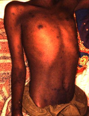
Boutonneuse fever is a fever as a result of a rickettsial infection caused by the bacterium Rickettsia conorii and transmitted by the dog tick Rhipicephalus sanguineus. Boutonneuse fever can be seen in many places around the world, although it is endemic in countries surrounding the Mediterranean Sea. This disease was first described in Tunisia in 1910 by Conor and Bruch and was named boutonneuse due to its papular skin-rash characteristics.

<i>Rickettsia rickettsii</i> Species of bacterium

Rickettsia rickettsii is a Gram-negative, intracellular, coccobacillus bacterium that was first discovered in 1902. R. rickettsii is the causative agent of Rocky Mountain Spotted Fever and is transferred to its host via a tick bite. It is one of the most pathogenic Rickettsia species and affects a large majority of the Western Hemisphere, most commonly the Americas.

<span class="mw-page-title-main">Robert Huebner</span> American physician

Robert Joseph Huebner, was an American physician and virologist whose research into viruses, their causes and treatment that led to his breakthrough insights into the connections between viruses and cancer, leading to new treatments, as well as his hypothesized oncogene, which was discovered to be a trigger for normal cells turning cancerous.

<span class="mw-page-title-main">Murine typhus</span> Medical condition

Murine typhus, also known as endemic typhus or flea-borne typhus, is a form of typhus transmitted by fleas, usually on rats, in contrast to epidemic typhus which is usually transmitted by lice. Murine typhus is an under-recognized entity, as it is often confused with viral illnesses. Most people who are infected do not realize that they have been bitten by fleas. Historically the term "hunger-typhus" was used in accounts by British POWs in Germany at the end of World War I when they described conditions in Germany.

<i>Orientia tsutsugamushi</i> Species of bacterium

Orientia tsutsugamushi is a mite-borne bacterium belonging to the family Rickettsiaceae and is responsible for a disease called scrub typhus in humans. It is a natural and an obligate intracellular parasite of mites belonging to the family Trombiculidae. With a genome of only 2.0–2.7 Mb, it has the most repeated DNA sequences among bacterial genomes sequenced so far. The disease, scrub typhus, occurs when infected mite larvae accidentally bite humans. Primarily indicated by undifferentiated febrile illnesses, the infection can be complicated and often fatal.

Rickettsia akari is a species of Rickettsia which causes rickettsialpox.

<span class="mw-page-title-main">Typhus</span> Group of infectious diseases

Typhus, also known as typhus fever, is a group of infectious diseases that include epidemic typhus, scrub typhus, and murine typhus. Common symptoms include fever, headache, and a rash. Typically these begin one to two weeks after exposure.

Rickettsia typhi is a small, aerobic, obligate intracellular, rod shaped gram negative bacterium. It belongs to the typhus group of the Rickettsia genus, along with R. prowazekii. R. typhi has an uncertain history, as it may have long gone shadowed by epidemic typhus. This bacterium is recognized as a biocontainment level 2/3 organism. R. typhi is a flea-borne disease that is best known to be the causative agent for the disease murine typhus, which is an endemic typhus in humans that is distributed worldwide. As with all rickettsial organisms, R. typhi is a zoonotic agent that causes the disease murine typhus, displaying non-specific mild symptoms of fevers, headaches, pains and rashes. There are two cycles of R. typhi transmission from animal reservoirs containing R. typhi to humans: a classic rat-flea-rat cycle that is most well studied and common, and a secondary periodomestic cycle that could involve cats, dogs, opossums, sheep, and their fleas.

Charles Pomerantz was a pest control expert and self-trained entomologist who played a pivotal role in identifying the etiology of a 1946 outbreak in New York City of what was later named rickettsialpox. In subsequent years, he spoke before audiences at colleges and other public forums about the menace from pests.

<span class="mw-page-title-main">African tick bite fever</span> Medical condition

African tick bite fever (ATBF) is a bacterial infection spread by the bite of a tick. Symptoms may include fever, headache, muscle pain, and a rash. At the site of the bite there is typically a red skin sore with a dark center. The onset of symptoms usually occurs 4–10 days after the bite. Complications are rare but may include joint inflammation. Some people do not develop symptoms.

Queensland tick typhus is a zoonotic disease caused by the bacterium Rickettsia australis. It is transmitted by the ticks Ixodes holocyclus and Ixodes tasmani.

Flea-borne spotted fever or California pseudotyphus is a condition characterized by a rash of maculopapules or furuncles.

<i>Rickettsia parkeri</i> Species of bacterium

Rickettsia parkeri is a gram-negative intracellular bacterium. The organism is found in the Western Hemisphere and is transmitted via the bite of hard ticks of the genus Amblyomma. R. parkeri causes mild spotted fever disease in humans, whose most common signs and symptoms are fever, an eschar at the site of tick attachment, rash, headache, and muscle aches. Doxycycline is the most common drug used to reduce the symptoms associated with disease.

<i>Ornithonyssus bacoti</i> Species of mite

Ornithonyssus bacoti is a hematophagous parasite. It feeds on blood and serum from many hosts. O. bacoti can be found and cause disease on rats and wild rodents most commonly, but also small mammals and humans when other hosts are scarce. Outbreaks tend to occur in older, less maintained buildings. The mite, however, can travel several hundred feet on its own if necessary to find a host and can survive for extended periods of time without a host. This, along with the nonspecific dermatitis it causes, can prevent accurate and fast diagnosis of rat mite dermatitis. The scarcity of reports, due in part to misdiagnosis and also the mildness of its symptoms, makes the disease seem less common than it is. The tropical rat mite can be found in both temperate and tropical regions or rather all continents except the Arctic and Antarctic.

Liponyssoides sanguineus is a species of mite that infests the house mouse.

Rickettsia felis is a species of bacterium, the pathogen that causes cat-flea typhus in humans, also known as flea-borne spotted fever. Rickettsia felis also is regarded as the causative organism of many cases of illnesses generally classed as fevers of unknown origin in humans in Africa.

Rodent mite dermatitis is an often unrecognized ectoparasitosis occurring after human contact with haematophagous mesostigmatid mites that infest rodents, such as house mice, rats and hamsters. The condition is associated with the tropical rat mite, spiny rat mite and house mouse mite which opportunistically feed on humans. Rodent mites are capable of surviving for long periods without feeding and travelling long distances when seeking hosts. Cases have been reported in homes, libraries, hospitals and care homes. A similar condition, known as gamasoidosis, is caused by avian mites.

References

  1. " Rickettsialpox " at Dorland's Medical Dictionary
  2. 1 2 Smith DS, Zhang E, Lopez FA, Go CH, Ake JA, Whitman TJ, Alam MJ, Mada PK (2019-07-10). Talavera F, Sanders CV, Brusch JL (eds.). "Rickettsialpox". EMedicine Infectious Diseases. WebMD LLC.
  3. Paddock, Christopher D. (2020-01-01), Ryan, Edward T.; Hill, David R.; Solomon, Tom; Aronson, Naomi E. (eds.), "70 - Rickettsialpox", Hunter's Tropical Medicine and Emerging Infectious Diseases (Tenth Edition), London: Elsevier, pp. 594–598, doi:10.1016/b978-0-323-55512-8.00070-3, ISBN   978-0-323-55512-8 , retrieved 2023-11-27
  4. Pedro-Pons A (1968). Patología y Clínica Médicas. Vol. 6 (3rd ed.). Barcelona: Salvat. p. 844. ISBN   84-345-1106-1.
  5. 1 2 3 4 5 6 Greenberg M, Pellitteri OJ, Jellison WL (July 1947). "Rickettsialpox-A Newly Recognized Rickettsial Disease : III. Epidemiology". American Journal of Public Health and the Nation's Health. 37 (7): 860–8. doi:10.2105/AJPH.37.7.860. PMC   1623794 . PMID   18016565.
  6. Beeman EA (2005). "Robert J. Huebner, M.D.:A Virologist's Odyssey" (PDF). National Institutes of Health . Retrieved 23 July 2009.
  7. Staff (4 October 1946). "NEW FEVER TRACED TO MITE ON MICE; U.S. Health Service Roots Out Cause of Spotted Ailment That Struck in Queens NO CURE IS FOUND AS YET Victim Made Ill by Bite of Insect". The New York Times . Retrieved 23 July 2009. Weinstein Urges War on Rodents Some Removed to Hospitals Blood of Patients Sampled
  8. Paddock CD, Eremeeva ME (2007). "Rickettsialpox". In Raoult D, Parola P (eds.). Rickettsial Diseases. Boca Raton, Florida: CRC Press. p. 63. ISBN   978-0-8493-7611-5.
  9. Freeman DW (16 November 2010). "Smallpox, Rickettsialpox Scare "House" Doctors: Is Threat Real?". www.cbsnews.com.
  10. "A Pox On Our House". House MD. Season 7. Episode 707. 15 November 2010.
  11. Bradley B (17 November 2014). "8 Rare 'House, M.D.' Cases That Can Actually Happen". HuffPost.