Rickettsia parkeri

Last updated

Rickettsia parkeri
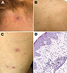
Rickettsia parkeri Rickettsiosis Argentina Eschar Papulovesicular Rash Histopathology - Romer et al 2011.jpg
Rickettsia parkeri rickettsiosis skin lesions: A - eschar after tick bite on neck; B, C - papulovesicular rash on back and leg; D - micrograph of biopsy specimen
Scientific classification OOjs UI icon edit-ltr.svg
Domain: Bacteria
Phylum: Pseudomonadota
Class: Alphaproteobacteria
Order: Rickettsiales
Family: Rickettsiaceae
Genus: Rickettsia
Species group: Spotted fever group
Species:
R. parkeri
Binomial name
Rickettsia parkeri
Lackman et al., 1965

Rickettsia parkeri (abbreviated R. parkeri) is a gram-negative intracellular bacterium. The organism is found in the Western Hemisphere and is transmitted via the bite of hard ticks of the genus Amblyomma . R. parkeri causes mild spotted fever disease in humans, whose most common signs and symptoms are fever, an eschar at the site of tick attachment, rash, headache, and muscle aches. Doxycycline is the most common drug used to reduce the symptoms associated with disease.

Contents

Biology

R. parkeri is classified in the spotted fever group of the genus Rickettsia . [1] [2] Genetically, its close relatives include R. africae , R. sibirica , R. conorii , R. rickettsii , R. peacockii , and R. honei . [1]

The organism has been isolated from numerous species of ticks in the genus Amblyomma: A. americanum in the United States; A. aureolatum in Brazil; A. maculatum in Mexico, Peru, and the United States; A. nodosum in Brazil; A. ovale in Brazil and Mexico; A. parvitarsum in Argentina and Chile; A. tigrinum in Argentina, Bolivia, Brazil, and Uruguay; and A. triste in Argentina, Brazil, the United States, and Uruguay. [2] [3] [4] [5] Different ticks may carry different strains of the organism. R. parkeri sensu stricto ("in the strict sense") is found in A. maculatum and A. triste; R. parkeri strain NOD, in A. nodosum; R. parkeri strain Parvitarsum, in A. parvitarsum; and R. parkeri strain Atlantic rainforest, in A. aureolatum and A. ovale. [2]

Human infections

The first report of a confirmed human case of infection with R. parkeri was published in 2004. [6] [7] The person was infected in the state of Virginia in the United States. [6] Other confirmed or probable human cases have been reported to have acquired infection elsewhere in the United States (e.g., Arizona, Georgia, and Mississippi), as well as in Argentina, Brazil, Colombia, Mexico, and Uruguay. [8] [9] Terms used to describe human infection with R. parkeri include "American boutonneuse fever" because of its similarity to boutonneuse fever caused by Rickettsia conorii ; [10] "American tick bite fever" because of its similarity to African tick bite fever caused by Rickettsia africae ; [11] "Tidewater spotted fever," after the Tidewater region in the eastern United States; [12] and "Rickettsia parkeri rickettsiosis" or "R. parkeri rickettsiosis." [7] [12]

Epidemiology

Of all human cases documented in the medical literature, 87% were 18-64 years of age, and most cases were male. [8] Brazil, Argentina, and the United States accounted for the majority of cases in the medical literature. [8] In the United States, most of the 40 cases reported to the Centers for Disease Control and Prevention (CDC) as of 2016 became infected between the months of July and September. [13] :5–6

Diagnosis

The CDC recommends polymerase chain reaction (PCR) of a biopsy or swab of an eschar, or PCR of a biopsy of a rash, for diagnosis of R. parkeri infection. [13] :27 In addition, indirect immunofluorescence antibody (IFA) assays using paired acute and convalescent sera can be used. [13] :27

Clinical manifestations

A 2008 study compared 12 R. parkeri cases with 208 Rocky Mountain spotted fever cases caused by R. rickettsii. [7] Although both R. parkeri and R. rickettsii caused fever, rash, myalgia, and headache, R. parkeri caused eschars and R. rickettsii did not. [7] Furthermore, the percentage of patients hospitalized was lower for R. parkeri than for R. rickettsii (33% vs 78%), and R. parkeri led to no deaths while R. rickettsii led to death in 7% of cases. [7]

A 2021 systematic review of 32 confirmed and 45 probable cases of human infection with R. parkeri determined that 94% of the confirmed cases had fever, 91% an eschar, 72% a rash, 56% headache, and 56% myalgia, with similar percentages among the probable cases. [8] The rash was most frequently described as papular or macular. [8] Among the confirmed and probable cases, the most common treatment was doxycycline, followed by tetracycline. [8] Although 9% of all the cases were hospitalized, there was a "100% rate of clinical recovery." [8]

History

In 1939, Ralph R. Parker, director of the Rocky Mountain Laboratory, and others published a paper on "a rickettsia-like infectious agent." [7] [14] The agent, found in Amblyomma maculatum ticks collected from cows in Texas, produced mild disease in guinea pigs. [7] [14] In 1965, Lackman and others named the rickettsial organism R. parkeri after Parker. [2] [15]

Related Research Articles

<span class="mw-page-title-main">Rocky Mountain spotted fever</span> Human disease

Rocky Mountain spotted fever (RMSF) is a bacterial disease spread by ticks. It typically begins with a fever and headache, which is followed a few days later with the development of a rash. The rash is generally made up of small spots of bleeding and starts on the wrists and ankles. Other symptoms may include muscle pains and vomiting. Long-term complications following recovery may include hearing loss or loss of part of an arm or leg.

<span class="mw-page-title-main">Boutonneuse fever</span> Medical condition

Boutonneuse fever is a fever as a result of a rickettsial infection caused by the bacterium Rickettsia conorii and transmitted by the dog tick Rhipicephalus sanguineus. Boutonneuse fever can be seen in many places around the world, although it is endemic in countries surrounding the Mediterranean Sea. This disease was first described in Tunisia in 1910 by Conor and Bruch and was named boutonneuse due to its papular skin-rash characteristics.

<i>Rickettsia rickettsii</i> Species of bacterium

Rickettsia rickettsii is a Gram-negative, intracellular, coccobacillus bacterium that was first discovered in 1902. R. rickettsii is the causative agent of Rocky Mountain Spotted Fever and is transferred to its host via a tick bite. It is one of the most pathogenic Rickettsia species and affects a large majority of the Western Hemisphere, most commonly the Americas.

<span class="mw-page-title-main">Eschar</span> Piece of dead tissue caused by some skin injuries

An eschar is a slough or piece of dead tissue that is cast off from the surface of the skin, particularly after a burn injury, but also seen in gangrene, ulcer, fungal infections, necrotizing spider bite wounds, tick bites associated with spotted fevers and exposure to cutaneous anthrax. The term ‘eschar’ is not interchangeable with ‘scab’. An eschar contains necrotic tissue whereas a scab is composed of dried blood and exudate.

Tick-borne diseases, which afflict humans and other animals, are caused by infectious agents transmitted by tick bites. They are caused by infection with a variety of pathogens, including rickettsia and other types of bacteria, viruses, and protozoa. The economic impact of tick-borne diseases is considered to be substantial in humans, and tick-borne diseases are estimated to affect ~80 % of cattle worldwide. Most of these pathogens require passage through vertebrate hosts as part of their life cycle. Tick-borne infections in humans, farm animals, and companion animals are primarily associated with wildlife animal reservoirs. many tick-borne infections in humans involve a complex cycle between wildlife animal reservoirs and tick vectors. The survival and transmission of these tick-borne viruses are closely linked to their interactions with tick vectors and host cells. These viruses are classified into different families, including Asfarviridae, Reoviridae, Rhabdoviridae, Orthomyxoviridae, Bunyaviridae, and Flaviviridae.

<span class="mw-page-title-main">Viral hemorrhagic fever</span> Type of illnesses

Viral hemorrhagic fevers (VHFs) are a diverse group of animal and human illnesses. VHFs may be caused by five distinct families of RNA viruses: the families Filoviridae, Flaviviridae, Rhabdoviridae, and several member families of the Bunyavirales order such as Arenaviridae, and Hantaviridae. All types of VHF are characterized by fever and bleeding disorders and all can progress to high fever, shock and death in many cases. Some of the VHF agents cause relatively mild illnesses, such as the Scandinavian nephropathia epidemica, while others, such as Ebola virus, can cause severe, life-threatening disease.

Relapsing fever is a vector-borne disease caused by infection with certain bacteria in the genus Borrelia, which is transmitted through the bites of lice or soft-bodied ticks.

A rickettsiosis is a disease caused by intracellular bacteria.

<i>Orientia tsutsugamushi</i> Species of bacterium

Orientia tsutsugamushi is a mite-borne bacterium belonging to the family Rickettsiaceae and is responsible for a disease called scrub typhus in humans. It is a natural and an obligate intracellular parasite of mites belonging to the family Trombiculidae. With a genome of only 2.0–2.7 Mb, it has the most repeated DNA sequences among bacterial genomes sequenced so far. The disease, scrub typhus, occurs when infected mite larvae accidentally bite humans. Primarily indicated by undifferentiated febrile illnesses, the infection can be complicated and often fatal.

<i>Rickettsia conorii</i> Species of bacterium

Rickettsia conorii is a Gram-negative, obligate intracellular bacterium of the genus Rickettsia that causes human disease called boutonneuse fever, Mediterranean spotted fever, Israeli tick typhus, Astrakhan spotted fever, Kenya tick typhus, Indian tick typhus, or other names that designate the locality of occurrence while having distinct clinical features. It is a member of the spotted fever group and the most geographically dispersed species in the group, recognized in most of the regions bordering on the Mediterranean Sea and Black Sea, Israel, Kenya, and other parts of North, Central, and South Africa, and India. The prevailing vector is the brown dog tick, Rhipicephalus sanguineus. The bacterium was isolated by Emile Brumpt in 1932 and named after A. Conor, who in collaboration with A. Bruch, provided the first description of boutonneuse fever in Tunisia in 1910.

<span class="mw-page-title-main">African tick bite fever</span> Medical condition

African tick bite fever (ATBF) is a bacterial infection spread by the bite of a tick. Symptoms may include fever, headache, muscle pain, and a rash. At the site of the bite there is typically a red skin sore with a dark center. The onset of symptoms usually occurs 4–10 days after the bite. Complications are rare but may include joint inflammation. Some people do not develop symptoms.

Queensland tick typhus is a zoonotic disease caused by the bacterium Rickettsia australis. It is transmitted by the ticks Ixodes holocyclus and Ixodes tasmani.

Flinders Island spotted fever is a condition characterized by a rash in approximately 85% of cases.

Rickettsia helvetica, previously known as the Swiss agent, is a bacterium found in Dermacentor reticulatus and other ticks, which has been implicated as a suspected but unconfirmed human pathogen. First recognized in 1979 in Ixodes ricinus ticks in Switzerland as a new member of the spotted fever group of Rickettsia, the R. helvetica bacterium was eventually isolated in 1993. Although R. helvetica was initially thought to be harmless in humans and many animal species, some individual case reports suggest that it may be capable of causing a nonspecific fever in humans. In 1997, a man living in eastern France seroconverted to Rickettsia 4 weeks after onset of an unexplained febrile illness. In 2010, a case report indicated that tick-borne R. helvetica can also cause meningitis in humans.

Rickettsia felis is a species of bacterium, the pathogen that causes cat-flea typhus in humans, also known as flea-borne spotted fever. Rickettsia felis also is regarded as the causative organism of many cases of illnesses generally classed as fevers of unknown origin in humans in Africa.

<i>Amblyomma maculatum</i> Species of tick

Amblyomma maculatum is a species of tick in the genus Amblyomma. Immatures usually infest small mammals and birds that dwell on the ground; cotton rats may be particularly favored hosts. Some recorded hosts include:

Rickettsia heilongjiangensis is a species of gram negative Alphaproteobacteria, within the spotted fever group, being carried by ticks. It is pathogenic.

The Asian monitor lizard tick, is a hard-bodied tick of the genus Amblyomma. It is found in India, Thailand, Taiwan and Sri Lanka. Adults parasitize various reptiles such as varanids and snakes. These ticks are potential vectors of spotted fever group (SFG) rickettsiae.

Pacific Coast tick fever is an infection caused by Rickettsia philipii. The disease is spread by the Pacific coast ticks. Symptoms may include an eschar. It is within a group known as spotted fever rickettsiosis together with Rickettsia parkeri rickettsiosis, Rocky Mountain spotted fever, and rickettsialpox. These infections can be difficult to tell apart.

<i>Amblyomma triste</i>

Amblyomma triste is a tick in the Amblyomma genus. The tick can be found in Venezuela, Argentina, Brasil, Colombia, Peru and Uruguay. Though not thought to be endemic to North America, a 2010 study found 27 specimens in 18 separate collections that had previously been misidentified in the United States.
References

  1. 1 2 Perlman SJ, Hunter MS, Zchori-Fein E (2006). "The emerging diversity of Rickettsia". Proc Biol Sci. 273 (1598): 2097–2106. doi:10.1098/rspb.2006.3541. PMC   1635513 . PMID   16901827.{{cite journal}}: CS1 maint: multiple names: authors list (link)
  2. 1 2 3 4 Nieri-Bastos FA, Marcili A, De Sousa R, Paddock CD, Labruna MB (2018). "Phylogenetic evidence for the existence of multiple strains of Rickettsia parkeri in the New World". Appl Environ Microbiol. 84 (8): e02872–17. Bibcode:2018ApEnM..84E2872N. doi:10.1128/AEM.02872-17. PMC   5881050 . PMID   29439989.{{cite journal}}: CS1 maint: multiple names: authors list (link)
  3. Cohen SB, Yabsley MJ, Garrison LE, Freye JD, Dunlap BG, Dunn JR; et al. (2009). "Rickettsia parkeri in Amblyomma americanum ticks, Tennessee and Georgia, USA". Emerg Infect Dis. 15 (9): 1471–3. doi:10.3201/eid1509.090330. PMC   2819870 . PMID   19788817.{{cite journal}}: CS1 maint: multiple names: authors list (link)
  4. Delgado-de la Mora J, Sánchez-Montes S, Licona-Enríquez JD, Delgado-de la Mora D, Paddock CD, Beati L; et al. (2019). "Rickettsia parkeri and Candidatus Rickettsia andeanae in Tick of the Amblyomma maculatum Group, Mexico". Emerg Infect Dis. 25 (4): 836–838. doi:10.3201/eid2504.181507. PMC   6433039 . PMID   30882330.{{cite journal}}: CS1 maint: multiple names: authors list (link)
  5. Sánchez-Montes S, Ballados-González GG, Hernández-Velasco A, Zazueta-Islas HM, Solis-Cortés M, Miranda-Ortiz H; et al. (2019). "Molecular Confirmation of Rickettsia parkeri in Amblyomma ovale Ticks, Veracruz, Mexico". Emerg Infect Dis. 25 (12): 2315–2317. doi:10.3201/eid2512.190964. PMC   6874242 . PMID   31742525.{{cite journal}}: CS1 maint: multiple names: authors list (link)
  6. 1 2 Paddock CD, Sumner JW, Comer JA, Zaki SR, Goldsmith CS, Goddard J; et al. (2004). "Rickettsia parkeri: a newly recognized cause of spotted fever rickettsiosis in the United States". Clin Infect Dis. 38 (6): 805–811. doi: 10.1086/381894 . PMID   14999622.{{cite journal}}: CS1 maint: multiple names: authors list (link)
  7. 1 2 3 4 5 6 7 Paddock CD, Finley RW, Wright CS, Robinson HN, Schrodt BJ, Lane CC; et al. (2008). "Rickettsia parkeri rickettsiosis and its clinical distinction from Rocky Mountain spotted fever". Clin Infect Dis. 47 (9): 1188–1196. doi: 10.1086/592254 . PMID   18808353.{{cite journal}}: CS1 maint: multiple names: authors list (link)
  8. 1 2 3 4 5 6 7 Silva-Ramos CR, Hidalgo M, Faccini-Martínez ÁA (2021). "Clinical, epidemiological, and laboratory features of Rickettsia parkeri rickettsiosis: A systematic review". Ticks Tick Borne Dis. 12 (4): 101734. doi:10.1016/j.ttbdis.2021.101734. PMID   33989945. S2CID   234596034.{{cite journal}}: CS1 maint: multiple names: authors list (link)
  9. Torres-Castro M, Sánchez-Montes S, Colunga-Salas P, Noh-Pech H, Reyes-Novelo E, Rodríguez-Vivas RI (2022). "Molecular confirmation of Rickettsia parkeri in humans from Southern Mexico". Zoonoses Public Health. 69 (4): 382–386. doi:10.1111/zph.12927. PMID   35142079. S2CID   246701684.{{cite journal}}: CS1 maint: multiple names: authors list (link)
  10. Goddard J, Paddock CD (2005). "Observations on distribution and seasonal activity of the Gulf Coast tick in Mississippi". J Med Entomol. 42 (2): 176–179. doi: 10.1093/jmedent/42.2.176 . PMID   15799527.
  11. Iweriebor BC, Nqoro A, Obi CL (2020). "Rickettsia africae an agent of African tick bite fever in ticks collected from domestic animals in Eastern Cape, South Africa". Pathogens. 9 (8): 631. doi: 10.3390/pathogens9080631 . PMC   7459594 . PMID   32748891.{{cite journal}}: CS1 maint: multiple names: authors list (link)
  12. 1 2 Wright CL, Nadolny RM, Jiang J, Richards AL, Sonenshine DE, Gaff HD; et al. (2011). "Rickettsia parkeri in Gulf Coast ticks, southeastern Virginia, USA". Emerg Infect Dis. 17 (5): 896–8. doi:10.3201/eid1705.101836. PMC   3321792 . PMID   21529406.{{cite journal}}: CS1 maint: multiple names: authors list (link)
  13. 1 2 3 Biggs HM, Behravesh CB, Bradley KK, Dahlgren FS, Drexler NA, Dumler JS; et al. (2016). "Diagnosis and management of tickborne rickettsial diseases: Rocky Mountain Spotted Fever and other spotted fever group rickettsioses, ehrlichioses, and anaplasmosis - United States". MMWR Recomm Rep. 65 (2): 1–44. doi: 10.15585/mmwr.rr6502a1 . PMID   27172113.{{cite journal}}: CS1 maint: multiple names: authors list (link)
  14. 1 2 Parker RR, Kohls GM, Cox GW, Davis GE (1939). "Observations on an infectious agent from Amblyomma maculatum". Public Health Rep. 54 (32): 1482–1484. doi: 10.1086/592254 . PMID   18808353.{{cite journal}}: CS1 maint: multiple names: authors list (link)
  15. Lackman DB, Bell EJ, Stoenner HG, Pickens EG (1965). "The Rocky Mountain spotted fever group of rickettsias". Health Lab Sci. 2: 135–141. PMID   14318051.{{cite journal}}: CS1 maint: multiple names: authors list (link)