Bullous impetigo

Last updated
Bullous impetigo
Bullous impetigo1.jpg
Bullous impetigo after rupture of the bullae
Specialty Infectious disease/dermatology

Bullous impetigo is a bacterial skin infection caused by Staphylococcus aureus that results in the formation of large blisters called bullae, usually in areas with skin folds like the armpit, groin, between the fingers or toes, beneath the breast, and between the buttocks. It accounts for 30% of cases of impetigo, the other 70% being non-bullous impetigo. [1]

Contents

The bullae are caused by exfoliative toxins produced by Staphylococcus aureus that cause the connections between cells in the uppermost layer of the skin to fall apart. [1] Bullous impetigo in newborns, children, or adults who are immunocompromised and/or are experiencing kidney failure, can develop into a more severe and generalized form called staphylococcal scalded skin syndrome (SSSS). The mortality rate is less than 3% for infected children, but up to 60% in adults. [2]

Signs and symptoms

Bullous impetigo on the arm Bullous impetigo2.jpg
Bullous impetigo on the arm
Bullous impetigo Impetigo.jpg
Bullous impetigo

Bullous impetigo can appear around the diaper region, axilla, or neck. The bacteria causes a toxin to be produced that reduces cell-to-cell stickiness (adhesion), causing for the top layer of skin (epidermis), and lower layer of skin (dermis) to separate. Vesicles rapidly enlarge and form the bullae which is a blister more than 5mm across. Bullae is also known as staphylococcal scalded skin syndrome. Other associated symptoms are itching, swelling of nearby glands, fever and diarrhea. Pain is very rare.[ citation needed ]

Long-term effects: once the scabs on the bullous have fallen off, scarring is minimal. Possible long-term effects are kidney disease.[ citation needed ]

Cause

Exposure is most commonly seen in hospital wards and nurseries, and can be passed from person to person in other settings, such as close contact sports. Therefore, the patient is advised to try to limit human contact as much as possible to minimize the risk of spreading the infection. [3]

Infectious period

After 48 hours the disease is considered no longer contagious assuming the proper antibiotic treatments have been administered.

Pathogenesis

Exfoliating toxins are serine proteases that specifically bind to and cleave desmoglein 1 (Dsg1). Previous studies suggested that exfoliating toxins bind to gangliosides, causing a release of protease by keratinocytes acting as superantigens in stimulating the skin's immune system. [2] A more recent proposal states there are three known exfoliating toxins; ETA, ETB, and ETD which act as a glutamic acid-specific serine protease with concentrated specificity. Which results in the cleavage of human Dsg1 at a unique site after glutamic acid residues causing deactivation. [2] Proteolysis of the peptide bond leading up to the dysfunction of Dsg1 and the desmosome allows for an understanding as to why the bullous forms, making the peptide bond crucial for proper function if Dsg1.[ citation needed ]

S. aureus

A pyogenic non-motile Gram-positive cocci which forms into grape like clusters. Just like other forms of staph, S. aureus has a variety of virulence factors which include surface proteins involved in adherence, secretion of enzymes that degrade proteins, and secrete toxins which damage the host's cells.[ citation needed ]

S. aureus expresses surface receptors for fibrinogen, fibronectin, and vitronectin. These surface receptors allow a bridge to be formed which binds to host endothelial cells. Lipases allow for the degradation of lipids on the skin surface and its expression can be directly correlated with its ability of the bacteria to produce abscesses. [4]

Diagnosis

Observing the skin's physical appearance, or swabbing a culture of the lesion for S. aureus. Nasal swabs from the patient's immediate family members are necessary to identify them as being asymptomatic nasal carriers of S. aureus.[ citation needed ]

Histology

The epidermis is composed of four layers, stratum basale, stratum spinosum, stratum granulosum, and stratum corneum. [5]

The cleavage plane can be found either subcorneally or within the upper stratum granulosum. The roof of the pustule is parakeratotic stratum corneum, and the floor is formed of keratinocytes, which may or may not be acantholytic. [6] Neutrophils begin to fill the pustule. Toxins are produced by S. aureus and target desmoglein, which is a desmosomal cell-cell adhesion molecule found in the upper levels of the epidermis. This correlates with the subcorneal localization of the bullae. [6]

Clinical Differential

Prevention

Since the common pathogens involved with impetigo are bacteria naturally found on the skin, most prevention (especially in children), is targeted towards appropriate hygiene, wound cleaning, and minimizing scratching (i.e. by keeping nails trimmed and short). Avoiding close contact and sharing of items such as towels with potentially infected individuals is also recommended.[ citation needed ]

Management

Antibiotic creams are the preferred treatment for mild cases of impetigo, despite their limited systemic absorption. Such prescribed ointments include neosporin, fusidic acid, chloramphenicol and mupirocin. More severe cases of impetigo however (especially bullous impetigo) will likely require oral agents with better systemic bioavailability, such as cephalexin. Cases that do not resolve with initial antibiotic therapy or require hospitalization may also be indicative an MRSA infection, which would require consultation with a local microbiologist. [7]

Antibiotic treatment typically last 7–10 days, and although highly effective some cases of methicillin resistant S. aureus (MRSA) may require longer therapy depending on the severity of infection and how much it has spread.

See also

Related Research Articles

<span class="mw-page-title-main">Integumentary system</span> Skin and other protective organs

The integumentary system is the set of organs forming the outermost layer of an animal's body. It comprises the skin and its appendages, which act as a physical barrier between the external environment and the internal environment that it serves to protect and maintain the body of the animal. Mainly it is the body's outer skin.

<span class="mw-page-title-main">Impetigo</span> Human disease (bacterial infection)

Impetigo is a bacterial infection that involves the superficial skin. The most common presentation is yellowish crusts on the face, arms, or legs. Less commonly there may be large blisters which affect the groin or armpits. The lesions may be painful or itchy. Fever is uncommon.

<i>Staphylococcus aureus</i> Species of Gram-positive bacterium

Staphylococcus aureus is a Gram-positive spherically shaped bacterium, a member of the Bacillota, and is a usual member of the microbiota of the body, frequently found in the upper respiratory tract and on the skin. It is often positive for catalase and nitrate reduction and is a facultative anaerobe that can grow without the need for oxygen. Although S. aureus usually acts as a commensal of the human microbiota, it can also become an opportunistic pathogen, being a common cause of skin infections including abscesses, respiratory infections such as sinusitis, and food poisoning. Pathogenic strains often promote infections by producing virulence factors such as potent protein toxins, and the expression of a cell-surface protein that binds and inactivates antibodies. S. aureus is one of the leading pathogens for deaths associated with antimicrobial resistance and the emergence of antibiotic-resistant strains, such as methicillin-resistant S. aureus (MRSA), is a worldwide problem in clinical medicine. Despite much research and development, no vaccine for S. aureus has been approved.

<span class="mw-page-title-main">Epidermis</span> Outermost of the three layers that make up the skin

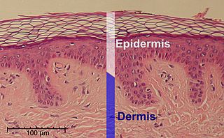
The epidermis is the outermost of the three layers that comprise the skin, the inner layers being the dermis and hypodermis. The epidermis layer provides a barrier to infection from environmental pathogens and regulates the amount of water released from the body into the atmosphere through transepidermal water loss.

<span class="mw-page-title-main">Desmosome</span> Cell junction involved in cell-to-cell adhesion

A desmosome, also known as a macula adherens, is a cell structure specialized for cell-to-cell adhesion. A type of junctional complex, they are localized spot-like adhesions randomly arranged on the lateral sides of plasma membranes. Desmosomes are one of the stronger cell-to-cell adhesion types and are found in tissue that experience intense mechanical stress, such as cardiac muscle tissue, bladder tissue, gastrointestinal mucosa, and epithelia.

<span class="mw-page-title-main">Skin condition</span> Any medical condition that affects the integumentary system

A skin condition, also known as cutaneous condition, is any medical condition that affects the integumentary system—the organ system that encloses the body and includes skin, nails, and related muscle and glands. The major function of this system is as a barrier against the external environment.

<span class="mw-page-title-main">Stratum corneum</span> Outermost layer of the epidermis

The stratum corneum is the outermost layer of the epidermis. The human stratum corneum comprises several levels of flattened corneocytes that are divided into two layers: the stratum disjunctum and stratum compactum. The skin's protective acid mantle and lipid barrier sit on top of the stratum disjunctum. The stratum disjunctum is the uppermost and loosest layer of skin. The stratum compactum is the comparatively deeper, more compacted and more cohesive part of the stratum corneum. The corneocytes of the stratum disjunctum are larger, more rigid and more hydrophobic than that of the stratum compactum.

<span class="mw-page-title-main">Pemphigus</span> Blistering autoimmune diseases

Pemphigus is a rare group of blistering autoimmune diseases that affect the skin and mucous membranes. The name is derived from the Greek root pemphix, meaning "pustule".

<span class="mw-page-title-main">Desquamation</span> Medical condition

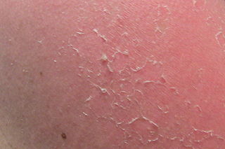
Desquamation occurs when the outermost layer of a tissue, such as the skin, is shed. The term is from Latin desquamare 'to scrape the scales off a fish'.

<span class="mw-page-title-main">Staphylococcal scalded skin syndrome</span> Medical condition

Staphylococcal scalded skin syndrome (SSSS) is a dermatological condition caused by Staphylococcus aureus.

<span class="mw-page-title-main">Stratum granulosum</span> Cell layer in the epidermis

The stratum granulosum is a thin layer of cells in the epidermis lying above the stratum spinosum and below the stratum corneum. Keratinocytes migrating from the underlying stratum spinosum become known as granular cells in this layer. These cells contain keratohyalin granules, which are filled with histidine- and cysteine-rich proteins that appear to bind the keratin filaments together. Therefore, the main function of keratohyalin granules is to bind intermediate keratin filaments together.

Nikolsky's sign is a clinical dermatological sign, named after Pyotr Nikolsky (1858–1940), a Russian physician who trained and worked in the Russian Empire. The sign is present when slight rubbing of the skin results in exfoliation of the outermost layer. A typical test would be to place the eraser of a pencil on the roof of a lesion and spin the pencil in a rolling motion between the thumb and forefinger. If the lesion is opened, then the Nikolsky's sign is present/positive.

A skin infection is an infection of the skin in humans and other animals, that can also affect the associated soft tissues such as loose connective tissue and mucous membranes. They comprise a category of infections termed skin and skin structure infections (SSSIs), or skin and soft tissue infections (SSTIs), and acute bacterial SSSIs (ABSSSIs). They are distinguished from dermatitis, although skin infections can result in skin inflammation.

<span class="mw-page-title-main">Pemphigus vulgaris</span> Medical condition

Pemphigus vulgaris is a rare chronic blistering skin disease and the most common form of pemphigus. Pemphigus was derived from the Greek word pemphix, meaning blister. It is classified as a type II hypersensitivity reaction in which antibodies are formed against desmosomes, components of the skin that function to keep certain layers of skin bound to each other. As desmosomes are attacked, the layers of skin separate and the clinical picture resembles a blister. These blisters are due to acantholysis, or breaking apart of intercellular connections through an autoantibody-mediated response. Over time the condition inevitably progresses without treatment: lesions increase in size and distribution throughout the body, behaving physiologically like a severe burn.

<span class="mw-page-title-main">Desmoglein-1</span> Protein found in humans

Desmoglein-1 is a protein that in humans is encoded by the DSG1 gene. Desmoglein-1 is expressed everywhere in the skin epidermis, but mainly it is expressed in the superficial upper layers of the skin epidermis.

<span class="mw-page-title-main">Lamellar bodies</span> Secretory organelles

In cell biology, lamellar bodies are secretory organelles found in type II alveolar cells in the lungs, and in keratinocytes in the skin. They are oblong structures, appearing about 300-400 nm in width and 100-150 nm in length in transmission electron microscopy images. Lamellar bodies in the alveoli of the lungs fuse with the cell membrane and release pulmonary surfactant into the extracellular space.

Corneocytes are terminally differentiated keratinocytes and compose most of the stratum corneum, the outermost layer of the epidermis. They are regularly replaced through desquamation and renewal from lower epidermal layers and are essential for its function as a skin barrier.

<span class="mw-page-title-main">Staphylococcal infection</span> Medical condition

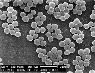
A staphylococcal infection or staph infection is an infection caused by members of the Staphylococcus genus of bacteria.

Exfoliatin is a Staphylococcus aureus exotoxin that causes a blistering of the skin known as staphylococcal scalded skin syndrome, usually in infants.

Pemphigus foliaceus is an autoimmune of the skin. Pemphigus foliaceus causes a characteristic inflammatory attack at the subcorneal layer of epidermis, which results in skin lesions that are scaly or crusted erosions with an erythematous (red) base. Mucosal involvement is absent even with widespread disease.

References

  1. 1 2 Hartman-Adams H, Banvard C, Juckett G (15 August 2014). "Impetigo: diagnosis and treatment". American Family Physician. 90 (4): 229–35. PMID   25250996.
  2. 1 2 3 Yasushi, Hanakawa. "Molecular mechanisms of blister formation in bullous impetigo and staphylococcal scalded skin syndrome." Journal of Clinical Investigation. 110.1 (2002): 53-60.
  3. Lucky, A. (2009, November 9). Blistering disorders in infancy. Retrieved from http://www.docstoc.com/docs/15402241/Blistering-Disorders-in-Infancy
  4. Kumar, V, A Abbas, and N Fausto. "Pathologic Basis of Disease." 7th. Chicago: Robbins and Cotran, 2004. 620
  5. Roy, S. (2009). Histology of the normal skin. Retrieved from http://www.histopathology-india.net/NH.htm Archived 2011-02-18 at the Wayback Machine
  6. 1 2 Carter, D, J Greenson, H Oberman, V Reuter, and StolerM. "Sternberg's Diagnostic Surgical Pathology." 4th. 1. New York City: Lippincott Williams & Wilkins, 2004. 17f
  7. "Impetigo: antimicrobial prescribing". NICE. Retrieved 8 July 2021.