Chlorovirus

Last updated

Chlorovirus
Virus classification OOjs UI icon edit-ltr.svg
(unranked): Virus
Realm: Varidnaviria
Kingdom: Bamfordvirae
Phylum: Nucleocytoviricota
Class: Megaviricetes
Order: Algavirales
Family: Phycodnaviridae
Genus:Chlorovirus

Chlorovirus, also known as Chlorella virus, is a genus of giant double-stranded DNA viruses, in the family Phycodnaviridae . This genus is found globally in freshwater environments [1] where freshwater microscopic algae serve as natural hosts. There are 19 species in this genus. [2] [3]

Contents

Chlorovirus was discovered in 1981 by Russel H. Meints, James L. Van Etten, Daniel Kuczmarski, Kit Lee, and Barbara Ang while attempting to culture Chlorella-like algae. During the attempted process viral particles were discovered in the cells 2 to 6 hours after being initially isolated, followed by lysis after 12 to 20 hours. This virus was initially called HVCV (Hydra viridis Chlorella virus) since it was first found to infect Chlorella-like algae. [4] [5]

Though relatively new to virologists and thus not extensively studied, one species, Chlorovirus ATCV-1, commonly found in lakes, has been recently found to infect humans. [6] New studies focusing on effects of infection in mouse model are currently emerging as well. [6] [7]

Taxonomy

Chlorovirus is a genus of giant double-stranded DNA (dsDNA) viruses in the family Phycodnaviridae , and Baltimore group 1: dsDNA viruses. The genus contains the following species: [3]

Ecology

Chloroviruses are widespread in freshwater environments in all parts of the globe and have been isolated from freshwater sources in Europe, Asia, Australia, as well as North and South America. [1] [8] Natural hosts of chloroviruses include various types of unicellular eukaryotic Chlorella-like algae, with individual virus species typically infecting only within a distinct strain. These algal hosts are known to establish endosymbiotic relationships with larger protists, such as Paramecium bursaria (a member of the ciliates), Acanthocystis turfacea (a centroheliozoan) and Hydra viridis (member of the hydrozoa). [9] While an individual protist can harbour up to several hundred algal cells at any given time, free-floating algae are highly susceptible to chloroviruses, indicating that such endosymbiosis serves to provide resistance from infection. [10]

Chlorovirus titers are variable by season and location, but typically fluctuate between 1 and 100 PFU/mL, although high abundances of up to 100,000 PFU/mL may occur in some environments. Due to the rich genetic diversity and high specialization of individual species with respect to infectious range, variations in their ecology are not unusual, resulting in unique spatio-temporal patterns, which ultimately depend on lifestyle and nature of the host. As such, previous survey data highlighted two prominent seasonal abundance peaks for both Chlorella variabilis NC64A and Chlorella variabilis Syngen viruses — one in late fall, and the other in late spring to mid-summer — which is likely attributed to the fact that they share a host species. Conversely, Chlorella heliozoae SAG viruses peaked at different times of the year and generally exhibited more variability in titers, as compared to the NC64A and Syngen viruses. [1] Additionally, studies revealed that chloroviruses demonstrate some resilience in response to decreased temperatures observed during the winter season, characterized by presence of infectious particles under ice layers in a stormwater management pond in Ontario, Canada. [11] Further, DeLong et al. (2016) suggest that predation by small crustaceans can play an indirect role in titer fluctuations, as degradation of protist cells passing through the digestive tract results in liberation of large numbers of unicellular algae that become susceptible to viral infection due to disruption of endosymbiosis. [10] Overall, seasonal abundance of chloroviruses depends not only on the host species, but also on the abundance of other microorganisms, general nutrient status and ecological conditions. [12]

Collectively, chloroviruses are able to mediate global biogeochemical cycles through phytoplankton turnover. Chlorella, in co-occurrence with other types of microscopic algae like Microcystis aeruginosa , are known to cause toxic algal blooms that typically last from February to June in the Northern hemisphere, resulting in oxygen depletion and deaths of larger organisms in freshwater habitats. [13] [14] Lytic infection of unicellular algae by chloroviruses results in termination of algal blooms and the subsequent release of carbon, nitrogen and phosphorus trapped in the cells, transporting them to lower trophic levels and, ultimately, fueling the food chain. [12]

Structure

Schematic drawing of a typical Phycodnaviridae virion (cross section and side view, not showing spike and vertex) Phycodnaviridae virion.jpg
Schematic drawing of a typical Phycodnaviridae virion (cross section and side view, not showing spike and vertex)

Viruses in the genus Chlorovirus are enveloped, with icosahedral and spherical geometries, and T=169 (triangulation number) symmetry. The diameter is around 100-220 nm. Genomes are linear, usually single-copy, composed of dsDNA (double-stranded DNA), and around 330 kb in length. The dsDNA is closed with a hairpin structure terminus. Genomes also often have several hundred open reading frames. [2] As a group, chloroviruses encode from 632 protein families; however, each individual virus only has 330 to 416 protein encoding genes. As part of the DNA modification systems, chloroviruses have methylated bases in specific sections of their DNA sequence. Some chloroviruses also contain introns and inteins, though this is rare within the genus. [9]

Paramecium bursaria Chlorella virus 1 (PBCV-1) have a 190 nm diameter [9] and a fivefold axis. [15] One face's juncture has a protruding spike, which is the first part of the virus to contact its host. [16] The outer capsid covers a single lipid bilayer membrane, which is obtained from the host's endoplasmic reticulum. [15] Some capsomers on the external shell have fibres extending away from the virus to aid in host attachment. [17] [16]

GenusStructureSymmetryCapsidGenomic arrangementGenomic segmentation
ChlorovirusIcosahedralT=169EnvelopedLinearMonopartite

Hosts

Chloroviruses infect certain unicellular, eukaryotic chlorella-like green algae, called zoochlorellae, and are very species and even strain specific. These zoochlorellae commonly establish endosymbiotic relationships with the protozoan Paramecium bursaria , the coelenterate Hydra viridis , the heliozoon Acanthocystis turfacea and other freshwater and marine invertebrates and protozoans. The viruses cannot infect zoochlorellae when they are in their symbiotic phase, and there is no evidence that zoochlorellae grow free of their hosts in indigenous waters. [18] Chloroviruses have also recently been found to infect people, leading to studies on infections in mice as well. [6]

Life cycle

Chlorella cells and chlorovirus Paramecium bursaria chlorella virus (PBCV-1) (A) PBCV-1 and its symbiotic chlorella cells. (B) Plaques formed as a result of PBCV-1 on Chlorella variabilis. ( C) 5 times averaged electron micrograph of PBCV-1 displayins a long narrow spike at one of its verticies with fibres extending. (D) PBCV-1 attached to the cell wall. (E) Surface view of PBCV-1 spike/fibres. (F) Initial attachment of PBCV-1 to a C. variabilis cell. (G) Digestion of the cell wall once PBCV-1 has attached (1-3 minutes postinfection). (H) Virion particles assembling within the cytoplasm, marking virus assembly centers approximately 4 hours post infection. (I) Depiction of PBCV-1 assembling into infectious particles. (J) Localized lysis of cell wall/plasma membrane, and release of progeny viruses approximately 8 hours postinfection. Chlorella cells and chlorovirus Paramecium bursaria chlorella virus (PBCV-1).png
Chlorella cells and chlorovirus Paramecium bursaria chlorella virus (PBCV-1) (A) PBCV-1 and its symbiotic chlorella cells. (B) Plaques formed as a result of PBCV-1 on Chlorella variabilis. ( C) 5 times averaged electron micrograph of PBCV-1 displayins a long narrow spike at one of its verticies with fibres extending. (D) PBCV-1 attached to the cell wall. (E) Surface view of PBCV-1 spike/fibres. (F) Initial attachment of PBCV-1 to a C. variabilis cell. (G) Digestion of the cell wall once PBCV-1 has attached (1-3 minutes postinfection). (H) Virion particles assembling within the cytoplasm, marking virus assembly centers approximately 4 hours post infection. (I) Depiction of PBCV-1 assembling into infectious particles. (J) Localized lysis of cell wall/plasma membrane, and release of progeny viruses approximately 8 hours postinfection.
Cross-section of a five-fold averaged cryo-EM of PBCV-1 as the virus is getting ready to release its DNA into the host cell. Viruses-12-00020-g001 (E) PBCV-1.png
Cross-section of a five-fold averaged cryo-EM of PBCV-1 as the virus is getting ready to release its DNA into the host cell.
PBCV-1 infected chlorella cells at 1.5-2 min p.i. were examined by Scanning-Transmission Electron Microscopy (STEM) tomography. The membrane-lined channel connecting the virus genome with the interior of the host is clearly visible. Viruses-12-00020-g007 PBCV-1 (frameless).png
PBCV-1 infected chlorella cells at 1.5–2 min p.i. were examined by Scanning-Transmission Electron Microscopy (STEM) tomography. The membrane-lined channel connecting the virus genome with the interior of the host is clearly visible.

Viral replication is nucleo-cytoplasmic. Replication follows the DNA strand displacement model, and DNA-templated transcription is the method of transcription. The virus exits the host cell by lysis via lytic phospholipids, with passive diffusion being the mechanism behind transmission routes.[ citation needed ]

In three dimensional recreations of PBCV-1, it is seen that the spike first contacts the host’s cell wall [21] and is aided by fibres in order to secure the virus to the host. The attachment of PBCV-1 to its receptor is very specific, and a major source of limitation with regards to viral host range. Virus-associated enzymes allow the host cell wall to degrade, and the viral internal membrane fuses with the host membrane. This fusion allows the transfer of viral DNA and virion-associated proteins into the host cell and also triggers depolarization of the host membrane. This is presumably occurring due to a virus encoded K+ channel. Studies predict this channel is within the virus, acting as an internal membrane releasing K+ from the cell, which may assist in the ejection of viral DNA and proteins from the viral cell to its host. The depolarization of the host’s cell membrane is also thought to prevent secondary infection from another virus or secondary transporters. [19]

Because PBCV-1 does not have an RNA polymerase gene, its DNA and viral-associated proteins move to the nucleus where transcription begins 5–10 minutes post infection. This rapid transcription is attributed to some component facilitating this transfer or viral DNA to the nucleus. This component is assumed to be a product of the PBCV-a443r gene, which obtains structures resembling proteins involved in nuclear trafficking in mammalian cells.

Host transcription rates decrease in this early phase of infection, and host transcription facilitators are reprogrammed to transcribe the new viral DNA. Minutes after infection, host chromosomal DNA degradation begins. This is presumed to occur through PBCV-1 encoded and packaged DNA restriction endonucleases. Degradation of the host chromosomal DNA inhibits host transcription. This results in 33-55% of the polyadenylated mRNAs in the infected cell being of viral origin by 20 minutes after initial infection. [22]

Viral DNA replication initiates after 60 to 90 minutes, which is then followed by the transcription of late genes within the host cell. Roughly 2–3 hours post infection, the assembly of virus capsids begins. This occurs within localized regions of the cytoplasm, with the virus capsids becoming prominent 3–4 hours after initial infection. 5–6 hours after PBCV-1 infection, the cytoplasm of the host cell fills with infectious progeny virus particles. Shortly after that (6–8 hours post infection), localized lysis of the host cell releases progeny. ~1000 particles are released from each infected cell, ~30% of which form plaques. [19]

Effects of infection

In algae infected with Cloroviruses the result is lysis, and thus death. As such, Chloroviruses are an important mechanism to the termination of algal blooms and play a vital role in the supply of nutrients to the water column [17] (See Ecology section for more information). Chloroviruses are also able to change the wall structure of infected cells. Some chloroviruses contain chitin synthase (CHS) genes while some others contain hyaluronan synthase (HAS) genes, respectively triggering the formation of chitin sensitive fibres or hyaluronan sensitive fibres. Though the function of producing a fibrous mat is not definitively known, it is believed that the fibres could: deter the uptake of the infected cell by symbiotic protozoans, which cause the digestion of the lysed cell; infect another host that takes up the fibre covered algae; or join with other infected and fibre covered cells. The ability to encode enzymes that trigger the synthesis of hyaluronan (hyaluronic acid) is found in no other viruses. [23]

Recently, chlorovirus ATCV-1 DNA has been found in human oropharyngeal samples. Prior to this is it was not known chlorovirus could infect humans, so there is limited knowledge about infections in people. People who were found to be infected had delayed memory and decreased attention. Humans found to be infected with ATCV-1 showed a decreased visual processing ability and reduced visual motor speed. This led to an overall decline in the ability to perform tasks based on vision and spatial reasoning. [6]

Studies infecting mice with ACTV-1 have been performed following the discovery chlorovirus can infect humans. The studies conducted on infected mice show changes in the Cdk5 pathway, which aids with learning and memory formation, as well as alterations in gene expression in the dopamine pathway. [6] Further, infected mice were found to be less social, interacting less with newly introduced companion mice than the control group. Infected mice also spent longer in a light-exposed portion of a test chamber, where the control mice tended to prefer the dark side and avoided the light. This indicates a decrease in anxiety with ACTV-1 infection. The test mice were also less able to recognize an object that had been moved from its previous location, showing a decrease in spatial reference memory. [7] As in humans, there is a decrease in vision spatial task ability. Within the hippocampus (area of brain responsible for memory and learning), changes in gene expression occur, and infection presents a change in the pathways of immune cell functioning and antigen processing. It has been suggested that this possibly indicates an immune system response to the ACTV-1 virus causing inflammation which may be the cause for the cognitive impairments. [6] The symptoms presented may also suggest hippocampus and medial prefrontal cortex interference from ACTV-1 infection. [7]

Evolution

Chloroviruses, as well as the remaining members of the family Phycodnaviridae, are considered part of the broader group of microbes called nucleocytoplasmic large DNA viruses (NCLDVs). Although phycodnaviruses are diverse genetically and infect different hosts, they display high levels of similarity on the structural level to each other and other NCLDVs. Phylogenetic analysis of the major capsid protein within the group indicates great likelihood of close relatedness, as well as prior divergence from a single common ancestor, which is believed to be a small DNA virus. [24] [25] Additionally, studies suggest that genome gigantism, characteristic of all chloroviruses, is a property which evolved early on in the history of NCLDVs, and subsequent adaptations towards respective hosts and particular habitats resulted in mutations and gene loss events, which ultimately shaped all currently existing chlorovirus species. [25]

Genome sequencing and functional screening of proteins from PBCV-1 and ATCV-1 revealed large number of horizontally transferred genes, which indicates a long history of co-evolution with the unicellular host and lateral gene transfer with other seemingly unrelated organisms. [25] Further, both viruses were found to encode several so-called "progenitor enzymes", which are smaller, but less specialized than their modern-day analogues. For example, one of the sugar-manipulating enzymes in PBCV-1 (GDP-d-mannose 4,6 dehydratase or GMD) was shown to mediate catalysis of not only the dehydration of GDP-d-mannose, but also reduction of the sugar molecule produced in the initially predicted process. Such dual functionality is uncommon among the currently existing sugar-manipulating enzymes, and possibly suggests the ancient nature of the PBCV-1 GMD. [26]

Infection cycle studies in PBCV-1 revealed that the virus relies on a unique capsid glycosylation process independent of the host's ER or Golgi machinery. This feature has not yet been observed in any other virus currently known to science and potentially represents an ancient and conserved pathway, which could have evolved before eukaryogenesis, which was estimated to occur around 2.0-2.7 billion years ago. [26]

Recent discovery regarding presence of DNA sequences homologous to ATCV-1 in the human oropharyngeal virome, as well as the subsequent studies demonstrating successful infection of mammalian animal model by ATCV-1, also point to the likelihood of ancient evolutionary history of chloroviruses, which possess structural features and utilize molecular mechanisms that potentially allow for replication within diverse animal hosts. [6] [27] [28]

Related Research Articles

A provirus is a virus genome that is integrated into the DNA of a host cell. In the case of bacterial viruses (bacteriophages), proviruses are often referred to as prophages. However, proviruses are distinctly different from prophages and these terms should not be used interchangeably. Unlike prophages, proviruses do not excise themselves from the host genome when the host cell is stressed.

Mouse mammary tumor virus (MMTV) is a milk-transmitted retrovirus like the HTL viruses, HI viruses, and BLV. It belongs to the genus Betaretrovirus. MMTV was formerly known as Bittner virus, and previously the "milk factor", referring to the extra-chromosomal vertical transmission of murine breast cancer by adoptive nursing, demonstrated in 1936, by John Joseph Bittner while working at the Jackson Laboratory in Bar Harbor, Maine. Bittner established the theory that a cancerous agent, or "milk factor", could be transmitted by cancerous mothers to young mice from a virus in their mother's milk. The majority of mammary tumors in mice are caused by mouse mammary tumor virus.

HHV Latency Associated Transcript is a length of RNA which accumulates in cells hosting long-term, or latent, Human Herpes Virus (HHV) infections. The LAT RNA is produced by genetic transcription from a certain region of the viral DNA. LAT regulates the viral genome and interferes with the normal activities of the infected host cell.

<i>Herpesviridae</i> Family of DNA viruses

Herpesviridae is a large family of DNA viruses that cause infections and certain diseases in animals, including humans. The members of this family are also known as herpesviruses. The family name is derived from the Greek word ἕρπειν, referring to spreading cutaneous lesions, usually involving blisters, seen in flares of herpes simplex 1, herpes simplex 2 and herpes zoster (shingles). In 1971, the International Committee on the Taxonomy of Viruses (ICTV) established Herpesvirus as a genus with 23 viruses among four groups. As of 2020, 115 species are recognized, all but one of which are in one of the three subfamilies. Herpesviruses can cause both latent and lytic infections.

<i>Paramecium bursaria</i> Species of single-celled organism

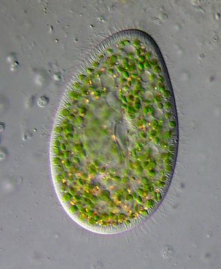
Paramecium bursaria is a species of ciliate found in marine and brackish waters. It has a mutualistic endosymbiotic relationship with green algae called Zoochlorella. About 700 Chlorella cells live inside the protist's cytoplasm and provide it with food, while the Paramecium provides the algae with movement and protection. P. bursaria is 80-150 μm long, with a wide oral groove, two contractile vacuoles, and a single micronucleus as well as a single macronucleus. P. bursaria is the only species of Paramecium that forms symbiotic relationships with algae, and it is often used in biology classrooms both as an example of a protozoan and also as an example of symbiosis.

The murine leukemia viruses are retroviruses named for their ability to cause cancer in murine (mouse) hosts. Some MLVs may infect other vertebrates. MLVs include both exogenous and endogenous viruses. Replicating MLVs have a positive sense, single-stranded RNA (ssRNA) genome that replicates through a DNA intermediate via the process of reverse transcription.

<span class="mw-page-title-main">Herpes simplex virus</span> Species of virus

Herpes simplex virus1 and 2, also known by their taxonomic names Human alphaherpesvirus 1 and Human alphaherpesvirus 2, are two members of the human Herpesviridae family, a set of viruses that produce viral infections in the majority of humans. Both HSV-1 and HSV-2 are very common and contagious. They can be spread when an infected person begins shedding the virus.

Phycodnaviridae is a family of large (100–560 kb) double-stranded DNA viruses that infect marine or freshwater eukaryotic algae. Viruses within this family have a similar morphology, with an icosahedral capsid. As of 2014, there were 33 species in this family, divided among 6 genera. This family belongs to a super-group of large viruses known as nucleocytoplasmic large DNA viruses. Evidence was published in 2014 suggesting that specific strains of Phycodnaviridae might infect humans rather than just algal species, as was previously believed. Most genera under this family enter the host cell by cell receptor endocytosis and replicate in the nucleus. Phycodnaviridae play important ecological roles by regulating the growth and productivity of their algal hosts. Algal species such Heterosigma akashiwo and the genus Chrysochromulina can form dense blooms which can be damaging to fisheries, resulting in losses in the aquaculture industry. Heterosigma akashiwo virus (HaV) has been suggested for use as a microbial agent to prevent the recurrence of toxic red tides produced by this algal species. Phycodnaviridae cause death and lysis of freshwater and marine algal species, liberating organic carbon, nitrogen and phosphorus into the water, providing nutrients for the microbial loop.

<span class="mw-page-title-main">Introduction to viruses</span> Non-technical introduction to viruses

A virus is a tiny infectious agent that reproduces inside the cells of living hosts. When infected, the host cell is forced to rapidly produce thousands of identical copies of the original virus. Unlike most living things, viruses do not have cells that divide; new viruses assemble in the infected host cell. But unlike simpler infectious agents like prions, they contain genes, which allow them to mutate and evolve. Over 4,800 species of viruses have been described in detail out of the millions in the environment. Their origin is unclear: some may have evolved from plasmids—pieces of DNA that can move between cells—while others may have evolved from bacteria.

<span class="mw-page-title-main">Virus</span> Infectious agent that replicates in cells

A virus is a submicroscopic infectious agent that replicates only inside the living cells of an organism. Viruses infect all life forms, from animals and plants to microorganisms, including bacteria and archaea. Viruses are found in almost every ecosystem on Earth and are the most numerous type of biological entity. Since Dmitri Ivanovsky's 1892 article describing a non-bacterial pathogen infecting tobacco plants and the discovery of the tobacco mosaic virus by Martinus Beijerinck in 1898, more than 11,000 of the millions of virus species have been described in detail. The study of viruses is known as virology, a subspeciality of microbiology.

<span class="mw-page-title-main">HBx</span>

HBx is a hepatitis B viral protein. It is 154 amino acids long and interferes with transcription, signal transduction, cell cycle progress, protein degradation, apoptosis and chromosomal stability in the host. It forms a heterodimeric complex with its cellular target protein, and this interaction dysregulates centrosome dynamics and mitotic spindle formation. It interacts with DDB1 redirecting the ubiquitin ligase activity of the CUL4-DDB1 E3 complexes, which are intimately involved in the intracellular regulation of DNA replication and repair, transcription and signal transduction.

<span class="mw-page-title-main">Nudivirus</span> Family of viruses

Nudiviruses are a family of animal viruses that constitute the family Nudiviridae. Insects and marine crustaceans serve as natural hosts. There are 11 species in this family, assigned to 4 genera. Diseases associated with this family include: death in larvae, chronic disease in adults.

Mamavirus is a large and complex virus in the Group I family Mimiviridae. The virus is exceptionally large, and larger than many bacteria. Mamavirus and other mimiviridae belong to nucleocytoplasmic large DNA virus (NCLDVs) family. Mamavirus can be compared to the similar complex virus mimivirus; mamavirus was so named because it is similar to but larger than mimivirus.

<i>Cafeteria roenbergensis virus</i> Species of virus

Cafeteria roenbergensis virus (CroV) is a giant virus that infects the marine bicosoecid flagellate Cafeteria roenbergensis, a member of the microzooplankton community.

A giant virus, sometimes referred to as a girus, is a very large virus, some of which are larger than typical bacteria. All known giant viruses belong to the phylum Nucleocytoviricota.

Acanthocystis turfacea chlorella virus 1 (ATCV-1), also called Chlorovirus ATCV-1 or Chlorella virus ATCV-1 is a species of giant double-stranded DNA virus in the genus Chlorovirus.

<span class="mw-page-title-main">Marine viruses</span> Viruses found in marine environments

Marine viruses are defined by their habitat as viruses that are found in marine environments, that is, in the saltwater of seas or oceans or the brackish water of coastal estuaries. Viruses are small infectious agents that can only replicate inside the living cells of a host organism, because they need the replication machinery of the host to do so. They can infect all types of life forms, from animals and plants to microorganisms, including bacteria and archaea.

<i>Varidnaviria</i> Realm of viruses

Varidnaviria is a realm of viruses that includes all DNA viruses that encode major capsid proteins that contain a vertical jelly roll fold. The major capsid proteins (MCP) form into pseudohexameric subunits of the viral capsid, which stores the viral deoxyribonucleic acid (DNA), and are perpendicular, or vertical, to the surface of the capsid. Apart from this, viruses in the realm also share many other characteristics, such as minor capsid proteins (mCP) with the vertical jelly roll fold, an ATPase that packages viral DNA into the capsid, and a DNA polymerase that replicates the viral genome.

Algal viruses are the viruses infecting photosynthetic single-celled eukaryotes, algae. As of 2020, there were 61 viruses known to infect algae. Algae are integral components of aquatic food webs and drive nutrient cycling, so the viruses infecting algal populations also impacts the organisms and nutrient cycling systems that depend on them. Thus, these viruses can have significant, worldwide economic and ecological effects. Their genomes varied between 4.4 to 560 kilobase pairs (kbp) long and used double-stranded Deoxyribonucleic Acid (dsDNA), double-stranded Ribonucleic Acid (dsRNA), single-stranded Deoxyribonucleic Acid (ssDNA), and single-stranded Ribonucleic Acid (ssRNA). The viruses ranged between 20 and 210 nm in diameter. Since the discovery of the first algae-infecting virus in 1979, several different techniques have been used to find new viruses infecting algae and it seems that there are many algae-infecting viruses left to be discovered

Virivore comes from the English prefix viro- meaning virus, derived from the Latin word for poison, and the suffix -vore from the Latin word vorare, meaning to eat, or to devour; therefore, a virivore is an organism that consumes viruses. Virivory is a well-described process in which organisms, primarily heterotrophic protists, but also some metazoans consume viruses.

References

  1. 1 2 3 Quispe CF, Sonderman O, Seng A, Rasmussen B, Weber G, Mueller C, Dunigan DD, Van Etten JL (July 2016). "Three-year survey of abundance, prevalence and genetic diversity of chlorovirus populations in a small urban lake". Archives of Virology. 161 (7): 1839–47. doi:10.1007/s00705-016-2853-4. PMID   27068168. S2CID   8751019.
  2. 1 2 "Viral Zone". ExPASy. Retrieved 15 June 2015.
  3. 1 2 "Virus Taxonomy: 2020 Release". International Committee on Taxonomy of Viruses (ICTV). March 2021. Retrieved 22 May 2021.
  4. Meints, Russel H.; Van Etten, James L.; Kuczmarski, Daniel; Lee, Kit; Ang, Barbara (September 1981). "Viral infection of the symbiotic chlorella-like alga present in Hydra viridis". Virology. 113 (2): 698–703. doi:10.1016/0042-6822(81)90198-7. PMID   18635088.
  5. Hoshina, Ryo; Shimizu, Mayumi; Makino, Yoichi; Haruyama, Yoshihiro; Ueda, Shin-ichiro; Kato, Yutaka; Kasahara, Masahiro; Ono, Bun-ichiro; Imamura, Nobutaka (13 September 2010). "Isolation and characterization of a virus (CvV-BW1) that infects symbiotic algae of Paramecium bursaria in Lake Biwa, Japan". Virology Journal. 7: 222. doi: 10.1186/1743-422X-7-222 . ISSN   1743-422X. PMC   2949830 . PMID   20831832.
  6. 1 2 3 4 5 6 7 Yolken RH, Jones-Brando L, Dunigan DD, Kannan G, Dickerson F, Severance E, Sabunciyan S, Talbot CC, Prandovszky E, Gurnon JR, Agarkova IV, Leister F, Gressitt KL, Chen O, Deuber B, Ma F, Pletnikov MV, Van Etten JL (November 2014). "Chlorovirus ATCV-1 is part of the human oropharyngeal virome and is associated with changes in cognitive functions in humans and mice". Proceedings of the National Academy of Sciences of the United States of America. 111 (45): 16106–11. Bibcode:2014PNAS..11116106Y. doi: 10.1073/pnas.1418895111 . PMC   4234575 . PMID   25349393.
  7. 1 2 3 Petro, Marilyn S.; Agarkova, Irina V.; Petro, Thomas M. (August 2016). "Effect of Chlorovirus ATCV-1 infection on behavior of C57Bl/6 mice". Journal of Neuroimmunology. 297: 46–55. doi:10.1016/j.jneuroim.2016.05.009. PMID   27397075. S2CID   38573451.
  8. Short SM (September 2012). "The ecology of viruses that infect eukaryotic algae". Environmental Microbiology. 14 (9): 2253–71. doi:10.1111/j.1462-2920.2012.02706.x. PMID   22360532.
  9. 1 2 3 Van Etten JL, Dunigan DD (August 2016). "Giant Chloroviruses: Five Easy Questions". PLOS Pathogens. 12 (8): e1005751. doi: 10.1371/journal.ppat.1005751 . PMC   4990331 . PMID   27536965.
  10. 1 2 DeLong JP, Al-Ameeli Z, Duncan G, Van Etten JL, Dunigan DD (November 2016). "Predators catalyze an increase in chloroviruses by foraging on the symbiotic hosts of zoochlorellae". Proceedings of the National Academy of Sciences of the United States of America. 113 (48): 13780–13784. doi: 10.1073/pnas.1613843113 . PMC   5137705 . PMID   27821770.
  11. Long AM, Short SM (July 2016). "Seasonal determinations of algal virus decay rates reveal overwintering in a temperate freshwater pond". The ISME Journal. 10 (7): 1602–12. doi:10.1038/ismej.2015.240. PMC   4918447 . PMID   26943625.
  12. 1 2 Yanai GM (2009). Transcription analysis of the chlorovirus Paramecium bursaria chlorella virus-1 (PhD). University of Nebraska at Lincoln.
  13. Song H, Lavoie M, Fan X, Tan H, Liu G, Xu P, Fu Z, Paerl HW, Qian H (August 2017). "Allelopathic interactions of linoleic acid and nitric oxide increase the competitive ability of Microcystis aeruginosa". The ISME Journal. 11 (8): 1865–1876. doi:10.1038/ismej.2017.45. PMC   5520033 . PMID   28398349.
  14. Rieper M (1 March 1976). "Investigations on the relationships between algal blooms and bacterial populations in the Schlei Fjord (western Baltic Sea)". Helgoländer Wissenschaftliche Meeresuntersuchungen. 28 (1): 1–18. Bibcode:1976HWM....28....1R. doi: 10.1007/bf01610792 . ISSN   0017-9957.
  15. 1 2 Quispe, Cristian F.; Esmael, Ahmed; Sonderman, Olivia; McQuinn, Michelle; Agarkova, Irina; Battah, Mohammed; Duncan, Garry A.; Dunigan, David D.; Smith, Timothy P.L.; De Castro, Cristina; Speciale, Immacolata; Ma, Fangrui; Van Etten, James L. (January 2017). "Characterization of a new chlorovirus type with permissive and non-permissive features on phylogenetically related algal strains". Virology. 500: 103–113. doi:10.1016/j.virol.2016.10.013. PMC   5127778 . PMID   27816636.
  16. 1 2 Van Etten, James L.; Dunigan, David D. (January 2012). "Chloroviruses: not your everyday plant virus". Trends in Plant Science. 17 (1): 1–8. doi:10.1016/j.tplants.2011.10.005. PMC   3259250 . PMID   22100667.
  17. 1 2 Van Etten, James L.; Dunigan, David D.; Condit, Richard C. (18 August 2016). "Giant Chloroviruses: Five Easy Questions". PLOS Pathogens. 12 (8): e1005751. doi: 10.1371/journal.ppat.1005751 . PMC   4990331 . PMID   27536965.
  18. Van Etten JL, Dunigan DD (January 2012). "Chloroviruses: not your everyday plant virus". Trends in Plant Science. 17 (1): 1–8. doi:10.1016/j.tplants.2011.10.005. PMC   3259250 . PMID   22100667.
  19. 1 2 3 Van Etten, James L.; Dunigan, David D. (2012). "Chloroviruses: not your everyday plant virus". Trends in Plant Science. 17 (1): 1–8. doi:10.1016/j.tplants.2011.10.005. PMC   3259250 . PMID   22100667.
  20. 1 2 James L. Van Etten, Irina V. Agarkova, David D. Dunigan: Chloroviruses. In: Viruses 2020, 12(1), 20. Special Issue Viruses Ten-Year Anniversary. doi:10.3390/v12010020. CC-BY icon.svg Material copied from this source, which is available under a Creative Commons Attribution 4.0 International License.
  21. Zhang, X; Xiang, Y; Dunigan, DD; Klose, T; Chipman, PR; Van Etten, JL; Rossmann, MG (2011). "Three dimensional structure and function of the Paramecium bursaria chlorella virus capsid". Proc. Natl. Acad. Sci. USA. 2011 (108): 14837–14842. Bibcode:2011PNAS..10814837Z. doi: 10.1073/pnas.1107847108 . PMC   3169150 . PMID   21873222.
  22. Blanc, G; Mozar, M; Agarkova, IV; Gurnon, JR; Yanai Balser, G; Rowe, JM; Xia, Y; Riethoven, JJ; Dunigan, DD; Van Etten, JL (2014). "Deep RNA sequencing reveals hidden features and dynamics of early gene transcription in Paramecium bursaria chlorella virus 1". PLOS ONE. 9 (3): e90989. Bibcode:2014PLoSO...990989B. doi: 10.1371/journal.pone.0090989 . PMC   3946568 . PMID   24608750.
  23. KANG, MING; DUNIGAN, DAVID D.; ETTEN, JAMES L. VAN (1 May 2005). "Chlorovirus: a genus of Phycodnaviridae that infects certain chlorella-like green algae". Molecular Plant Pathology. 6 (3): 213–224. doi: 10.1111/j.1364-3703.2005.00281.x . PMID   20565652.
  24. Yutin N, Wolf YI, Koonin EV (October 2014). "Origin of giant viruses from smaller DNA viruses not from a fourth domain of cellular life". Virology. 466–467: 38–52. doi:10.1016/j.virol.2014.06.032. PMC   4325995 . PMID   25042053.
  25. 1 2 3 Dunigan DD, Fitzgerald LA, Van Etten JL (April 2006). "Phycodnaviruses: a peek at genetic diversity". Virus Research. 117 (1): 119–32. doi:10.1016/j.virusres.2006.01.024. PMID   16516998.
  26. 1 2 Van Etten JL, Agarkova I, Dunigan DD, Tonetti M, De Castro C, Duncan GA (April 2017). "Chloroviruses Have a Sweet Tooth". Viruses. 9 (4): 88. doi: 10.3390/v9040088 . PMC   5408694 . PMID   28441734.
  27. Petro TM, Agarkova IV, Zhou Y, Yolken RH, Van Etten JL, Dunigan DD (December 2015). "Response of Mammalian Macrophages to Challenge with the Chlorovirus Acanthocystis turfacea Chlorella Virus 1". Journal of Virology. 89 (23): 12096–107. doi:10.1128/JVI.01254-15. PMC   4645302 . PMID   26401040.
  28. Petro MS, Agarkova IV, Petro TM (August 2016). "Effect of Chlorovirus ATCV-1 infection on behavior of C57Bl/6 mice". Journal of Neuroimmunology. 297: 46–55. doi:10.1016/j.jneuroim.2016.05.009. PMID   27397075. S2CID   38573451.