DNA nanotechnology

Last updated

DNA nanotechnology involves forming artificial, designed nanostructures out of nucleic acids, such as this DNA tetrahedron. Each edge of the tetrahedron is a 20 base pair DNA double helix, and each vertex is a three-arm junction. The 4 DNA strands that form the 4 tetrahedral faces are color-coded. DNA tetrahedron white.png
DNA nanotechnology involves forming artificial, designed nanostructures out of nucleic acids, such as this DNA tetrahedron. Each edge of the tetrahedron is a 20 base pair DNA double helix, and each vertex is a three-arm junction. The 4 DNA strands that form the 4 tetrahedral faces are color-coded.

DNA nanotechnology is the design and manufacture of artificial nucleic acid structures for technological uses. In this field, nucleic acids are used as non-biological engineering materials for nanotechnology rather than as the carriers of genetic information in living cells. Researchers in the field have created static structures such as two- and three-dimensional crystal lattices, nanotubes, polyhedra, and arbitrary shapes, and functional devices such as molecular machines and DNA computers. The field is beginning to be used as a tool to solve basic science problems in structural biology and biophysics, including applications in X-ray crystallography and nuclear magnetic resonance spectroscopy of proteins to determine structures. Potential applications in molecular scale electronics and nanomedicine are also being investigated.

Contents

The conceptual foundation for DNA nanotechnology was first laid out by Nadrian Seeman in the early 1980s, and the field began to attract widespread interest in the mid-2000s. This use of nucleic acids is enabled by their strict base pairing rules, which cause only portions of strands with complementary base sequences to bind together to form strong, rigid double helix structures. This allows for the rational design of base sequences that will selectively assemble to form complex target structures with precisely controlled nanoscale features. Several assembly methods are used to make these structures, including tile-based structures that assemble from smaller structures, folding structures using the DNA origami method, and dynamically reconfigurable structures using strand displacement methods. The field's name specifically references DNA, but the same principles have been used with other types of nucleic acids as well, leading to the occasional use of the alternative name nucleic acid nanotechnology.

History

The conceptual foundation for DNA nanotechnology was first laid out by Nadrian Seeman in the early 1980s. [2] Seeman's original motivation was to create a three-dimensional DNA lattice for orienting other large molecules, which would simplify their crystallographic study by eliminating the difficult process of obtaining pure crystals. This idea had reportedly come to him in late 1980, after realizing the similarity between the woodcut Depth by M. C. Escher and an array of DNA six-arm junctions. [3] [4] Several natural branched DNA structures were known at the time, including the DNA replication fork and the mobile Holliday junction, but Seeman's insight was that immobile nucleic acid junctions could be created by properly designing the strand sequences to remove symmetry in the assembled molecule, and that these immobile junctions could in principle be combined into rigid crystalline lattices. The first theoretical paper proposing this scheme was published in 1982, and the first experimental demonstration of an immobile DNA junction was published the following year. [5] [6]

The woodcut Depth (pictured) by M. C. Escher reportedly inspired Nadrian Seeman to consider using three-dimensional lattices of DNA to orient hard-to-crystallize molecules. This led to the beginning of the field of DNA nanotechnology. Escher Depth.jpg
The woodcut Depth (pictured) by M. C. Escher reportedly inspired Nadrian Seeman to consider using three-dimensional lattices of DNA to orient hard-to-crystallize molecules. This led to the beginning of the field of DNA nanotechnology.

In 1991, Seeman's laboratory published a report on the synthesis of a cube made of DNA, the first synthetic three-dimensional nucleic acid nanostructure, for which he received the 1995 Feynman Prize in Nanotechnology. This was followed by a DNA truncated octahedron. It soon became clear that these structures, polygonal shapes with flexible junctions as their vertices, were not rigid enough to form extended three-dimensional lattices. Seeman developed the more rigid double-crossover (DX) structural motif, and in 1998, in collaboration with Erik Winfree, published the creation of two-dimensional lattices of DX tiles. [3] [2] [7] These tile-based structures had the advantage that they provided the ability to implement DNA computing, which was demonstrated by Winfree and Paul Rothemund in their 2004 paper on the algorithmic self-assembly of a Sierpinski gasket structure, and for which they shared the 2006 Feynman Prize in Nanotechnology. Winfree's key insight was that the DX tiles could be used as Wang tiles, meaning that their assembly could perform computation. [2] The synthesis of a three-dimensional lattice was finally published by Seeman in 2009, nearly thirty years after he had set out to achieve it. [8]

New abilities continued to be discovered for designed DNA structures throughout the 2000s. The first DNA nanomachine—a motif that changes its structure in response to an input—was demonstrated in 1999 by Seeman. An improved system, which was the first nucleic acid device to make use of toehold-mediated strand displacement, was demonstrated by Bernard Yurke in 2000. [9] The next advance was to translate this into mechanical motion, and in 2004 and 2005, several DNA walker systems were demonstrated by the groups of Seeman, Niles Pierce, Andrew Turberfield, and Chengde Mao. [10] The idea of using DNA arrays to template the assembly of other molecules such as nanoparticles and proteins, first suggested by Bruche Robinson and Seeman in 1987, [11] was demonstrated in 2002 by Seeman, Kiehl et al. [12] and subsequently by many other groups.

In 2006, Rothemund first demonstrated the DNA origami method for easily and robustly forming folded DNA structures of arbitrary shape. Rothemund had conceived of this method as being conceptually intermediate between Seeman's DX lattices, which used many short strands, and William Shih's DNA octahedron, which consisted mostly of one very long strand. Rothemund's DNA origami contains a long strand which folding is assisted by several short strands. This method allowed forming much larger structures than formerly possible, and which are less technically demanding to design and synthesize. [7] DNA origami was the cover story of Nature on March 15, 2006. [13] Rothemund's research demonstrating two-dimensional DNA origami structures was followed by the demonstration of solid three-dimensional DNA origami by Douglas et al. in 2009, [14] while the labs of Jørgen Kjems and Yan demonstrated hollow three-dimensional structures made out of two-dimensional faces. [8]

DNA nanotechnology was initially met with some skepticism due to the unusual non-biological use of nucleic acids as materials for building structures and doing computation, and the preponderance of proof of principle experiments that extended the abilities of the field but were far from actual applications. Seeman's 1991 paper on the synthesis of the DNA cube was rejected by the journal Science after one reviewer praised its originality while another criticized it for its lack of biological relevance. [15] By the early 2010s the field was considered to have increased its abilities to the point that applications for basic science research were beginning to be realized, and practical applications in medicine and other fields were beginning to be considered feasible. [8] [16] The field had grown from very few active laboratories in 2001 to at least 60 in 2010, which increased the talent pool and thus the number of scientific advances in the field during that decade. [17]


Fundamental concepts

These four strands associate into a DNA four-arm junction because this structure maximizes the number of correct base pairs, with A matched to T and C matched to G. See this image for a more realistic model of the four-arm junction showing its tertiary structure. Nepodvizhnaia struktura Khollideia (angl.).svg
These four strands associate into a DNA four-arm junction because this structure maximizes the number of correct base pairs, with A matched to T and C matched to G. See this image for a more realistic model of the four-arm junction showing its tertiary structure.
This double-crossover (DX) supramolecular complex consists of five DNA single strands that form two double-helical domains, on the top and the bottom in this image. There are two crossover points where the strands cross from one domain into the other. Mao-DX-schematic-2.svg
This double-crossover (DX) supramolecular complex consists of five DNA single strands that form two double-helical domains, on the top and the bottom in this image. There are two crossover points where the strands cross from one domain into the other.

Properties of nucleic acids

Nanotechnology is often defined as the study of materials and devices with features on a scale below 100 nanometers. DNA nanotechnology, specifically, is an example of bottom-up molecular self-assembly, in which molecular components spontaneously organize into stable structures; the particular form of these structures is induced by the physical and chemical properties of the components selected by the designers. [19] In DNA nanotechnology, the component materials are strands of nucleic acids such as DNA; these strands are often synthetic and are almost always used outside the context of a living cell. DNA is well-suited to nanoscale construction because the binding between two nucleic acid strands depends on simple base pairing rules which are well understood, and form the specific nanoscale structure of the nucleic acid double helix. These qualities make the assembly of nucleic acid structures easy to control through nucleic acid design. This property is absent in other materials used in nanotechnology, including proteins, for which protein design is very difficult, and nanoparticles, which lack the capability for specific assembly on their own. [5]

The structure of a nucleic acid molecule consists of a sequence of nucleotides distinguished by which nucleobase they contain. In DNA, the four bases present are adenine (A), cytosine (C), guanine (G), and thymine (T). Nucleic acids have the property that two molecules will only bind to each other to form a double helix if the two sequences are complementary, meaning that they form matching sequences of base pairs, with A only binding to T, and C only to G. [5] [20] Because the formation of correctly matched base pairs is energetically favorable, nucleic acid strands are expected in most cases to bind to each other in the conformation that maximizes the number of correctly paired bases. The sequences of bases in a system of strands thus determine the pattern of binding and the overall structure in an easily controllable way. In DNA nanotechnology, the base sequences of strands are rationally designed by researchers so that the base pairing interactions cause the strands to assemble in the desired conformation. [3] [5] While DNA is the dominant material used, structures incorporating other nucleic acids such as RNA and peptide nucleic acid (PNA) have also been constructed. [21] [22]

Subfields

DNA nanotechnology is sometimes divided into two overlapping subfields: structural DNA nanotechnology and dynamic DNA nanotechnology. Structural DNA nanotechnology, sometimes abbreviated as SDN, focuses on synthesizing and characterizing nucleic acid complexes and materials that assemble into a static, equilibrium end state. On the other hand, dynamic DNA nanotechnology focuses on complexes with useful non-equilibrium behavior such as the ability to reconfigure based on a chemical or physical stimulus. Some complexes, such as nucleic acid nanomechanical devices, combine features of both the structural and dynamic subfields. [23] [24]

The complexes constructed in structural DNA nanotechnology use topologically branched nucleic acid structures containing junctions. (In contrast, most biological DNA exists as an unbranched double helix.) One of the simplest branched structures is a four-arm junction that consists of four individual DNA strands, portions of which are complementary in a specific pattern. Unlike in natural Holliday junctions, each arm in the artificial immobile four-arm junction has a different base sequence, causing the junction point to be fixed at a certain position. Multiple junctions can be combined in the same complex, such as in the widely used double-crossover (DX) structural motif, which contains two parallel double helical domains with individual strands crossing between the domains at two crossover points. Each crossover point is, topologically, a four-arm junction, but is constrained to one orientation, in contrast to the flexible single four-arm junction, providing a rigidity that makes the DX motif suitable as a structural building block for larger DNA complexes. [3] [5]

Dynamic DNA nanotechnology uses a mechanism called toehold-mediated strand displacement to allow the nucleic acid complexes to reconfigure in response to the addition of a new nucleic acid strand. In this reaction, the incoming strand binds to a single-stranded toehold region of a double-stranded complex, and then displaces one of the strands bound in the original complex through a branch migration process. The overall effect is that one of the strands in the complex is replaced with another one. [23] In addition, reconfigurable structures and devices can be made using functional nucleic acids such as deoxyribozymes and ribozymes, which can perform chemical reactions, and aptamers, which can bind to specific proteins or small molecules. [25]

Structural DNA nanotechnology

Structural DNA nanotechnology, sometimes abbreviated as SDN, focuses on synthesizing and characterizing nucleic acid complexes and materials where the assembly has a static, equilibrium endpoint. The nucleic acid double helix has a robust, defined three-dimensional geometry that makes it possible to simulate, [26] predict and design the structures of more complicated nucleic acid complexes. Many such structures have been created, including two- and three-dimensional structures, and periodic, aperiodic, and discrete structures. [24]

Extended lattices

Mao-DXarray-schematic-small.gif
DX DNA array.png
The assembly of a DX array. Left, schematic diagram. Each bar represents a double-helical domain of DNA, with the shapes representing complementary sticky ends. The DX complex at top will combine with other DX complexes into the two-dimensional array shown at bottom. [18] Right, an atomic force microscopy image of the assembled array. The individual DX tiles are clearly visible within the assembled structure. The field is 150  nm across.
Left, a model of a DNA tile used to make another two-dimensional periodic lattice. Right, an atomic force micrograph of the assembled lattice. DNA nanostructures.png
Left, a model of a DNA tile used to make another two-dimensional periodic lattice. Right, an atomic force micrograph of the assembled lattice.
SierpinskiTriangle.svg
Rothemund-DNA-SierpinskiGasket.jpg
An example of an aperiodic two-dimensional lattice that assembles into a fractal pattern. Left, the Sierpinski gasket fractal. Right, DNA arrays that display a representation of the Sierpinski gasket on their surfaces [29]

Small nucleic acid complexes can be equipped with sticky ends and combined into larger two-dimensional periodic lattices containing a specific tessellated pattern of the individual molecular tiles. [24] The earliest example of this used double-crossover (DX) complexes as the basic tiles, each containing four sticky ends designed with sequences that caused the DX units to combine into periodic two-dimensional flat sheets that are essentially rigid two-dimensional crystals of DNA. [30] [31] Two-dimensional arrays have been made from other motifs as well, including the Holliday junction rhombus lattice, [32] and various DX-based arrays making use of a double-cohesion scheme. [33] [34] The top two images at right show examples of tile-based periodic lattices.

Two-dimensional arrays can be made to exhibit aperiodic structures whose assembly implements a specific algorithm, exhibiting one form of DNA computing. [17] The DX tiles can have their sticky end sequences chosen so that they act as Wang tiles, allowing them to perform computation. A DX array whose assembly encodes an XOR operation has been demonstrated; this allows the DNA array to implement a cellular automaton that generates a fractal known as the Sierpinski gasket. The third image at right shows this type of array. [29] Another system has the function of a binary counter, displaying a representation of increasing binary numbers as it grows. These results show that computation can be incorporated into the assembly of DNA arrays. [35]

DX arrays have been made to form hollow nanotubes 4–20  nm in diameter, essentially two-dimensional lattices which curve back upon themselves. [36] These DNA nanotubes are somewhat similar in size and shape to carbon nanotubes, and while they lack the electrical conductance of carbon nanotubes, DNA nanotubes are more easily modified and connected to other structures. One of many schemes for constructing DNA nanotubes uses a lattice of curved DX tiles that curls around itself and closes into a tube. [37] In an alternative method that allows the circumference to be specified in a simple, modular fashion using single-stranded tiles, the rigidity of the tube is an emergent property. [38]

Forming three-dimensional lattices of DNA was the earliest goal of DNA nanotechnology, but this proved to be one of the most difficult to realize. Success using a motif based on the concept of tensegrity, a balance between tension and compression forces, was finally reported in 2009. [17] [39]

Discrete structures

Researchers have synthesized many three-dimensional DNA complexes that each have the connectivity of a polyhedron, such as a cube or octahedron, meaning that the DNA duplexes trace the edges of a polyhedron with a DNA junction at each vertex. [6] The earliest demonstrations of DNA polyhedra were very work-intensive, requiring multiple ligations and solid-phase synthesis steps to create catenated polyhedra. [40] Subsequent work yielded polyhedra whose synthesis was much easier. These include a DNA octahedron made from a long single strand designed to fold into the correct conformation, [41] and a tetrahedron that can be produced from four DNA strands in one step, pictured at the top of this article. [1]

Nanostructures of arbitrary, non-regular shapes are usually made using the DNA origami method. These structures consist of a long, natural virus strand as a "scaffold", which is made to fold into the desired shape by computationally designed short "staple" strands. This method has the advantages of being easy to design, as the base sequence is predetermined by the scaffold strand sequence, and not requiring high strand purity and accurate stoichiometry, as most other DNA nanotechnology methods do. DNA origami was first demonstrated for two-dimensional shapes, such as a smiley face, a coarse map of the Western Hemisphere, and the Mona Lisa painting. [6] [13] [42] Solid three-dimensional structures can be made by using parallel DNA helices arranged in a honeycomb pattern, [14] and structures with two-dimensional faces can be made to fold into a hollow overall three-dimensional shape, akin to a cardboard box. These can be programmed to open and reveal or release a molecular cargo in response to a stimulus, making them potentially useful as programmable molecular cages. [43] [44]

Templated assembly

Nucleic acid structures can be made to incorporate molecules other than nucleic acids, sometimes called heteroelements, including proteins, metallic nanoparticles, quantum dots, amines, [45] and fullerenes. This allows the construction of materials and devices with a range of functionalities much greater than is possible with nucleic acids alone. The goal is to use the self-assembly of the nucleic acid structures to template the assembly of the nanoparticles hosted on them, controlling their position and in some cases orientation. [6] [46] Many of these schemes use a covalent attachment scheme, using oligonucleotides with amide or thiol functional groups as a chemical handle to bind the heteroelements. This covalent binding scheme has been used to arrange gold nanoparticles on a DX-based array, [47] and to arrange streptavidin protein molecules into specific patterns on a DX array. [48] A non-covalent hosting scheme using Dervan polyamides on a DX array was used to arrange streptavidin proteins in a specific pattern on a DX array. [49] Carbon nanotubes have been hosted on DNA arrays in a pattern allowing the assembly to act as a molecular electronic device, a carbon nanotube field-effect transistor. [50] In addition, there are nucleic acid metallization methods, in which the nucleic acid is replaced by a metal which assumes the general shape of the original nucleic acid structure, [51] and schemes for using nucleic acid nanostructures as lithography masks, transferring their pattern into a solid surface. [52]

Dynamic DNA nanotechnology

Dynamic DNA nanotechnology often makes use of toehold-mediated strand displacement reactions. In this example, the red strand binds to the single stranded toehold region on the green strand (region 1), and then in a branch migration process across region 2, the blue strand is displaced and freed from the complex. Reactions like these are used to dynamically reconfigure or assemble nucleic acid nanostructures. In addition, the red and blue strands can be used as signals in a molecular logic gate. Strand displacement.svg
Dynamic DNA nanotechnology often makes use of toehold-mediated strand displacement reactions. In this example, the red strand binds to the single stranded toehold region on the green strand (region 1), and then in a branch migration process across region 2, the blue strand is displaced and freed from the complex. Reactions like these are used to dynamically reconfigure or assemble nucleic acid nanostructures. In addition, the red and blue strands can be used as signals in a molecular logic gate.

Dynamic DNA nanotechnology focuses on forming nucleic acid systems with designed dynamic functionalities related to their overall structures, such as computation and mechanical motion. There is some overlap between structural and dynamic DNA nanotechnology, as structures can be formed through annealing and then reconfigured dynamically, or can be made to form dynamically in the first place. [6] [10]

Nanomechanical devices

DNA complexes have been made that change their conformation upon some stimulus, making them one form of nanorobotics. These structures are initially formed in the same way as the static structures made in structural DNA nanotechnology, but are designed so that dynamic reconfiguration is possible after the initial assembly. [23] [10] The earliest such device made use of the transition between the B-DNA and Z-DNA forms to respond to a change in buffer conditions by undergoing a twisting motion. [53] This reliance on buffer conditions caused all devices to change state at the same time. Subsequent systems could change states based upon the presence of control strands, allowing multiple devices to be independently operated in solution. Some examples of such systems are a "molecular tweezers" design that has an open and a closed state, [54] a device that could switch from a paranemic-crossover (PX) conformation to a (JX2) conformation with two non-junction juxtapositions of the DNA backbone, undergoing rotational motion in the process, [55] and a two-dimensional array that could dynamically expand and contract in response to control strands. [56] Structures have also been made that dynamically open or close, potentially acting as a molecular cage to release or reveal a functional cargo upon opening. [43] [57] [58] In another example, a DNA origami nanostructure was coupled to T7 RNA polymerase and could thus be operated as a chemical energy-driven motor that can be coupled to a passive follower, which it then drives. [59]

DNA walkers are a class of nucleic acid nanomachines that exhibit directional motion along a linear track. A large number of schemes have been demonstrated. [10] One strategy is to control the motion of the walker along the track using control strands that need to be manually added in sequence. [60] [61] It is also possible to control individual steps of a DNA walker by irradiation with light of different wavelengths. [62] Another approach is to make use of restriction enzymes or deoxyribozymes to cleave the strands and cause the walker to move forward, which has the advantage of running autonomously. [63] [64] A later system could walk upon a two-dimensional surface rather than a linear track, and demonstrated the ability to selectively pick up and move molecular cargo. [65] In 2018, a catenated DNA that uses rolling circle transcription by an attached T7 RNA polymerase was shown to walk along a DNA-path, guided by the generated RNA strand. [66] Additionally, a linear walker has been demonstrated that performs DNA-templated synthesis as the walker advances along the track, allowing autonomous multistep chemical synthesis directed by the walker. [67] The synthetic DNA walkers' function is similar to that of the proteins dynein and kinesin. [68]

Strand displacement cascades

Cascades of strand displacement reactions can be used for either computational or structural purposes. An individual strand displacement reaction involves revealing a new sequence in response to the presence of some initiator strand. Many such reactions can be linked into a cascade where the newly revealed output sequence of one reaction can initiate another strand displacement reaction elsewhere. This in turn allows for the construction of chemical reaction networks with many components, exhibiting complex computational and information processing abilities. These cascades are made energetically favorable through the formation of new base pairs, and the entropy gain from disassembly reactions. Strand displacement cascades allow isothermal operation of the assembly or computational process, in contrast to traditional nucleic acid assembly's requirement for a thermal annealing step, where the temperature is raised and then slowly lowered to ensure proper formation of the desired structure. They can also support catalytic function of the initiator species, where less than one equivalent of the initiator can cause the reaction to go to completion. [23] [69]

Strand displacement complexes can be used to make molecular logic gates capable of complex computation. [70] Unlike traditional electronic computers, which use electric current as inputs and outputs, molecular computers use the concentrations of specific chemical species as signals. In the case of nucleic acid strand displacement circuits, the signal is the presence of nucleic acid strands that are released or consumed by binding and unbinding events to other strands in displacement complexes. This approach has been used to make logic gates such as AND, OR, and NOT gates. [71] More recently, a four-bit circuit was demonstrated that can compute the square root of the integers 0–15, using a system of gates containing 130 DNA strands. [72]

Another use of strand displacement cascades is to make dynamically assembled structures. These use a hairpin structure for the reactants, so that when the input strand binds, the newly revealed sequence is on the same molecule rather than disassembling. This allows new opened hairpins to be added to a growing complex. This approach has been used to make simple structures such as three- and four-arm junctions and dendrimers. [69]

Applications

DNA nanotechnology provides one of the few ways to form designed, complex structures with precise control over nanoscale features. The field is beginning to see application to solve basic science problems in structural biology and biophysics. The earliest such application envisaged for the field, and one still in development, is in crystallography, where molecules that are difficult to crystallize in isolation could be arranged within a three-dimensional nucleic acid lattice, allowing determination of their structure. Another application is the use of DNA origami rods to replace liquid crystals in residual dipolar coupling experiments in protein NMR spectroscopy; using DNA origami is advantageous because, unlike liquid crystals, they are tolerant of the detergents needed to suspend membrane proteins in solution. DNA walkers have been used as nanoscale assembly lines to move nanoparticles and direct chemical synthesis. Further, DNA origami structures have aided in the biophysical studies of enzyme function and protein folding. [24] [8]

DNA nanotechnology is moving toward potential real-world applications. The ability of nucleic acid arrays to arrange other molecules indicates its potential applications in molecular scale electronics. The assembly of a nucleic acid structure could be used to template the assembly of molecular electronic elements such as molecular wires, providing a method for nanometer-scale control of the placement and overall architecture of the device analogous to a molecular breadboard. [24] [6] DNA nanotechnology has been compared to the concept of programmable matter because of the coupling of computation to its material properties. [73]

In a study conducted by a group of scientists from iNANO and CDNA centers in Aarhus University, researchers were able to construct a small multi-switchable 3D DNA Box Origami. The proposed nanoparticle was characterized by atomic force microscopy (AFM), transmission electron microscopy (TEM) and Förster resonance energy transfer (FRET). The constructed box was shown to have a unique reclosing mechanism, which enabled it to repeatedly open and close in response to a unique set of DNA or RNA keys. The authors proposed that this "DNA device can potentially be used for a broad range of applications such as controlling the function of single molecules, controlled drug delivery, and molecular computing." [74]

There are potential applications for DNA nanotechnology in nanomedicine, making use of its ability to perform computation in a biocompatible format to make "smart drugs" for targeted drug delivery, as well as for diagnostic applications. One such system being investigated uses a hollow DNA box containing proteins that induce apoptosis, or cell death, that will only open when in proximity to a cancer cell. [8] [75] There has additionally been interest in expressing these artificial structures in engineered living bacterial cells, most likely using the transcribed RNA for the assembly, although it is unknown whether these complex structures are able to efficiently fold or assemble in the cell's cytoplasm. If successful, this could enable directed evolution of nucleic acid nanostructures. [6] Scientists at Oxford University reported the self-assembly of four short strands of synthetic DNA into a cage which can enter cells and survive for at least 48 hours. The fluorescently labeled DNA tetrahedra were found to remain intact in the laboratory cultured human kidney cells despite the attack by cellular enzymes after two days. This experiment showed the potential of drug delivery inside the living cells using the DNA ‘cage’. [76] [77] A DNA tetrahedron was used to deliver RNA Interference (RNAi) in a mouse model, reported a team of researchers in MIT. Delivery of the interfering RNA for treatment has showed some success using polymer or lipid, but there are limits of safety and imprecise targeting, in addition to short shelf life in the blood stream. The DNA nanostructure created by the team consists of six strands of DNA to form a tetrahedron, with one strand of RNA affixed to each of the six edges. The tetrahedron is further equipped with targeting protein, three folate molecules, which lead the DNA nanoparticles to the abundant folate receptors found on some tumors. The result showed that the gene expression targeted by the RNAi, luciferase, dropped by more than half. This study shows promise in using DNA nanotechnology as an effective tool to deliver treatment using the emerging RNA Interference technology. [78] [79] The DNA tetrahedron was also used in an effort to overcome the phenomena multidrug resistance. Doxorubicin (DOX) was conjugated with the tetrahedron and was loaded into MCF-7 breast cancer cells that contained the P-glycoprotein drug efflux pump. The results of the experiment showed the DOX was not being pumped out and apoptosis of the cancer cells was achieved. The tetrahedron without DOX was loaded into cells to test its biocompatibility, and the structure showed no cytotoxicity itself. [80] The DNA tetrahedron was also used as barcode for profiling the subcellular expression and distribution of proteins in cells for diagnostic purposes. The tetrahedral-nanostructured showed enhanced signal due to higher labeling efficiency and stability. [81]

Applications for DNA nanotechnology in nanomedicine also focus on mimicking the structure and function of naturally occurring membrane proteins with designed DNA nanostructures. In 2012, Langecker et al. [82] introduced a pore-shaped DNA origami structure that can self-insert into lipid membranes via hydrophobic cholesterol modifications and induce ionic currents across the membrane. This first demonstration of a synthetic DNA ion channel was followed by a variety of pore-inducing designs ranging from a single DNA duplex, [83] to small tile-based structures, [84] [85] [86] [87] [88] and large DNA origami transmembrane porins. [89] Similar to naturally occurring protein ion channels, this ensemble of synthetic DNA-made counterparts thereby spans multiple orders of magnitude in conductance. The study of the membrane-inserting single DNA duplex showed that current must also flow on the DNA-lipid interface as no central channel lumen is present in the design that lets ions pass across the lipid bilayer. This indicated that the DNA-induced lipid pore has a toroidal shape, rather than cylindrical, as lipid headgroups reorient to face towards the membrane-inserted part of the DNA. [83] Researchers from the University of Cambridge and the University of Illinois at Urbana-Champaign then demonstrated that such a DNA-induced toroidal pore can facilitate rapid lipid flip-flop between the lipid bilayer leaflets. Utilizing this effect, they designed a synthetic DNA-built enzyme that flips lipids in biological membranes orders of magnitudes faster than naturally occurring proteins called scramblases. [90] This development highlights the potential of synthetic DNA nanostructures for personalized drugs and therapeutics.

Design

DNA nanostructures must be rationally designed so that individual nucleic acid strands will assemble into the desired structures. This process usually begins with specification of a desired target structure or function. Then, the overall secondary structure of the target complex is determined, specifying the arrangement of nucleic acid strands within the structure, and which portions of those strands should be bound to each other. The last step is the primary structure design, which is the specification of the actual base sequences of each nucleic acid strand. [36] [91]

Structural design

The first step in designing a nucleic acid nanostructure is to decide how a given structure should be represented by a specific arrangement of nucleic acid strands. This design step determines the secondary structure, or the positions of the base pairs that hold the individual strands together in the desired shape. [36] Several approaches have been demonstrated:

Sequence design

After any of the above approaches are used to design the secondary structure of a target complex, an actual sequence of nucleotides that will form into the desired structure must be devised. Nucleic acid design is the process of assigning a specific nucleic acid base sequence to each of a structure's constituent strands so that they will associate into a desired conformation. Most methods have the goal of designing sequences so that the target structure has the lowest energy, and is thus the most thermodynamically favorable, while incorrectly assembled structures have higher energies and are thus disfavored. This is done either through simple, faster heuristic methods such as sequence symmetry minimization, or by using a full nearest-neighbor thermodynamic model, which is more accurate but slower and more computationally intensive. Geometric models are used to examine tertiary structure of the nanostructures and to ensure that the complexes are not overly strained. [91] [93]

Nucleic acid design has similar goals to protein design. In both, the sequence of monomers is designed to favor the desired target structure and to disfavor other structures. Nucleic acid design has the advantage of being much computationally easier than protein design, because the simple base pairing rules are sufficient to predict a structure's energetic favorability, and detailed information about the overall three-dimensional folding of the structure is not required. This allows the use of simple heuristic methods that yield experimentally robust designs. Nucleic acid structures are less versatile than proteins in their function because of proteins' increased ability to fold into complex structures, and the limited chemical diversity of the four nucleotides as compared to the twenty proteinogenic amino acids. [93]

Materials and methods

Gel electrophoresis methods, such as this formation assay on a DX complex, are used to ascertain whether the desired structures are forming properly. Each vertical lane contains a series of bands, where each band is characteristic of a particular reaction intermediate. DX formation gel.png
Gel electrophoresis methods, such as this formation assay on a DX complex, are used to ascertain whether the desired structures are forming properly. Each vertical lane contains a series of bands, where each band is characteristic of a particular reaction intermediate.

The sequences of the DNA strands making up a target structure are designed computationally, using molecular modeling and thermodynamic modeling software. [91] [93] The nucleic acids themselves are then synthesized using standard oligonucleotide synthesis methods, usually automated in an oligonucleotide synthesizer, and strands of custom sequences are commercially available. [94] Strands can be purified by denaturing gel electrophoresis if needed, [95] and precise concentrations determined via any of several nucleic acid quantitation methods using ultraviolet absorbance spectroscopy. [96]

The fully formed target structures can be verified using native gel electrophoresis, which gives size and shape information for the nucleic acid complexes. An electrophoretic mobility shift assay can assess whether a structure incorporates all desired strands. [97] Fluorescent labeling and Förster resonance energy transfer (FRET) are sometimes used to characterize the structure of the complexes. [98]

Nucleic acid structures can be directly imaged by atomic force microscopy, which is well suited to extended two-dimensional structures, but less useful for discrete three-dimensional structures because of the microscope tip's interaction with the fragile nucleic acid structure; transmission electron microscopy and cryo-electron microscopy are often used in this case. Extended three-dimensional lattices are analyzed by X-ray crystallography. [99] [100]

See also

Related Research Articles

<span class="mw-page-title-main">DNA</span> Molecule that carries genetic information

Deoxyribonucleic acid is a polymer composed of two polynucleotide chains that coil around each other to form a double helix. The polymer carries genetic instructions for the development, functioning, growth and reproduction of all known organisms and many viruses. DNA and ribonucleic acid (RNA) are nucleic acids. Alongside proteins, lipids and complex carbohydrates (polysaccharides), nucleic acids are one of the four major types of macromolecules that are essential for all known forms of life.

<span class="mw-page-title-main">Peptide nucleic acid</span> Biological molecule

Peptide nucleic acid (PNA) is an artificially synthesized polymer similar to DNA or RNA.

<span class="mw-page-title-main">Zinc finger</span> Small structural protein motif found mostly in transcriptional proteins

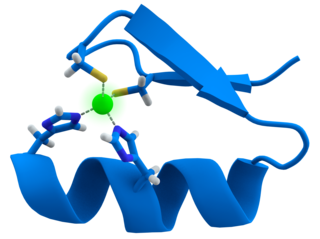
A zinc finger is a small protein structural motif that is characterized by the coordination of one or more zinc ions (Zn2+) which stabilizes the fold. It was originally coined to describe the finger-like appearance of a hypothesized structure from the African clawed frog (Xenopus laevis) transcription factor IIIA. However, it has been found to encompass a wide variety of differing protein structures in eukaryotic cells. Xenopus laevis TFIIIA was originally demonstrated to contain zinc and require the metal for function in 1983, the first such reported zinc requirement for a gene regulatory protein followed soon thereafter by the Krüppel factor in Drosophila. It often appears as a metal-binding domain in multi-domain proteins.

<span class="mw-page-title-main">DNA computing</span> Computing using molecular biology hardware

DNA computing is an emerging branch of unconventional computing which uses DNA, biochemistry, and molecular biology hardware, instead of the traditional electronic computing. Research and development in this area concerns theory, experiments, and applications of DNA computing. Although the field originally started with the demonstration of a computing application by Len Adleman in 1994, it has now been expanded to several other avenues such as the development of storage technologies, nanoscale imaging modalities, synthetic controllers and reaction networks, etc.

<span class="mw-page-title-main">Nanorobotics</span> Emerging technology field

Nanoid robotics, or for short, nanorobotics or nanobotics, is an emerging technology field creating machines or robots, which are called nanorobots or simply nanobots, whose components are at or near the scale of a nanometer. More specifically, nanorobotics refers to the nanotechnology engineering discipline of designing and building nanorobots with devices ranging in size from 0.1 to 10 micrometres and constructed of nanoscale or molecular components. The terms nanobot, nanoid, nanite, nanomachine and nanomite have also been used to describe such devices currently under research and development.

A nanoruler is a tool or a method used within the subfield of "nanometrology" to achieve precise control and measurements at the nanoscale. Measurements of extremely tiny proportions require more complicated procedures, such as manipulating the properties of light (plasmonic) or DNA to determine distances. At the nanoscale, materials and devices exhibit unique properties that can significantly influence their behavior. In fields like electronics, medicine, and biotechnology, where advancements come from manipulating matter at the atomic and molecular levels, nanoscale measurements become essential.

<span class="mw-page-title-main">M13 bacteriophage</span> Species of virus

M13 is one of the Ff phages, a member of the family filamentous bacteriophage (inovirus). Ff phages are composed of circular single-stranded DNA (ssDNA), which in the case of the m13 phage is 6407 nucleotides long and is encapsulated in approximately 2700 copies of the major coat protein p8, and capped with about 5 copies each of four different minor coat proteins. The minor coat protein p3 attaches to the receptor at the tip of the F pilus of the host Escherichia coli. The life cycle is relatively short, with the early phage progeny exiting the cell ten minutes after infection. Ff phages are chronic phage, releasing their progeny without killing the host cells. The infection causes turbid plaques in E. coli lawns, of intermediate opacity in comparison to regular lysis plaques. However, a decrease in the rate of cell growth is seen in the infected cells. The replicative form of M13 is circular double-stranded DNA similar to plasmids that are used for many recombinant DNA processes, and the virus has also been used for phage display, directed evolution, nanostructures and nanotechnology applications.

<span class="mw-page-title-main">DNA origami</span> Folding of DNA to create two- and three-dimensional shapes at the nanoscale

DNA origami is the nanoscale folding of DNA to create arbitrary two- and three-dimensional shapes at the nanoscale. The specificity of the interactions between complementary base pairs make DNA a useful construction material, through design of its base sequences. DNA is a well-understood material that is suitable for creating scaffolds that hold other molecules in place or to create structures all on its own.

<span class="mw-page-title-main">Holliday junction</span> Branched nucleic acid structure

A Holliday junction is a branched nucleic acid structure that contains four double-stranded arms joined. These arms may adopt one of several conformations depending on buffer salt concentrations and the sequence of nucleobases closest to the junction. The structure is named after Robin Holliday, the molecular biologist who proposed its existence in 1964.

<span class="mw-page-title-main">Molecular self-assembly</span> Movement of molecules into a defined arrangement without outside influence

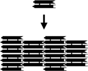
In chemistry and materials science, molecular self-assembly is the process by which molecules adopt a defined arrangement without guidance or management from an outside source. There are two types of self-assembly: intermolecular and intramolecular. Commonly, the term molecular self-assembly refers to the former, while the latter is more commonly called folding.

<span class="mw-page-title-main">Nucleic acid design</span>

Nucleic acid design is the process of generating a set of nucleic acid base sequences that will associate into a desired conformation. Nucleic acid design is central to the fields of DNA nanotechnology and DNA computing. It is necessary because there are many possible sequences of nucleic acid strands that will fold into a given secondary structure, but many of these sequences will have undesired additional interactions which must be avoided. In addition, there are many tertiary structure considerations which affect the choice of a secondary structure for a given design.

<span class="mw-page-title-main">Molecular models of DNA</span>

Molecular models of DNA structures are representations of the molecular geometry and topology of deoxyribonucleic acid (DNA) molecules using one of several means, with the aim of simplifying and presenting the essential, physical and chemical, properties of DNA molecular structures either in vivo or in vitro. These representations include closely packed spheres made of plastic, metal wires for skeletal models, graphic computations and animations by computers, artistic rendering. Computer molecular models also allow animations and molecular dynamics simulations that are very important for understanding how DNA functions in vivo.

<span class="mw-page-title-main">Nucleic acid tertiary structure</span> Three-dimensional shape of a nucleic acid polymer

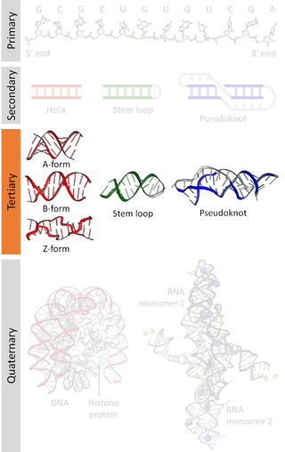
Nucleic acid tertiary structure is the three-dimensional shape of a nucleic acid polymer. RNA and DNA molecules are capable of diverse functions ranging from molecular recognition to catalysis. Such functions require a precise three-dimensional structure. While such structures are diverse and seemingly complex, they are composed of recurring, easily recognizable tertiary structural motifs that serve as molecular building blocks. Some of the most common motifs for RNA and DNA tertiary structure are described below, but this information is based on a limited number of solved structures. Many more tertiary structural motifs will be revealed as new RNA and DNA molecules are structurally characterized.

<span class="mw-page-title-main">Nucleic acid secondary structure</span>

Nucleic acid secondary structure is the basepairing interactions within a single nucleic acid polymer or between two polymers. It can be represented as a list of bases which are paired in a nucleic acid molecule. The secondary structures of biological DNAs and RNAs tend to be different: biological DNA mostly exists as fully base paired double helices, while biological RNA is single stranded and often forms complex and intricate base-pairing interactions due to its increased ability to form hydrogen bonds stemming from the extra hydroxyl group in the ribose sugar.

<span class="mw-page-title-main">Nadrian Seeman</span> American physicist (1945–2021)

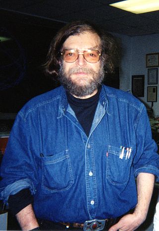
Nadrian C. "Ned" Seeman was an American nanotechnologist and crystallographer known for inventing the field of DNA nanotechnology.

<span class="mw-page-title-main">Spherical nucleic acid</span>

Spherical nucleic acids (SNAs) are nanostructures that consist of a densely packed, highly oriented arrangement of linear nucleic acids in a three-dimensional, spherical geometry. This novel three-dimensional architecture is responsible for many of the SNA's novel chemical, biological, and physical properties that make it useful in biomedicine and materials synthesis. SNAs were first introduced in 1996 by Chad Mirkin’s group at Northwestern University.

<span class="mw-page-title-main">Robert Dirks</span> American computational chemist killed in 2015 train wreck

Robert Dirks was an American chemist known for his theoretical and experimental work in DNA nanotechnology. Born in Thailand to a Thai Chinese mother and American father, he moved to Spokane, Washington at a young age. Dirks was the first graduate student in Niles Pierce's research group at the California Institute of Technology, where his dissertation work was on algorithms and computational tools to analyze nucleic acid thermodynamics and predict their structure. He also performed experimental work developing a biochemical chain reaction to self-assemble nucleic acid devices. Dirks later worked at D. E. Shaw Research on algorithms for protein folding that could be used to design new pharmaceuticals.

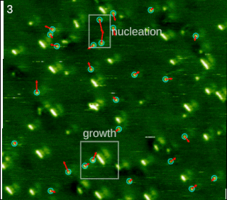
A DNA walker is a class of nucleic acid nanomachines where a nucleic acid "walker" is able to move along a nucleic acid "track". The concept of a DNA walker was first defined and named by John H. Reif in 2003. A nonautonomous DNA walker requires external changes for each step, whereas an autonomous DNA walker progresses without any external changes. Various nonautonomous DNA walkers were developed, for example Shin controlled the motion of DNA walker by using 'control strands' which needed to be manually added in a specific order according to the template's sequence in order to get the desired path of motion. In 2004 the first autonomous DNA walker, which did not require external changes for each step, was experimentally demonstrated by the Reif group.

<span class="mw-page-title-main">RNA origami</span> Nanoscale folding of RNA

RNA origami is the nanoscale folding of RNA, enabling the RNA to create particular shapes to organize these molecules. It is a new method that was developed by researchers from Aarhus University and California Institute of Technology. RNA origami is synthesized by enzymes that fold RNA into particular shapes. The folding of the RNA occurs in living cells under natural conditions. RNA origami is represented as a DNA gene, which within cells can be transcribed into RNA by RNA polymerase. Many computer algorithms are present to help with RNA folding, but none can fully predict the folding of RNA of a singular sequence.

TectoRNAs are modular RNA units able to self-assemble into larger nanostructures in a programmable fashion. They are generated by rational design through an approach called RNA architectonics, which make use of RNA structural modules identified in natural RNA molecules to form pre-defined 3D structures spontaneously.

References

  1. 1 2 DNA polyhedra: Goodman RP, Schaap IA, Tardin CF, Erben CM, Berry RM, Schmidt CF, Turberfield AJ (December 2005). "Rapid chiral assembly of rigid DNA building blocks for molecular nanofabrication". Science. 310 (5754): 1661–1665. Bibcode:2005Sci...310.1661G. doi:10.1126/science.1120367. PMID   16339440. S2CID   13678773.
  2. 1 2 3 History: Pelesko JA (2007). Self-assembly: the science of things that put themselves together. New York: Chapman & Hall/CRC. pp. 201, 242, 259. ISBN   978-1-58488-687-7.
  3. 1 2 3 4 5 Overview: Seeman NC (June 2004). "Nanotechnology and the double helix". Scientific American. 290 (6): 64–75. Bibcode:2004SciAm.290f..64S. doi:10.1038/scientificamerican0604-64. PMID   15195395.
  4. History: See "Current crystallization protocol". Nadrian Seeman Lab. for a statement of the problem, and "DNA cages containing oriented guests". Nadrian Seeman Laboratory. for the proposed solution.
  5. 1 2 3 4 5 Overview: Seeman NC (2010). "Nanomaterials based on DNA". Annual Review of Biochemistry. 79: 65–87. doi:10.1146/annurev-biochem-060308-102244. PMC   3454582 . PMID   20222824.
  6. 1 2 3 4 5 6 7 8 9 Overview: Pinheiro AV, Han D, Shih WM, Yan H (November 2011). "Challenges and opportunities for structural DNA nanotechnology". Nature Nanotechnology. 6 (12): 763–772. Bibcode:2011NatNa...6..763P. doi:10.1038/nnano.2011.187. PMC   3334823 . PMID   22056726.
  7. 1 2 DNA origami: Rothemund PW (2006). "Scaffolded DNA origami: from generalized multicrossovers to polygonal networks". In Chen J, Jonoska N, Rozenberg G (eds.). Nanotechnology: science and computation. Natural Computing Series. New York: Springer. pp. 3–21. CiteSeerX   10.1.1.144.1380 . doi:10.1007/3-540-30296-4_1. ISBN   978-3-540-30295-7.
  8. 1 2 3 4 5 History/applications: Service RF (June 2011). "DNA nanotechnology. DNA nanotechnology grows up". Science. 332 (6034): 1140–1, 1143. Bibcode:2011Sci...332.1140S. doi:10.1126/science.332.6034.1140. PMID   21636754.
  9. Yurke, Bernard; Turberfield, Andrew J.; Mills, Allen P.; Simmel, Friedrich C.; Neumann, Jennifer L. (August 2000). "A DNA-fuelled molecular machine made of DNA". Nature. 406 (6796): 605–608. Bibcode:2000Natur.406..605Y. doi:10.1038/35020524. ISSN   1476-4687. S2CID   2064216.
  10. 1 2 3 4 DNA machines: Bath J, Turberfield AJ (May 2007). "DNA nanomachines". Nature Nanotechnology. 2 (5): 275–284. Bibcode:2007NatNa...2..275B. doi:10.1038/nnano.2007.104. PMID   18654284.
  11. Nanoarchitecture: Robinson BH, Seeman NC (August 1987). "The design of a biochip: a self-assembling molecular-scale memory device". Protein Engineering. 1 (4): 295–300. doi:10.1093/protein/1.4.295. PMID   3508280.
  12. Nanoarchitecture: Xiao S, Liu F, Rosen AE, Hainfeld JF, Seeman NC, Musier-Forsyth K, Kiehl RA (August 2002). "Selfassembly of metallic nanoparticle arrays by DNA scaffolding". Journal of Nanoparticle Research. 4 (4): 313–317. Bibcode:2002JNR.....4..313X. doi:10.1023/A:1021145208328. S2CID   2257083.
  13. 1 2 3 DNA origami: Rothemund PW (March 2006). "Folding DNA to create nanoscale shapes and patterns" (PDF). Nature. 440 (7082): 297–302. Bibcode:2006Natur.440..297R. doi:10.1038/nature04586. PMID   16541064. S2CID   4316391.
  14. 1 2 DNA origami: Douglas SM, Dietz H, Liedl T, Högberg B, Graf F, Shih WM (May 2009). "Self-assembly of DNA into nanoscale three-dimensional shapes". Nature. 459 (7245): 414–418. Bibcode:2009Natur.459..414D. doi:10.1038/nature08016. PMC   2688462 . PMID   19458720.
  15. Service RF (June 2011). "DNA nanotechnology. DNA nanotechnology grows up". Science. 332 (6034): 1140–1, 1143. Bibcode:2011Sci...332.1140S. doi:10.1126/science.332.6034.1140. PMID   21636754.
  16. History: Hopkin K (August 2011). "Profile: 3-D seer". The Scientist. Archived from the original on 10 October 2011. Retrieved 8 August 2011.
  17. 1 2 3 History: Seeman NC (June 2010). "Structural DNA nanotechnology: growing along with Nano Letters". Nano Letters. 10 (6): 1971–1978. Bibcode:2010NanoL..10.1971S. doi:10.1021/nl101262u. PMC   2901229 . PMID   20486672.
  18. 1 2 3 Overview: Mao C (December 2004). "The emergence of complexity: lessons from DNA". PLOS Biology. 2 (12): e431. doi: 10.1371/journal.pbio.0020431 . PMC   535573 . PMID   15597116.
  19. Background: Pelesko JA (2007). Self-assembly: the science of things that put themselves together. New York: Chapman & Hall/CRC. pp. 5, 7. ISBN   978-1-58488-687-7.
  20. Background: Long EC (1996). "Fundamentals of nucleic acids". In Hecht SM (ed.). Bioorganic chemistry: nucleic acids. New York: Oxford University Press. pp. 4–10. ISBN   978-0-19-508467-2.
  21. RNA nanotechnology: Chworos A, Severcan I, Koyfman AY, Weinkam P, Oroudjev E, Hansma HG, Jaeger L (December 2004). "Building programmable jigsaw puzzles with RNA". Science. 306 (5704): 2068–2072. Bibcode:2004Sci...306.2068C. doi:10.1126/science.1104686. PMID   15604402. S2CID   9296608.
  22. RNA nanotechnology: Guo P (December 2010). "The emerging field of RNA nanotechnology". Nature Nanotechnology. 5 (12): 833–842. Bibcode:2010NatNa...5..833G. doi:10.1038/nnano.2010.231. PMC   3149862 . PMID   21102465.
  23. 1 2 3 4 Dynamic DNA nanotechnology: Zhang DY, Seelig G (February 2011). "Dynamic DNA nanotechnology using strand-displacement reactions". Nature Chemistry. 3 (2): 103–113. Bibcode:2011NatCh...3..103Z. doi:10.1038/nchem.957. PMID   21258382.
  24. 1 2 3 4 5 Structural DNA nanotechnology: Seeman NC (November 2007). "An overview of structural DNA nanotechnology". Molecular Biotechnology. 37 (3): 246–257. doi:10.1007/s12033-007-0059-4. PMC   3479651 . PMID   17952671.
  25. Dynamic DNA nanotechnology: Lu Y, Liu J (December 2006). "Functional DNA nanotechnology: emerging applications of DNAzymes and aptamers". Current Opinion in Biotechnology. 17 (6): 580–588. doi:10.1016/j.copbio.2006.10.004. PMID   17056247.
  26. Simulation of DNA structures: Doye JP, Ouldridge TE, Louis AA, Romano F, Šulc P, Matek C, et al. (December 2013). "Coarse-graining DNA for simulations of DNA nanotechnology". Physical Chemistry Chemical Physics. 15 (47): 20395–20414. arXiv: 1308.3843 . Bibcode:2013PCCP...1520395D. doi:10.1039/C3CP53545B. PMID   24121860. S2CID   15324396.
  27. Other arrays: Strong M (March 2004). "Protein nanomachines". PLOS Biology. 2 (3): E73. doi: 10.1371/journal.pbio.0020073 . PMC   368168 . PMID   15024422.
  28. Yan H, Park SH, Finkelstein G, Reif JH, LaBean TH (September 2003). "DNA-templated self-assembly of protein arrays and highly conductive nanowires". Science. 301 (5641): 1882–1884. Bibcode:2003Sci...301.1882Y. doi:10.1126/science.1089389. PMID   14512621. S2CID   137635908.
  29. 1 2 Algorithmic self-assembly: Rothemund PW, Papadakis N, Winfree E (December 2004). "Algorithmic self-assembly of DNA Sierpinski triangles". PLOS Biology. 2 (12): e424. doi: 10.1371/journal.pbio.0020424 . PMC   534809 . PMID   15583715.
  30. DX arrays: Winfree E, Liu F, Wenzler LA, Seeman NC (August 1998). "Design and self-assembly of two-dimensional DNA crystals". Nature. 394 (6693): 539–544. Bibcode:1998Natur.394..539W. doi:10.1038/28998. PMID   9707114. S2CID   4385579.
  31. DX arrays: Liu F, Sha R, Seeman NC (10 February 1999). "Modifying the surface features of two-dimensional DNA crystals". Journal of the American Chemical Society. 121 (5): 917–922. doi:10.1021/ja982824a.
  32. Other arrays: Mao C, Sun W, Seeman NC (16 June 1999). "Designed two-dimensional DNA Holliday junction arrays visualized by atomic force microscopy". Journal of the American Chemical Society. 121 (23): 5437–5443. doi:10.1021/ja9900398.
  33. Other arrays: Constantinou PE, Wang T, Kopatsch J, Israel LB, Zhang X, Ding B, et al. (September 2006). "Double cohesion in structural DNA nanotechnology". Organic & Biomolecular Chemistry. 4 (18): 3414–3419. doi:10.1039/b605212f. PMC   3491902 . PMID   17036134.
  34. Other arrays: Mathieu F, Liao S, Kopatsch J, Wang T, Mao C, Seeman NC (April 2005). "Six-helix bundles designed from DNA". Nano Letters. 5 (4): 661–665. Bibcode:2005NanoL...5..661M. doi:10.1021/nl050084f. PMC   3464188 . PMID   15826105.
  35. Algorithmic self-assembly: Barish RD, Rothemund PW, Winfree E (December 2005). "Two computational primitives for algorithmic self-assembly: copying and counting". Nano Letters. 5 (12): 2586–2592. Bibcode:2005NanoL...5.2586B. CiteSeerX   10.1.1.155.676 . doi:10.1021/nl052038l. PMID   16351220.
  36. 1 2 3 4 Design: Feldkamp U, Niemeyer CM (March 2006). "Rational design of DNA nanoarchitectures". Angewandte Chemie. 45 (12): 1856–1876. doi:10.1002/anie.200502358. PMID   16470892.
  37. DNA nanotubes: Rothemund PW, Ekani-Nkodo A, Papadakis N, Kumar A, Fygenson DK, Winfree E (December 2004). "Design and characterization of programmable DNA nanotubes". Journal of the American Chemical Society. 126 (50): 16344–16352. doi:10.1021/ja044319l. PMID   15600335.
  38. DNA nanotubes: Yin P, Hariadi RF, Sahu S, Choi HM, Park SH, Labean TH, Reif JH (August 2008). "Programming DNA tube circumferences". Science. 321 (5890): 824–826. Bibcode:2008Sci...321..824Y. doi:10.1126/science.1157312. PMID   18687961. S2CID   12100380.
  39. Three-dimensional arrays: Zheng J, Birktoft JJ, Chen Y, Wang T, Sha R, Constantinou PE, et al. (September 2009). "From molecular to macroscopic via the rational design of a self-assembled 3D DNA crystal". Nature. 461 (7260): 74–77. Bibcode:2009Natur.461...74Z. doi:10.1038/nature08274. PMC   2764300 . PMID   19727196.
  40. DNA polyhedra: Zhang Y, Seeman NC (1 March 1994). "Construction of a DNA-truncated octahedron". Journal of the American Chemical Society. 116 (5): 1661–1669. doi:10.1021/ja00084a006.
  41. DNA polyhedra: Shih WM, Quispe JD, Joyce GF (February 2004). "A 1.7-kilobase single-stranded DNA that folds into a nanoscale octahedron". Nature. 427 (6975): 618–621. Bibcode:2004Natur.427..618S. doi:10.1038/nature02307. PMID   14961116. S2CID   4419579.
  42. Tikhomirov G, Petersen P, Qian L (December 2017). "Fractal assembly of micrometre-scale DNA origami arrays with arbitrary patterns". Nature. 552 (7683): 67–71. Bibcode:2017Natur.552...67T. doi:10.1038/nature24655. PMID   29219965. S2CID   4455780.
  43. 1 2 DNA boxes: Andersen ES, Dong M, Nielsen MM, Jahn K, Subramani R, Mamdouh W, et al. (May 2009). "Self-assembly of a nanoscale DNA box with a controllable lid". Nature. 459 (7243): 73–76. Bibcode:2009Natur.459...73A. doi:10.1038/nature07971. hdl: 11858/00-001M-0000-0010-9363-9 . PMID   19424153. S2CID   4430815.
  44. DNA boxes: Ke Y, Sharma J, Liu M, Jahn K, Liu Y, Yan H (June 2009). "Scaffolded DNA origami of a DNA tetrahedron molecular container". Nano Letters. 9 (6): 2445–2447. Bibcode:2009NanoL...9.2445K. doi:10.1021/nl901165f. PMID   19419184.
  45. Zaborova, O. V.; Voinova, A. D.; Shmykov, B. D.; Sergeyev, V. G. (2021). "Solid Lipid Nanoparticles for the Nucleic Acid Encapsulation". Reviews and Advances in Chemistry. 11 (3–4): 178–188. doi:10.1134/S2079978021030055. ISSN   2634-8276. S2CID   246946068.
  46. Overview: Endo M, Sugiyama H (October 2009). "Chemical approaches to DNA nanotechnology". ChemBioChem. 10 (15): 2420–2443. doi:10.1002/cbic.200900286. PMID   19714700. S2CID   205554125.
  47. Nanoarchitecture: Zheng J, Constantinou PE, Micheel C, Alivisatos AP, Kiehl RA, Seeman NC (July 2006). "Two-dimensional nanoparticle arrays show the organizational power of robust DNA motifs". Nano Letters. 6 (7): 1502–1504. Bibcode:2006NanoL...6.1502Z. doi:10.1021/nl060994c. PMC   3465979 . PMID   16834438.
  48. Nanoarchitecture: Park SH, Pistol C, Ahn SJ, Reif JH, Lebeck AR, Dwyer C, LaBean TH (January 2006). "Finite-size, fully addressable DNA tile lattices formed by hierarchical assembly procedures". Angewandte Chemie. 45 (5): 735–739. Bibcode:2006AngCh.118.6759P. doi: 10.1002/ange.200690141 . PMID   16374784.
  49. Nanoarchitecture: Cohen JD, Sadowski JP, Dervan PB (22 October 2007). "Addressing single molecules on DNA nanostructures". Angewandte Chemie. 46 (42): 7956–7959. doi:10.1002/anie.200702767. PMID   17763481.
  50. Nanoarchitecture: Maune HT, Han SP, Barish RD, Bockrath M, Goddard WA, Rothemund PW, Winfree E (January 2010). "Self-assembly of carbon nanotubes into two-dimensional geometries using DNA origami templates". Nature Nanotechnology. 5 (1): 61–66. Bibcode:2010NatNa...5...61M. doi:10.1038/nnano.2009.311. PMID   19898497.
  51. Nanoarchitecture: Liu J, Geng Y, Pound E, Gyawali S, Ashton JR, Hickey J, et al. (March 2011). "Metallization of branched DNA origami for nanoelectronic circuit fabrication". ACS Nano. 5 (3): 2240–2247. doi:10.1021/nn1035075. PMID   21323323.
  52. Nanoarchitecture: Deng Z, Mao C (August 2004). "Molecular lithography with DNA nanostructures". Angewandte Chemie. 43 (31): 4068–4070. doi:10.1002/anie.200460257. PMID   15300697.
  53. DNA machines: Mao C, Sun W, Shen Z, Seeman NC (January 1999). "A nanomechanical device based on the B-Z transition of DNA". Nature. 397 (6715): 144–146. Bibcode:1999Natur.397..144M. doi:10.1038/16437. PMID   9923675. S2CID   4406177.
  54. DNA machines: Yurke B, Turberfield AJ, Mills AP, Simmel FC, Neumann JL (August 2000). "A DNA-fuelled molecular machine made of DNA". Nature. 406 (6796): 605–608. Bibcode:2000Natur.406..605Y. doi:10.1038/35020524. PMID   10949296. S2CID   2064216.
  55. DNA machines: Yan H, Zhang X, Shen Z, Seeman NC (January 2002). "A robust DNA mechanical device controlled by hybridization topology". Nature. 415 (6867): 62–65. Bibcode:2002Natur.415...62Y. doi:10.1038/415062a. PMID   11780115. S2CID   52801697.
  56. DNA machines: Feng L, Park SH, Reif JH, Yan H (September 2003). "A two-state DNA lattice switched by DNA nanoactuator". Angewandte Chemie. 42 (36): 4342–4346. Bibcode:2003AngCh.115.4478F. doi:10.1002/ange.200351818. PMID   14502706.
  57. DNA machines: Goodman RP, Heilemann M, Doose S, Erben CM, Kapanidis AN, Turberfield AJ (February 2008). "Reconfigurable, braced, three-dimensional DNA nanostructures". Nature Nanotechnology. 3 (2): 93–96. Bibcode:2008NatNa...3...93G. doi:10.1038/nnano.2008.3. PMID   18654468.
  58. Applications: Douglas SM, Bachelet I, Church GM (February 2012). "A logic-gated nanorobot for targeted transport of molecular payloads". Science. 335 (6070): 831–834. Bibcode:2012Sci...335..831D. doi:10.1126/science.1214081. PMID   22344439. S2CID   9866509.
  59. Centola, Mathias; Poppleton, Erik; Ray, Sujay; Centola, Martin; Welty, Robb; Valero, Julián; Walter, Nils G.; Šulc, Petr; Famulok, Michael (2023-10-19). "A rhythmically pulsing leaf-spring DNA-origami nanoengine that drives a passive follower". Nature Nanotechnology: 1–11. doi: 10.1038/s41565-023-01516-x . ISSN   1748-3395. PMC   10873200 . PMID   37857824.
  60. DNA walkers: Shin JS, Pierce NA (September 2004). "A synthetic DNA walker for molecular transport". Journal of the American Chemical Society. 126 (35): 10834–10835. doi:10.1021/ja047543j. PMID   15339155.
  61. DNA walkers: Sherman WB, Seeman NC (July 2004). "A precisely controlled DNA biped walking device". Nano Letters. 4 (7): 1203–1207. Bibcode:2004NanoL...4.1203S. doi:10.1021/nl049527q.
  62. DNA walkers: Škugor M, Valero J, Murayama K, Centola M, Asanuma H, Famulok M (May 2019). "Orthogonally Photocontrolled Non-Autonomous DNA Walker". Angewandte Chemie. 58 (21): 6948–6951. doi:10.1002/anie.201901272. PMID   30897257. S2CID   85446523.
  63. DNA walkers: Tian Y, He Y, Chen Y, Yin P, Mao C (July 2005). "A DNAzyme that walks processively and autonomously along a one-dimensional track". Angewandte Chemie. 44 (28): 4355–4358. Bibcode:2005AngCh.117.4429T. doi:10.1002/ange.200500703. PMID   15945114.
  64. DNA walkers: Bath J, Green SJ, Turberfield AJ (July 2005). "A free-running DNA motor powered by a nicking enzyme". Angewandte Chemie. 44 (28): 4358–4361. doi:10.1002/anie.200501262. PMID   15959864.
  65. Functional DNA walkers: Lund K, Manzo AJ, Dabby N, Michelotti N, Johnson-Buck A, Nangreave J, et al. (May 2010). "Molecular robots guided by prescriptive landscapes". Nature. 465 (7295): 206–210. Bibcode:2010Natur.465..206L. doi:10.1038/nature09012. PMC   2907518 . PMID   20463735.
  66. Functional DNA walkers: Valero J, Pal N, Dhakal S, Walter NG, Famulok M (June 2018). "A bio-hybrid DNA rotor-stator nanoengine that moves along predefined tracks". Nature Nanotechnology. 13 (6): 496–503. Bibcode:2018NatNa..13..496V. doi:10.1038/s41565-018-0109-z. PMC   5994166 . PMID   29632399.
  67. Functional DNA walkers: He Y, Liu DR (November 2010). "Autonomous multistep organic synthesis in a single isothermal solution mediated by a DNA walker". Nature Nanotechnology. 5 (11): 778–782. Bibcode:2010NatNa...5..778H. doi:10.1038/nnano.2010.190. PMC   2974042 . PMID   20935654.
  68. Pan J, Li F, Cha TG, Chen H, Choi JH (August 2015). "Recent progress on DNA based walkers". Current Opinion in Biotechnology. 34: 56–64. doi:10.1016/j.copbio.2014.11.017. PMID   25498478.
  69. 1 2 3 Kinetic assembly: Yin P, Choi HM, Calvert CR, Pierce NA (January 2008). "Programming biomolecular self-assembly pathways". Nature. 451 (7176): 318–322. Bibcode:2008Natur.451..318Y. doi:10.1038/nature06451. PMID   18202654. S2CID   4354536.
  70. Fuzzy and Boolean logic gates based on DNA: Zadegan RM, Jepsen MD, Hildebrandt LL, Birkedal V, Kjems J (April 2015). "Construction of a fuzzy and Boolean logic gates based on DNA". Small. 11 (15): 1811–1817. doi:10.1002/smll.201402755. PMID   25565140.
  71. Strand displacement cascades: Seelig G, Soloveichik D, Zhang DY, Winfree E (December 2006). "Enzyme-free nucleic acid logic circuits". Science. 314 (5805): 1585–1588. Bibcode:2006Sci...314.1585S. doi:10.1126/science.1132493. PMID   17158324. S2CID   10966324.
  72. Strand displacement cascades: Qian L, Winfree E (June 2011). "Scaling up digital circuit computation with DNA strand displacement cascades". Science. 332 (6034): 1196–1201. Bibcode:2011Sci...332.1196Q. doi:10.1126/science.1200520. PMID   21636773. S2CID   10053541.
  73. Applications: Rietman EA (2001). Molecular engineering of nanosystems. Springer. pp. 209–212. ISBN   978-0-387-98988-4 . Retrieved 17 April 2011.
  74. Zadegan RM, Jepsen MD, Thomsen KE, Okholm AH, Schaffert DH, Andersen ES, et al. (November 2012). "Construction of a 4 zeptoliters switchable 3D DNA box origami". ACS Nano. 6 (11): 10050–10053. doi:10.1021/nn303767b. PMID   23030709.
  75. Applications: Jungmann R, Renner S, Simmel FC (April 2008). "From DNA nanotechnology to synthetic biology". HFSP Journal. 2 (2): 99–109. doi:10.2976/1.2896331. PMC   2645571 . PMID   19404476.
  76. Lovy, Howard (5 July 2011). "DNA cages can unleash meds inside cells". fiercedrugdelivery.com. Retrieved 22 September 2013.[ permanent dead link ]
  77. Walsh AS, Yin H, Erben CM, Wood MJ, Turberfield AJ (July 2011). "DNA cage delivery to mammalian cells". ACS Nano. 5 (7): 5427–5432. doi:10.1021/nn2005574. PMID   21696187.
  78. Trafton, Anne (4 June 2012). "Researchers achieve RNA interference, in a lighter package". MIT News. Retrieved 22 September 2013.
  79. Lee H, Lytton-Jean AK, Chen Y, Love KT, Park AI, Karagiannis ED, et al. (June 2012). "Molecularly self-assembled nucleic acid nanoparticles for targeted in vivo siRNA delivery". Nature Nanotechnology. 7 (6): 389–393. Bibcode:2012NatNa...7..389L. doi:10.1038/NNANO.2012.73. PMC   3898745 . PMID   22659608.
  80. Kim KR, Kim DR, Lee T, Yhee JY, Kim BS, Kwon IC, Ahn DR (March 2013). "Drug delivery by a self-assembled DNA tetrahedron for overcoming drug resistance in breast cancer cells". Chemical Communications. 49 (20): 2010–2012. doi:10.1039/c3cc38693g. PMID   23380739.
  81. Sundah NR, Ho NR, Lim GS, Natalia A, Ding X, Liu Y, et al. (September 2019). "Barcoded DNA nanostructures for the multiplexed profiling of subcellular protein distribution". Nature Biomedical Engineering. 3 (9): 684–694. doi:10.1038/s41551-019-0417-0. PMID   31285580. S2CID   195825879.
  82. DNA ion channels: Langecker M, Arnaut V, Martin TG, List J, Renner S, Mayer M, et al. (November 2012). "Synthetic lipid membrane channels formed by designed DNA nanostructures". Science. 338 (6109): 932–936. Bibcode:2012Sci...338..932L. doi:10.1126/science.1225624. PMC   3716461 . PMID   23161995.
  83. 1 2 DNA ion channels: Göpfrich K, Li CY, Mames I, Bhamidimarri SP, Ricci M, Yoo J, et al. (July 2016). "Ion Channels Made from a Single Membrane-Spanning DNA Duplex". Nano Letters. 16 (7): 4665–4669. Bibcode:2016NanoL..16.4665G. doi:10.1021/acs.nanolett.6b02039. PMC   4948918 . PMID   27324157.
  84. DNA ion channels: Burns JR, Stulz E, Howorka S (June 2013). "Self-assembled DNA nanopores that span lipid bilayers". Nano Letters. 13 (6): 2351–2356. Bibcode:2013NanoL..13.2351B. CiteSeerX   10.1.1.659.7660 . doi:10.1021/nl304147f. PMID   23611515.
  85. DNA ion channels: Burns JR, Göpfrich K, Wood JW, Thacker VV, Stulz E, Keyser UF, Howorka S (November 2013). "Lipid-bilayer-spanning DNA nanopores with a bifunctional porphyrin anchor". Angewandte Chemie. 52 (46): 12069–12072. doi:10.1002/anie.201305765. PMC   4016739 . PMID   24014236.
  86. DNA ion channels: Seifert A, Göpfrich K, Burns JR, Fertig N, Keyser UF, Howorka S (February 2015). "Bilayer-spanning DNA nanopores with voltage-switching between open and closed state". ACS Nano. 9 (2): 1117–1126. doi:10.1021/nn5039433. PMC   4508203 . PMID   25338165.
  87. DNA ion channels: Göpfrich K, Zettl T, Meijering AE, Hernández-Ainsa S, Kocabey S, Liedl T, Keyser UF (May 2015). "DNA-Tile Structures Induce Ionic Currents through Lipid Membranes". Nano Letters. 15 (5): 3134–3138. Bibcode:2015NanoL..15.3134G. doi:10.1021/acs.nanolett.5b00189. PMID   25816075.
  88. DNA ion channels: Burns JR, Seifert A, Fertig N, Howorka S (February 2016). "A biomimetic DNA-based channel for the ligand-controlled transport of charged molecular cargo across a biological membrane". Nature Nanotechnology. 11 (2): 152–156. Bibcode:2016NatNa..11..152B. doi:10.1038/nnano.2015.279. PMID   26751170.
  89. DNA ion channels: Göpfrich K, Li CY, Ricci M, Bhamidimarri SP, Yoo J, Gyenes B, et al. (September 2016). "Large-Conductance Transmembrane Porin Made from DNA Origami". ACS Nano. 10 (9): 8207–8214. doi:10.1021/acsnano.6b03759. PMC   5043419 . PMID   27504755.
  90. DNA scramblase: Ohmann A, Li CY, Maffeo C, Al Nahas K, Baumann KN, Göpfrich K, et al. (June 2018). "A synthetic enzyme built from DNA flips 107 lipids per second in biological membranes". Nature Communications. 9 (1): 2426. Bibcode:2018NatCo...9.2426O. doi:10.1038/s41467-018-04821-5. PMC   6013447 . PMID   29930243.
  91. 1 2 3 Design: Brenneman A, Condon A (25 September 2002). "Strand design for biomolecular computation". Theoretical Computer Science. 287: 39–58. doi: 10.1016/S0304-3975(02)00135-4 .
  92. Overview: Lin C, Liu Y, Rinker S, Yan H (August 2006). "DNA tile based self-assembly: building complex nanoarchitectures". ChemPhysChem. 7 (8): 1641–1647. doi:10.1002/cphc.200600260. PMID   16832805.
  93. 1 2 3 Design: Dirks RM, Lin M, Winfree E, Pierce NA (15 February 2004). "Paradigms for computational nucleic acid design". Nucleic Acids Research. 32 (4): 1392–1403. doi:10.1093/nar/gkh291. PMC   390280 . PMID   14990744.
  94. Methods: Ellington A, Pollard JD (1 May 2001). "Synthesis and Purification of Oligonucleotides". Current Protocols in Molecular Biology. 42: 2.11.1–2.11.25. doi:10.1002/0471142727.mb0211s42. ISBN   978-0471142720. PMID   18265179. S2CID   205152989.
  95. Methods: Ellington A, Pollard JD (1 May 2001). "Purification of Oligonucleotides Using Denaturing Polyacrylamide Gel Electrophoresis". Current Protocols in Molecular Biology. 42: Unit2.12. doi:10.1002/0471142727.mb0212s42. ISBN   978-0471142720. PMID   18265180. S2CID   27187583.
  96. Methods: Gallagher SR, Desjardins P (1 July 2011). "Quantitation of nucleic acids and proteins". Current Protocols Essential Laboratory Techniques. Vol. 5. doi:10.1002/9780470089941.et0202s5. ISBN   978-0470089934. S2CID   94329398.
  97. Methods: Chory J, Pollard JD (1 May 2001). "Separation of Small DNA Fragments by Conventional Gel Electrophoresis". Current Protocols in Molecular Biology. 47: Unit2.7. doi:10.1002/0471142727.mb0207s47. ISBN   978-0471142720. PMID   18265187. S2CID   43406338.
  98. Methods: Walter NG (1 February 2003). "Probing RNA Structural Dynamics and Function by Fluorescence Resonance Energy Transfer (FRET)". Current Protocols in Nucleic Acid Chemistry. 11: 11.10.1–11.10.23. doi:10.1002/0471142700.nc1110s11. ISBN   978-0471142706. PMID   18428904. S2CID   9978415.
  99. Methods: Lin C, Ke Y, Chhabra R, Sharma J, Liu Y, Yan H (2011). "Synthesis and Characterization of Self-Assembled DNA Nanostructures". In Zuccheri G, Samorì B (eds.). DNA Nanotechnology. Methods in Molecular Biology. Vol. 749. pp. 1–11. doi:10.1007/978-1-61779-142-0_1. ISBN   978-1-61779-141-3. PMID   21674361.
  100. Methods: Bloomfield VA, Crothers DM, Tinoco Jr I (2000). Nucleic acids: structures, properties, and functions. Sausalito, Calif: University Science Books. pp. 84–86, 396–407. ISBN   978-0-935702-49-1.

Further reading

General:

Specific subfields: