Tuberculosis diagnosis

Last updated
Tuberculosis diagnosis
TB in sputum.png
M. tuberculosis (stained red) in sputum
Purposediagnosed by finding Mycobacterium tuberculosis

Tuberculosis is diagnosed by finding Mycobacterium tuberculosis bacteria in a clinical specimen taken from the patient. While other investigations may strongly suggest tuberculosis as the diagnosis, they cannot confirm it.

Contents

A complete medical evaluation for tuberculosis (TB) must include a medical history, a physical examination, a chest X-ray and microbiological examination (of sputum or some other appropriate sample). It may also include a tuberculin skin test, other scans and X-rays, surgical biopsy.

Medical history

The medical history includes obtaining the symptoms of pulmonary TB: productive, prolonged cough of three or more weeks, chest pain, and hemoptysis. Systemic symptoms include low grade remittent fever, chills, night sweats, appetite loss, weight loss, easy fatiguability, and production of sputum that starts out mucoid but changes to purulent. [1] Other parts of the medical history include prior TB exposure, infection or disease and medical conditions that increase risk for TB disease such as HIV infection. Depending on the sort of patient population surveyed, as few as 20%, or as many as 75% of pulmonary tuberculosis cases may be without symptoms. [2]

Tuberculosis should be suspected in adults when a pneumonia-like illness has persisted longer than three weeks, or when a respiratory illness in an otherwise healthy individual does not respond to regular antibiotics.[ citation needed ]

The diagnosis is particularly challenging in children because they more commonly have extrapulmonary disease and present with less mycobacteria (paucibacillary disease). [3]

Physical examination

A physical examination is done to assess the patient's general health. It cannot be used to confirm or rule out TB. However, certain findings are suggestive of TB. For example, blood in the sputum, significant weight loss and drenching night sweats may be due to TB.[ citation needed ]

Tuberculosis classification (Class 0 to 5) used in the US is based on the pathogenesis of the disease. The U.S. Citizenship and Immigration Services has an additional TB classification (Class A, B1, or B2) for immigrants and refugees developed by the Centers for Disease Control and Prevention (CDC). The (Class) B notification program is an important screening strategy to identify new arrivals who have a high risk for TB.[ citation needed ]

Microbiological studies

Lab findings
TB Culture.jpg
Distinctive clusters of colorless Mycobacterium tuberculosis form in this culture.
Gram +
Shape rods

A definitive diagnosis of tuberculosis can only be made by culturing Mycobacterium tuberculosis organisms from a specimen taken from the patient (most often sputum, but may also include pus, CSF, biopsied tissue, etc.). [1] A diagnosis made other than by culture may only be classified as "probable" or "presumed". For a diagnosis negating the possibility of tuberculosis infection, most protocols require that two separate cultures both test negative. [1]

Sputum

Sputum smears and cultures should be done for acid-fast bacilli if the patient is producing sputum. [1] The preferred method for this is fluorescence microscopy (auramine-rhodamine staining), which is more sensitive than conventional Ziehl–Neelsen staining. [4] In cases where there is no spontaneous sputum production, a sample can be induced, usually by inhalation of a nebulized saline or saline with bronchodilator solution. A comparative study found that inducing three sputum samples is more sensitive than three gastric washings. [5]

Alternative sampling

In patients incapable of producing a sputum sample, common alternative sample sources for diagnosing pulmonary tuberculosis include gastric washings, laryngeal swab, bronchoscopy (with bronchoalveolar lavage, bronchial washings, and/or transbronchial biopsy), and fine needle aspiration (transtracheal or transbronchial). In some cases, a more invasive technique is necessary, including tissue biopsy during mediastinoscopy or thoracoscopy.[ citation needed ]

Nucleic acid amplification tests (NAAT)

Other mycobacteria are also acid-fast. If the smear is positive, PCR or gene probe tests can distinguish M. tuberculosis from other mycobacteria. Even if sputum smear is negative, tuberculosis must be considered and is only excluded after negative cultures.[ citation needed ]

Nucleic acid amplification tests (NAAT) for TB are a heterogeneous group of tests that use either the polymerase chain reaction (PCR) technique or transcription-mediated amplification (TMA) or other forms of nucleic acid amplification methods to detect mycobacterial nucleic acid. These tests vary in which nucleic acid sequence they detect and vary in their accuracy. In the decade of the 2000s, the two most common commercially available tests were the amplified mycobacterium tuberculosis direct test (MTD, Gen-Probe) and Amplicor (Roche Diagnostics). In 2007, a systematic review of NAAT by the NHS Health Technology Assessment Programme concluded that "NAAT test accuracy to be far superior when applied to respiratory samples as opposed to other specimens. Although the results were not statistically significant, the AMTD test appears to perform better than other currently available commercial tests." [6] Xpert ® MTB/RIF and Xpert MTB/RIF Ultra has high specificity in diagnosing extrapulmonary tuberculosis and is accurate in detecting rifampicin resistance. However, clinicians should rely on clinical judgement to diagnose TB meningitis when culture results are negative. [7]

A 2007 before-after observational study found that use of the MTD test reduced inappropriate tuberculosis therapy. The study found the accuracy of the MTD test as follows: [8] Overall sensitivity 92%, specificity 99%. In Smear positive patients high sensitivity 99%, specificity 98%. In smear negative patients low sensitivity 62%, but high specificity 99%.

In 2010 the Xpert MTB/RIF test, another NAAT for TB, became commercially available and, as the CDC said in 2015, [9] it began "revolutionizing tuberculosis (TB) control by contributing to the rapid diagnosis of TB disease and drug resistance. The test simultaneously detects Mycobacterium tuberculosis complex (MTBC) and resistance to rifampin (RIF) in less than 2 hours. In comparison, standard cultures can take 2 to 6 weeks for MTBC to grow and conventional drug resistance tests can add 3 more weeks." [9] The Xpert MTB/RIF Ultra improves upon the Xpert MTB/RIF test. [10]

Culture

Many types of microbiological culture are available. [11] Traditionally, cultures have used the Löwenstein-Jensen (LJ), Kirchner, or Middlebrook media (7H9, 7H10, and 7H11). A culture of the AFB can distinguish the various forms of mycobacteria, although results from this may take four to eight weeks for a conclusive answer. New automated systems that are faster include the MB/BacT, BACTEC 9000, VersaTREK, and the Mycobacterial Growth Indicator Tube (MGIT). The Microscopic Observation Drug Susceptibility assay culture may be a faster and more accurate method. [12]

Radiography

Chest X-ray and CT

Tuberculosis creates cavities visible in x-rays like this one in the patient's right upper lobe. TB CXR.jpg
Tuberculosis creates cavities visible in x-rays like this one in the patient's right upper lobe.
CT scan of peritoneal tuberculosis, a form of extrapulmonary tuberculosis. The omentum and peritoneal surfaces are thickened (arrows). CT of peritoneal tuberculosis.jpg
CT scan of peritoneal tuberculosis, a form of extrapulmonary tuberculosis. The omentum and peritoneal surfaces are thickened (arrows).

In active pulmonary TB, infiltrates or consolidations and/or cavities are often seen in the upper lungs with or without mediastinal or hilar lymphadenopathy or pleural effusions ( tuberculous pleurisy). However, lesions may appear anywhere in the lungs. In disseminated TB a pattern of many tiny nodules throughout the lung fields is common - the so-called miliary TB. In HIV and other immunosuppressed persons, any abnormality may indicate TB or the chest X-ray may even appear entirely normal.[ citation needed ]

Abnormalities on chest radiographs may be suggestive of, but are not necessarily diagnostic of, TB. However, chest radiographs may be used to rule out the possibility of pulmonary TB in a person who has a positive reaction to the tuberculin skin test and no symptoms of the disease.[ citation needed ]

Cavitation or consolidation of the apexes of the upper lobes of the lung or the tree-in-bud sign [14] may be visible on an affected patient's chest X-ray. [1] The tree-in-bud sign may appear on the chest CTs of some patients affected by tuberculosis, but it is not specific to tuberculosis. [14]

FDG PET/CT

FDG PET/CT can play several useful roles in patients with confirmed or suspected TB. [15] These roles include detection of active TB lesions, assessment of disease activity, differentiation between active and latent disease, assessment of disease extent (staging), monitoring response to treatment, and identification of potential biopsy target. [15]

Abreugraphy

A variant of the chest X-Ray, abreugraphy (from the name of its inventor, Dr. Manuel Dias de Abreu) was a small radiographic image, also called miniature mass radiography (MMR) or miniature chest radiograph. Though its resolution is limited (it doesn't allow the diagnosis of lung cancer, for example) it is sufficiently accurate for diagnosis of tuberculosis.[ citation needed ]

Much less expensive than traditional X-Ray, MMR was quickly adopted and extensively utilized in some countries, in the 1950s. For example, in Brazil and in Japan, tuberculosis prevention laws went into effect, obligating ca. 60% of the population to undergo MMR screening.[ citation needed ]

The procedure went out of favor, as the incidence of tuberculosis dramatically decreased, but is still used in certain situations, such as the screening of prisoners and immigration applicants.[ citation needed ]

Immunological tests

Tuberculin skin test

Two tests are available: the Mantoux and Heaf tests.

Mantoux skin test

Injecting a Mantoux skin test Mantoux tuberculin skin test.jpg
Injecting a Mantoux skin test
The Mantoux test for TB involves intradermally injecting PPD (Purified Protein Derivative) tuberculin and measuring the size of induration 48-72 hours later. Mantoux test.jpg
The Mantoux test for TB involves intradermally injecting PPD (Purified Protein Derivative) tuberculin and measuring the size of induration 48-72 hours later.

The Mantoux skin test is used in the United States and is endorsed by the American Thoracic Society and Centers for Disease Control and Prevention (CDC).[ citation needed ]

If a person has had a history of a positive tuberculin skin test, another skin test is not needed.[ citation needed ]

Heaf test

The Heaf test was used in the United Kingdom until 2005, and is graded on a four-point scale. The Mantoux test is now used.

The equivalent Mantoux test positive levels done with 10 TU (0.1 ml 100 TU/ml, 1:1000) are

  • 0–4 mm induration (Heaf 0 to 1)
  • 5–14 mm induration (Heaf 2)
  • Greater than 15 mm induration (Heaf 3 to 5)

CDC classification of tuberculin reaction

An induration (palpable raised hardened area of skin) of more than 5–15 mm (depending upon the person's risk factors) to 10 Mantoux units is considered a positive result, indicating TB infection.[ citation needed ]

  • 5 mm or more is positive in
    • HIV-positive person
    • Recent contacts of TB case
    • Persons with nodular or fibrotic changes on CXR consistent with old healed TB
    • Patients with organ transplants and other immunosuppressed patients
  • 10 mm or more is positive in
    • Recent arrivals (less than 5 years) from high-prevalent countries
    • Injection drug users
    • Residents and employees of high-risk congregate settings (e.g., prisons, nursing homes, hospitals, homeless shelters, etc.)
    • Mycobacteriology lab personnel
    • Persons with clinical conditions that place them at high risk (e.g., diabetes, prolonged corticosteroid therapy, leukemia, end-stage renal disease, chronic malabsorption syndromes, low body weight, etc.)
    • Children less than 4 years of age, or children and adolescents exposed to adults in high-risk categories
  • 15 mm or more is positive in
    • Persons with no known risk factors for TB
    • (Note: Targeted skin testing programs should only be conducted among high-risk groups)

A tuberculin test conversion is defined as an increase of 10 mm or more within a 2-year period, regardless of age.[ citation needed ]

Tuberculin skin test after BCG vaccine

There is disagreement on the use of the Mantoux test on people who have been immunized with BCG. The US recommendation is that in administering and interpreting the Mantoux test, previous BCG vaccination should be ignored; the UK recommendation is that interferon-γ tests should be used to help interpret positive tuberculin tests, also, the UK does not recommend serial tuberculin skin testing in people who have had BCG (a key part of the US strategy). In their guidelines on the use of QuantiFERON Gold the US Centers for Disease Control and Prevention state that whereas Quantiferon Gold is not affected by BCG inoculation tuberculin tests can be affected. [16] In general the US approach is likely to result in more false positives and more unnecessary treatment with potentially toxic drugs; the UK approach is as sensitive in theory and should also be more specific, because of the use of interferon-γ tests.[ citation needed ]

Under the US recommendations, diagnosis and treatment of latent tuberculosis infection (LTBI) is considered for any BCG-vaccinated person whose skin test is 10 mm or greater, if any of these circumstances are present:[ citation needed ]

  • Was in contact with another person with infectious TB
  • Was born or has resided in a high TB prevalence country
  • Is continually exposed to populations where TB prevalence is high.

These have been reviewed in detail. [11] [6]

Interferon-γ release assays

Interferon-γ (interferon-gamma) release assays (IGRAs) are 21st century tests for tuberculosis. Guidelines for the use of the FDA approved QuantiFERON-TB Gold were released by the CDC in December 2005. In October 2007, the FDA gave approval of QuantiFERON-TB Gold In Tube for use in the United States.For diagnosing latent TB, three systematic reviews of IGRAs concluded the tests noted excellent specificity for the tests to distinguish latent TB from prior vaccination. [6] [17] IGRAs are based on the ability of the Mycobacterium tuberculosis antigens for early secretory antigen target 6 (ESAT-6) and culture filtrate protein 10 (CFP-10) to stimulate host production of interferon-gamma. Because these antigens are only present in few non-tuberculous mycobacteria or not in any BCG vaccine strain, these tests are thought to be more specific than the tuberculin skin test.[ citation needed ]

The blood tests QuantiFERON-TB Gold In-Tube and T-SPOT.TB use these antigens to detect people with tuberculosis. Lymphocytes from the patient's blood are incubated with the antigens. These tests are called interferon γ tests and are not equivalent. [18] If the patient has been exposed to tuberculosis before, T lymphocytes produce interferon γ in response. The QuantiFERON-TB Gold In-Tube uses an ELISA format to detect the whole blood production of interferon γ. The distinction between the tests is that QuantiFERON-TB Gold quantifies the total amount of interferon γ when whole blood is exposed to the antigens(ESAT-6, CFP-10 and TB 7.7(p4)), whereas

As of 2003, the enzyme-linked immunospot assay (ELISPOT) has been another blood test available in the UK that may replace the skin test for diagnosis. [19] [20] [21] T-SPOT.TB, [22] a type of ELISpot Assay, [23] counts the number of activated T lymphocytes that secrete interferon γ.

According to a 2007 study from Korea, a high prevalence country of LTBI, QuantiFERON-TB Gold and T-SPOT.TB have good sensitivity but reduced specificity for diagnosing active TB, due to their ability to detect latent TB. [24] In a recently published metaanalysis, [25] with data from both developed and developing countries, QuantiFERON-TB Gold In Tube had a pooled sensitivity for active TB of 81% and specificity of 99.2%, whereas T-SPOT.TB had a pooled sensitivity of 87.5% and specificity of 86.3%. In head-to-head comparisons, the sensitivity of IGRAs surpassed TST. However, several subsequent studies have reported higher sensitivity for TST than for IGRAs in patients with active TB; one large 2017 study reported a sensitivity of 90% for TST and only of 81% for the QuantiFERON-TB Gold assay. [26]

A 2012 study at Stanford University confirmed that addition of immune boosters can make the IGRA more reliable in terms of separating positive from negative individuals. [27] A study from the University of Southampton shows that variations in environmental temperatures can have a profound effect on the performance of IGRA. [28] A recently published study from the same group also provided evidence that immunosuppressive agents significantly impair the performance of IGRAs, raising concerns about their reliability in immunosuppressed patients. [29] Although IGRA replaced the TST in most of clinical settings its variability as of 2013 was a concern while reading the result [30]

Lipoarabinomannan (LAM) detection assays

In 2014, tests based on the detection of mycobacterial lipoarabinomannan (LAM) antigen in urine emerged as point-of-care tests for tuberculosis (TB). LAM antigen is a lipopolysaccharide present in mycobacterial cell walls, which is released from metabolically active or degenerating bacterial cells and appears to be present only in people with active TB disease. Urine-based testing have advantages over sputum-based testing because urine is easy to collect and store, and lacks the infection control risks associated with sputum collection. [31]

In 2015, World Health Organization recommended the use of the Alere Determine TB LAM Ag assay for people with HIV and a CD4 count below 100 cells/μL and in those defined as seriously ill according to WHO criteria (respiratory rate >30 breaths per min, body temperature >39 °C, heart rate >120 beats per min, or unable to walk unaided). [32] This recommendation was informed by a Cochrane systematic review and meta-analysis of 12 cross-sectional or cohort studies that showed a relatively low pooled sensitivity of 45% and specificity of 92% against a microbiological reference standard. [33] Despite the limited sensitivity, test-guided initiation of anti-TB treatment reduced mortality in immunocompromised, hospitalized PLHIV. [34] [35]

In 2019, an international R&D consortium including Foundation for Innovative New Diagnostics, Fujifilm, University of Cape Town, Rutgers University, University of Alberta and Otsuka funded by Global Health Innovative Technology Fund (GHIT) completed the development and a first clinical study of the Fujifilm SILVAMP TB point-of-care LAM assay. [36] Compared with the Alere Determine TB LAM Ag assay, the Fujifilm SILVAMP TB LAM assay includes novel monoclonal antibodies directed towards the 5-methylthio-d-xylofuranose (MTX) epitope and a silver amplification technology to enable higher diagnostic sensitivity at high specificity. [37] [38] A 2019 study with 968 HIV+ hospital inpatients found the Fujifilm SILVAMP TB LAM test to have a 28% higher sensitivity than the Alere Determine TB LAM Ag and the Fujifilm SILVAMP TB LAM could diagnose 65% of patients with active TB within 24 h. [38] A meta-analysis with 1,595 inpatients and outpatients showed 70% sensitivity and 90% specificity for TB diagnosis in people living with HIV for Fujifilm SILVAMP TB LAM. [39] As of 2020, the test showed a high positive predictive value (95.2%) in HIV-negative outpatients. [40] Large prospective studies are on the way. [41]

Transcriptomic host response, polygenic risk score

In 2022, a study reported the use of a fingerstick blood point-of-care triage test by Cepheid (company) looking for messenger RNA (mRNA) expression of 3 genes in response to TB. The Xpert MTB Host Response test calculated a polygenic risk score based on the expression levels of guanylate-binding protein 5 [GBP5], dual-specificity phosphatase 3 [DUSP3], and Krüppel-like factor 2 [KLF2] genes to differentiate between active TB and other diseases. [42]

Adenosine deaminase

In 2007, a systematic review of adenosine deaminase by the NHS Health Technology Assessment Programme concluded "There is no evidence to support the use of ADA tests for the diagnosis of pulmonary TB. However, there is considerable evidence to support their use in pleural fluid samples for diagnosis of pleural TB, where sensitivity was very high, and to a slightly lesser extent for TB meningitis. In both pleural TB and TB meningitis, ADA tests had higher sensitivity than any other tests." [6]

Transdermal Patch

The transdermal patch is an experimental method" of detecting active M.tuberculosis circulating within blood vessels of a patient. The skin patch contains antibodies recognizing the secreted bacterial protein MPB-64 passing through the blood capillaries of the skin creating an immunological response. [43] If the patch detects this secreted bacterial protein, the surrounding skin will redden. [43]

Using trained rats

In 2018, the international nonprofit organization APOPO was working with Sokoine University of Agriculture in Tanzania to train African giant pouched rats (Cricetomys ansorgei) to detect the "scent" of tuberculosis. [44] A recent study shows that "rats increased pediatric tuberculosis detection by 67.6%" and that training these creatures could help address the current challenges related to the diagnosis of this illness in children. [45]

Related Research Articles

<span class="mw-page-title-main">Tuberculosis</span> Infectious disease

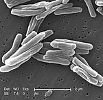
Tuberculosis (TB), also known colloquially as the "white death", or historically as consumption, is an infectious disease usually caused by Mycobacterium tuberculosis (MTB) bacteria. Tuberculosis generally affects the lungs, but it can also affect other parts of the body. Most infections show no symptoms, in which case it is known as latent tuberculosis. Around 10% of latent infections progress to active disease which, if left untreated, kill about half of those affected. Typical symptoms of active TB are chronic cough with blood-containing mucus, fever, night sweats, and weight loss. Infection of other organs can cause a wide range of symptoms.

<span class="mw-page-title-main">Mantoux test</span> Immunological method to test for tuberculosis

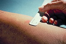
The Mantoux test or Mendel–Mantoux test is a tool for screening for tuberculosis (TB) and for tuberculosis diagnosis. It is one of the major tuberculin skin tests used around the world, largely replacing multiple-puncture tests such as the tine test. The Heaf test, a form of tine test, was used until 2005 in the UK, when it was replaced by the Mantoux test. The Mantoux test is endorsed by the American Thoracic Society and Centers for Disease Control and Prevention. It was also used in the USSR and is now prevalent in most of the post-Soviet states, although Soviet mantoux produced many false positives due to children's allergic reaction.

Radiology (X-rays) is used in the diagnosis of tuberculosis. Abnormalities on chest radiographs may be suggestive of, but are never diagnostic of TB, but can be used to rule out pulmonary TB.

<span class="mw-page-title-main">Ziehl–Neelsen stain</span> Bacteriological technique

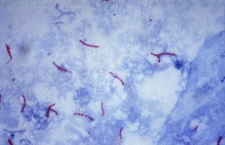
The Ziehl-Neelsen stain, also known as the acid-fast stain, is a bacteriological staining technique used in cytopathology and microbiology to identify acid-fast bacteria under microscopy, particularly members of the Mycobacterium genus. This staining method was initially introduced by Paul Ehrlich (1854–1915) and subsequently modified by the German bacteriologists Franz Ziehl (1859–1926) and Friedrich Neelsen (1854–1898) during the late 19th century.

<span class="mw-page-title-main">Tuberculosis management</span>

Tuberculosis management describes the techniques and procedures utilized for treating tuberculosis (TB) or simply a treatment plan for TB.

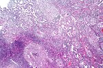
Peripheral tuberculous lymphadenitis is a form of tuberculosis infection occurring outside of the lungs. In general, it describes tuberculosis infection of the lymph nodes, leading to lymphadenopathy. When cervical lymph nodes are affected, it is commonly referred to as "Scrofula." A majority of tuberculosis infections affect the lungs, and extra-pulmonary tuberculosis infections account for the remainder; these most commonly involve the lymphatic system. Although the cervical region is most commonly affected, tuberculous lymphadenitis can occur all around the body, including the axillary and inguinal regions.

<span class="mw-page-title-main">Miliary tuberculosis</span> Medical condition

Miliary tuberculosis is a form of tuberculosis that is characterized by a wide dissemination into the human body and by the tiny size of the lesions (1–5 mm). Its name comes from a distinctive pattern seen on a chest radiograph of many tiny spots distributed throughout the lung fields with the appearance similar to millet seeds—thus the term "miliary" tuberculosis. Miliary TB may infect any number of organs, including the lungs, liver, and spleen. Miliary tuberculosis is present in about 2% of all reported cases of tuberculosis and accounts for up to 20% of all extra-pulmonary tuberculosis cases.

<i>Mycobacterium avium-intracellulare</i> infection Medical condition

Mycobacterium avium-intracellulare infection (MAI) is an atypical mycobacterial infection, i.e. one with nontuberculous mycobacteria or NTM, caused by Mycobacterium avium complex (MAC), which is made of two Mycobacterium species, M. avium and M. intracellulare. This infection causes respiratory illness in birds, pigs, and humans, especially in immunocompromised people. In the later stages of AIDS, it can be very severe. It usually first presents as a persistent cough. It is typically treated with a series of three antibiotics for a period of at least six months.

Latent tuberculosis (LTB), also called latent tuberculosis infection (LTBI) is when a person is infected with Mycobacterium tuberculosis, but does not have active tuberculosis (TB). Active tuberculosis can be contagious while latent tuberculosis is not, and it is therefore not possible to get TB from someone with latent tuberculosis. The main risk is that approximately 10% of these people will go on to develop active tuberculosis. This is particularly true, and there is added risk, in particular situations such as medication that suppresses the immune system or advancing age.

<span class="mw-page-title-main">Tuberculous meningitis</span> Medical condition

Tuberculous meningitis, also known as TB meningitis or tubercular meningitis, is a specific type of bacterial meningitis caused by the Mycobacterium tuberculosis infection of the meninges—the system of membranes which envelop the central nervous system.

Lipoarabinomannan, also called LAM, is a glycolipid, and a virulence factor associated with Mycobacterium tuberculosis, the bacteria responsible for tuberculosis. Its primary function is to inactivate macrophages and scavenge oxidative radicals.

Interferon-γ release assays (IGRA) are medical tests used in the diagnosis of some infectious diseases, especially tuberculosis. Interferon-γ (IFN-γ) release assays rely on the fact that T-lymphocytes will release IFN-γ when exposed to specific antigens. These tests are mostly developed for the field of tuberculosis diagnosis, but in theory, may be used in the diagnosis of other diseases that rely on cell-mediated immunity, e.g. cytomegalovirus and leishmaniasis and COVID-19. For example, in patients with cutaneous adverse drug reactions, the challenge of peripheral blood lymphocytes with the drug causing the reaction produced a positive test result for half of the drugs tested.

<span class="mw-page-title-main">Extensively drug-resistant tuberculosis</span> Tuberculosis that is resistant to the most effective drugs

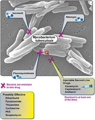
Extensively drug-resistant tuberculosis (XDR-TB) is a form of tuberculosis caused by bacteria that are resistant to some of the most effective anti-TB drugs. XDR-TB strains have arisen after the mismanagement of individuals with multidrug-resistant TB (MDR-TB).

The National Tuberculosis Elimination Programme (NTEP), earlier known as the Revised National Tuberculosis Control Programme (RNTCP), is the Public Health initiative of the Government of India that organizes its anti-Tuberculosis efforts. It functions as a flagship component of the National Health Mission (NHM) and provides technical and managerial leadership to anti-tuberculosis activities in the country. As per the National Strategic Plan 2017–25, the program has a vision of achieving a "TB free India",with a strategies under the broad themes of "Prevent, Detect,Treat and Build pillars for universal coverage and social protection". The program provides, various free of cost, quality tuberculosis diagnosis and treatment services across the country through the government health system.

Interferon-gamma release assays (IGRAs) are diagnostic tools for latent tuberculosis infection (LTBI). They are surrogate markers of Mycobacterium tuberculosis infection and indicate a cellular immune response to M. tuberculosis if the latter is present.

T-SPOT.TB is a type of ELISpot assay used for tuberculosis diagnosis, which belongs to the group of interferon gamma release assays. The test is manufactured by Oxford Immunotec in the UK. It is available in most European countries, the United States as well as various other countries. It was developed by researchers at the University of Oxford in England.

Tuberculous pericarditis is a form of pericarditis. It is a condition in which the pericardium surrounding the heart is infected by the bacterial species Mycobacterium tuberculosis. Tuberculous pericarditis accounts for a significant percentage of presentations of tuberculosis worldwide. The condition has four stages of disease which manifests with clinical presentations ranging from acute pericarditis to overt heart failure. Tuberculous pericarditis is an under-diagnosed condition. Diagnosis often requires a range of diagnostic tools, including pericardiocentesis, biochemical tests, and imaging. Treatment of this disease is similar to treatment of pulmonary tuberculosis. Alternative treatment options to reduce cardiac complications are also available.

The Xpert MTB/RIF is a cartridge-based nucleic acid amplification test (NAAT) for simultaneous rapid tuberculosis diagnosis and rapid antibiotic sensitivity test. It is an automated diagnostic test that can identify Mycobacterium tuberculosis (MTB) DNA and resistance to rifampicin (RIF). It was co-developed by the laboratory of Professor David Alland at the University of Medicine and Dentistry of New Jersey (UMDNJ), Cepheid Inc. and Foundation for Innovative New Diagnostics, with additional financial support from the US National Institutes of Health (NIH).

<span class="mw-page-title-main">National Tuberculosis Institute</span> Indian Government institute

The National Tuberculosis Institute (NTIB) is a Government of India institute, under the Directorate General of Health Services, Ministry of Health and Family Welfare, dedicated to advanced research on Tuberculosis. The Institute is located along Bellary Road, in Bengaluru, Karnataka state, India.

Diagnostic delay is the time interval between the onset of symptoms and confirmed diagnosis of a disease. For a variety of reasons, including the mitigation of disease severity and financial expense, it is desirable for this delay to be minimized.

References

  1. 1 2 3 4 5 Kumar, Vinay; Abbas, Abul K.; Fausto, Nelson; & Mitchell, Richard N. (2007). Robbins Basic Pathology (8th ed.). Saunders Elsevier. pp. 516-522 ISBN   978-1-4160-2973-1
  2. Burke and Parnell. Minimal Pulmonary Tuberculosis. 1948. 59:348 Canadian Medical Association Journal.
  3. Togun TO, Kampmann B (February 2024). "Diagnostics for childhood tuberculosis: a marathon rather than a sprint". The Lancet Infectious Diseases. 24 (2): 110–112. doi:10.1016/s1473-3099(23)00517-0. ISSN   1473-3099. PMID   37918415. S2CID   264822650.
  4. Steingart KR, Henry M, Ng V, Hopewell PC, Ramsay A, Cunningham J, et al. (September 2006). "Fluorescence versus conventional sputum smear microscopy for tuberculosis: a systematic review". The Lancet. Infectious Diseases. 6 (9): 570–81. doi:10.1016/S1473-3099(06)70578-3. PMID   16931408.
  5. Brown M, Varia H, Bassett P, Davidson RN, Wall R, Pasvol G (June 2007). "Prospective study of sputum induction, gastric washing, and bronchoalveolar lavage for the diagnosis of pulmonary tuberculosis in patients who are unable to expectorate". Clinical Infectious Diseases. 44 (11): 1415–20. doi: 10.1086/516782 . PMID   17479935.
  6. 1 2 3 4 Dinnes J, Deeks J, Kunst H, Gibson A, Cummins E, Waugh N, et al. (January 2007). "A systematic review of rapid diagnostic tests for the detection of tuberculosis infection". Health Technology Assessment. 11 (3): 1–196. doi: 10.3310/hta11030 . PMID   17266837.
  7. Kohli M, Schiller I, Dendukuri N, Yao M, Dheda K, Denkinger CM, et al. (January 15, 2021). "Xpert MTB/RIF Ultra and Xpert MTB/RIF assays for extrapulmonary tuberculosis and rifampicin resistance in adults". The Cochrane Database of Systematic Reviews. 1 (1): CD012768. doi:10.1002/14651858.CD012768.pub3. ISSN   1469-493X. PMC   8078545 . PMID   33448348.
  8. Guerra RL, Hooper NM, Baker JF, Alborz R, Armstrong DT, Maltas G, et al. (September 2007). "Use of the amplified mycobacterium tuberculosis direct test in a public health laboratory: test performance and impact on clinical care". Chest. 132 (3): 946–51. doi:10.1378/chest.06-2959. PMID   17573496.
  9. 1 2 Centers for Disease Control and Prevention (2015), "A New Tool to Diagnose Tuberculosis: The Xpert MTB/RIF Assay" (PDF), CDC website, archived from the original (PDF) on 2017-12-17, retrieved 2018-05-25.
  10. Chakravorty S, Simmons AM, Rowneki M, Parmar H, Cao Y, Ryan J, et al. (August 29, 2017). "The New Xpert MTB/RIF Ultra: Improving Detection of Mycobacterium tuberculosis and Resistance to Rifampin in an Assay Suitable for Point-of-Care Testing". mBio. 8 (4). doi:10.1128/mBio.00812-17. ISSN   2150-7511. PMC   5574709 . PMID   28851844.
  11. 1 2 Drobniewski FA, Caws M, Gibson A, Young D (March 2003). "Modern laboratory diagnosis of tuberculosis". The Lancet. Infectious Diseases. 3 (3): 141–7. doi:10.1016/S1473-3099(03)00544-9. PMID   12614730.
  12. Moore DA, Evans CA, Gilman RH, Caviedes L, Coronel J, Vivar A, et al. (October 2006). "Microscopic-observation drug-susceptibility assay for the diagnosis of TB". The New England Journal of Medicine. 355 (15): 1539–50. doi:10.1056/NEJMoa055524. PMC   1780278 . PMID   17035648.
  13. Akce M, Bonner S, Liu E, Daniel R (2014). "Peritoneal tuberculosis mimicking peritoneal carcinomatosis". Case Reports in Medicine. 2014: 436568. doi: 10.1155/2014/436568 . PMC   3970461 . PMID   24715911. CC-BY 3.0
  14. 1 2 Rossi SE, Franquet T, Volpacchio M, Giménez A, Aguilar G (1 May 2005). "Tree-in-bud pattern at thin-section CT of the lungs: radiologic-pathologic overview". Radiographics. 25 (3): 789–801. doi:10.1148/rg.253045115. PMID   15888626.
  15. 1 2 Pelletier-Galarneau M, Martineau P, Zuckier LS, Pham X, Lambert R, Turpin S (May 2017). "18F-FDG-PET/CT Imaging of Thoracic and Extrathoracic Tuberculosis in Children". Seminars in Nuclear Medicine. 47 (3): 304–318. doi:10.1053/j.semnuclmed.2016.12.003. PMID   28417858.
  16. CDC - Guidelines for Using the QuantiFERON-TB Gold Test for Detecting Mycobacterium tuberculosis Infection, United States
  17. Menzies D, Pai M, Comstock G (March 2007). "Meta-analysis: new tests for the diagnosis of latent tuberculosis infection: areas of uncertainty and recommendations for research". Annals of Internal Medicine. 146 (5): 340–54. doi:10.7326/0003-4819-146-5-200703060-00006. PMID   17339619. S2CID   20820188.
  18. Ferrara G, Losi M, D'Amico R, Roversi P, Piro R, Meacci M, et al. (April 2006). "Use in routine clinical practice of two commercial blood tests for diagnosis of infection with Mycobacterium tuberculosis: a prospective study". Lancet. 367 (9519): 1328–34. doi:10.1016/S0140-6736(06)68579-6. PMID   16631911. S2CID   21218187.
  19. Lalvani A (November 2003). "Spotting latent infection: the path to better tuberculosis control". Thorax. 58 (11): 916–8. doi:10.1136/thorax.58.11.916. PMC   1746498 . PMID   14586040.
  20. Ewer K, Deeks J, Alvarez L, Bryant G, Waller S, Andersen P, et al. (April 2003). "Comparison of T-cell-based assay with tuberculin skin test for diagnosis of Mycobacterium tuberculosis infection in a school tuberculosis outbreak". Lancet. 361 (9364): 1168–73. doi:10.1016/S0140-6736(03)12950-9. PMID   12686038. S2CID   144795.
  21. Lalvani A, Pathan AA, Durkan H, Wilkinson KA, Whelan A, Deeks JJ, et al. (June 2001). "Enhanced contact tracing and spatial tracking of Mycobacterium tuberculosis infection by enumeration of antigen-specific T cells". Lancet. 357 (9273): 2017–21. doi:10.1016/S0140-6736(00)05115-1. PMID   11438135. S2CID   24678475.
  22. "The Science behind T-SPOT.TB Technology".
  23. "How T-SPOT.TB Works".
  24. Kang YA, Lee HW, Hwang SS, Um SW, Han SK, Shim YS, et al. (September 2007). "Usefulness of whole-blood interferon-gamma assay and interferon-gamma enzyme-linked immunospot assay in the diagnosis of active pulmonary tuberculosis". Chest. 132 (3): 959–65. doi:10.1378/chest.06-2805. PMID   17505029.
  25. Diel R, Loddenkemper R, Nienhaus A (April 2010). "Evidence-based comparison of commercial interferon-gamma release assays for detecting active TB: a metaanalysis". Chest. 137 (4): 952–68. doi:10.1378/chest.09-2350. PMID   20022968.
  26. Ruhwald M, Aggerbeck H, Gallardo RV, Hoff ST, Villate JI, Borregaard B, et al. (April 2017). "Safety and efficacy of the C-Tb skin test to diagnose Mycobacterium tuberculosis infection, compared with an interferon γ release assay and the tuberculin skin test: a phase 3, double-blind, randomised, controlled trial". The Lancet. Respiratory Medicine. 5 (4): 259–268. doi:10.1016/S2213-2600(16)30436-2. PMID   28159608.
  27. Gaur RL, Suhosk MM, Banaei N (2012). "In vitro immunomodulation of a whole blood IFN-γ release assay enhances T cell responses in subjects with latent tuberculosis infection". PLOS ONE. 7 (10): e48027. Bibcode:2012PLoSO...748027G. doi: 10.1371/journal.pone.0048027 . PMC   3483295 . PMID   23144722.
  28. Jarvis J, Gao Y, de Graaf H, Hughes S, Allan RN, Williams A, et al. (August 2015). "Environmental temperature impacts on the performance of QuantiFERON-TB Gold In-Tube assays". The Journal of Infection. 71 (2): 276–80. doi:10.1016/j.jinf.2015.04.004. PMID   25869537.
  29. Edwards A, Gao Y, Allan RN, Ball D, de Graaf H, Coelho T, et al. (October 2017). "Corticosteroids and infliximab impair the performance of interferon-γ release assays used for diagnosis of latent tuberculosis" (PDF). Thorax. 72 (10): 946–949. doi: 10.1136/thoraxjnl-2016-209397 . PMID   28159773. S2CID   46880941.
  30. Gaur RL, Pai M, Banaei N (November 2013). "Impact of blood volume, tube shaking, and incubation time on reproducibility of QuantiFERON-TB gold in-tube assay". Journal of Clinical Microbiology. 51 (11): 3521–6. doi:10.1128/JCM.01627-13. PMC   3889728 . PMID   23966505.
  31. Kik SV, Denkinger CM, Casenghi M, Vadnais C, Pai M (August 2014). "Tuberculosis diagnostics: which target product profiles should be prioritised?". The European Respiratory Journal. 44 (2): 537–40. doi: 10.1183/09031936.00027714 . PMID   24696110.
  32. "The use of lateral flow urine lipoarabinomannan assay (LF-LAM) for the diagnosis and screening of active tuberculosis in people living with HIV" (PDF). Retrieved 2019-06-10.
  33. Shah M, Hanrahan C, Wang ZY, Dendukuri N, Lawn SD, Denkinger CM, et al. (May 2016). "Lateral flow urine lipoarabinomannan assay for detecting active tuberculosis in HIV-positive adults". The Cochrane Database of Systematic Reviews. 2016 (5): CD011420. doi:10.1002/14651858.cd011420.pub2. PMC   4916932 . PMID   27163343.
  34. Peter JG, Zijenah LS, Chanda D, Clowes P, Lesosky M, Gina P, et al. (March 2016). "Effect on mortality of point-of-care, urine-based lipoarabinomannan testing to guide tuberculosis treatment initiation in HIV-positive hospital inpatients: a pragmatic, parallel-group, multicountry, open-label, randomised controlled trial". Lancet. 387 (10024): 1187–97. doi:10.1016/s0140-6736(15)01092-2. PMID   26970721. S2CID   6075068.
  35. Gupta-Wright A, Corbett EL, van Oosterhout JJ, Wilson D, Grint D, Alufandika-Moyo M, et al. (July 2018). "Rapid urine-based screening for tuberculosis in HIV-positive patients admitted to hospital in Africa (STAMP): a pragmatic, multicentre, parallel-group, double-blind, randomised controlled trial". Lancet. 392 (10144): 292–301. doi:10.1016/s0140-6736(18)31267-4. PMC   6078909 . PMID   30032978.
  36. "Diagnostic accuracy study published on next-generation urine test to detect tuberculosis in HIV-positive people". FIND. Archived from the original on 2022-10-05. Retrieved 2019-06-10.
  37. Sigal GB, Pinter A, Lowary TL, Kawasaki M, Li A, Mathew A, et al. (December 2018). Miller MB (ed.). "A Novel Sensitive Immunoassay Targeting the 5-Methylthio-d-Xylofuranose-Lipoarabinomannan Epitope Meets the WHO's Performance Target for Tuberculosis Diagnosis". Journal of Clinical Microbiology. 56 (12): e01338–18, /jcm/56/12/e01338–18.atom. doi:10.1128/JCM.01338-18. PMC   6258851 . PMID   30257899.
  38. 1 2 Broger T, Sossen B, du Toit E, Kerkhoff AD, Schutz C, Ivanova Reipold E, et al. (August 2019). "Novel lipoarabinomannan point-of-care tuberculosis test for people with HIV: a diagnostic accuracy study". The Lancet. Infectious Diseases. 19 (8): 852–861. doi:10.1016/S1473-3099(19)30001-5. PMC   6656794 . PMID   31155318.
  39. Broger T, Nicol MP, Székely R, Bjerrum S, Sossen B, Schutz C, et al. (May 2020). "Diagnostic accuracy of a novel tuberculosis point-of-care urine lipoarabinomannan assay for people living with HIV: A meta-analysis of individual in- and outpatient data". PLOS Medicine. 17 (5): e1003113. doi: 10.1371/journal.pmed.1003113 . PMC   7194366 . PMID   32357197.
  40. Broger T, Nicol M, Sigal G, Gotuzzo E, Zimmer AJ, Surtie S, et al. (July 2020). "Diagnostic accuracy of three urine lipoarabinomannan tuberculosis assays in HIV-negative outpatients". The Journal of Clinical Investigation. 130 (11): 5756–5764. doi: 10.1172/JCI140461 . PMC   7598043 . PMID   32692731.
  41. "FujiLAM Prospective Evaluation Trial - Full Text View - ClinicalTrials.gov". clinicaltrials.gov. Retrieved 2020-08-23.
  42. Sutherland JS, van der Spuy G, Gindeh A, Thuong NT, Namuganga A, Owolabi O, et al. (2021-09-22). "Diagnostic Accuracy of the Cepheid 3-gene Host Response Fingerstick Blood Test in a Prospective, Multi-site Study: Interim Results". Clinical Infectious Diseases. 74 (12): 2136–2141. doi:10.1093/cid/ciab839. ISSN   1058-4838. PMC   9258935 . PMID   34550342.
  43. 1 2 Nakamura RM, Einck L, Velmonte MA, Kawajiri K, Ang CF, Delasllagas CE, et al. (2001-01-01). "Detection of active tuberculosis by an MPB-64 transdermal patch: a field study". Scandinavian Journal of Infectious Diseases. 33 (6): 405–7. doi:10.1080/00365540152029846. PMID   11450857. S2CID   218880088.
  44. Law YH (2018-05-16). "With a little convincing, rats can detect tuberculosis". Science News. Retrieved 2018-05-18.
  45. Mgode GF, Cox CL, Mwimanzi S, Mulder C (July 2018). "Pediatric tuberculosis detection using trained African giant pouched rats". Pediatric Research. 84 (1): 99–103. doi: 10.1038/pr.2018.40 . PMID   29617007.

Further reading

Notes