Viroplasm

Last updated
Viroplasms (green) in cells infected with rotavirus (top), and uninfected cells (bottom). (Immunofluorescent stain) Virus Infected Cells.jpg
Viroplasms (green) in cells infected with rotavirus (top), and uninfected cells (bottom). (Immunofluorescent stain)

A viroplasm, sometimes called "virus factory" or "virus inclusion", [1] is an inclusion body in a cell where viral replication and assembly occurs. They may be thought of as viral factories in the cell. There are many viroplasms in one infected cell, where they appear dense to electron microscopy. Very little is understood about the mechanism of viroplasm formation.

Contents

Definition

A viroplasm is a perinuclear or a cytoplasmic large compartment where viral replication and assembly occurs. [2] The viroplasm formation is caused by the interactions between the virus and the infected cell, where viral products and cell elements are confined. [2]

Groups of viruses that form viroplasms

Viroplasms have been reported in many unrelated groups of Eukaryotic viruses that replicate in cytoplasm, however, viroplasms from plant viruses have not been as studied as viroplasms from animal viruses. [2] Viroplasms have been found in the cauliflower mosaic virus, [3] rotavirus, [4] vaccinia virus [5] and the rice dwarf virus. [6] These appear electron-dense under an electron microscope and are insoluble. [2]

Baltimore's classificationFamilySpecies
I: dsDNA viruses Poxviridae
Asfarviridae
Iridoviridae
Mimiviridae

Herpesviridae

vaccinia virus [7]

African swine fever virus [8]
frog virus [2]
Acanthamoeba polyphaga mimivirus [9]

Pithovirus [10]

Herpes simplex virus [2]

II: ssDNA viruses
III: dsRNA viruses Reoviridae Avian reovirus [11]
IV: (+)ssRNA viruses Togaviridae
Flaviviridae
Rubella virus [12]

Flavivirus [2]

V: (−)ssRNA viruses Rhabdoviridae

Peribunyaviridae

Rabies virus [13]

Orthobunyavirus [14]

VI: ssRNA-RT viruses Retroviridae Human immunodeficiency virus [15]
VII: dsDNA-RT viruses Caulimoviridae Cauliflower mosaic virus [16]

Structure and formation

Viroplasms are localized in the perinuclear area or in the cytoplasm of infected cells and are formed early in the infection cycle. [2] [17] The number and the size of viroplasms depend on the virus, the virus isolate, hosts species, and the stage of the infection. [18] For example, viroplasms of mimivirus have a similar size to the nucleus of its host, the amoeba Acanthamoeba polyphaga. [9]

A virus can induce changes in composition and organization of host cell cytoskeletal and membrane compartments, depending on the step of the viral replication cycle. [1] This process involves a number of complex interactions and signaling events between viral and host cell factors.

Viroplasms are formed early during the infection; in many cases, the cellular rearrangements caused during virus infection lead to the construction of sophisticated inclusions —viroplasms— in the cell where the factory will be assembled. The viroplasm is where components such as replicase enzymes, virus genetic material, and host proteins required for replication concentrate, and thereby increase the efficiency of replication. [1] At the same time, large amounts of ribosomes, protein-synthesis components, protein folding chaperones, and mitochondria are recruited. Some of the membrane components are used for viral replication while some others will be modified to produce viral envelopes, when the viruses are enveloped. The viral replication, protein synthesis and assembly require a considerable amount of energy, provided by large clusters of mitochondria at the periphery of viroplasms. The virus factory is often enclosed by a membrane derived from the rough endoplasmic reticulum or by cytoskeletal elements. [2] [17]

In animal cells, virus particles are gathered by the microtubule-dependent aggregation of toxic or misfolded protein near the microtubule organizing center (MTOC), so the viroplasms of animal viruses are generally localized near the MTOC. [2] [19] MTOCs are not found in plant cells. Plant viruses induce the rearrangement of membranes structures to form the viroplasm. This is mostly shown for plant RNA viruses. [17]

Functions

Viroplasm is the location within the infected cell where viral replication and assembly take place. [2] Wrapping the viroplasm with a membrane, concentrates the viral components required for the genome replication and the morphogenesis of new virus particles, so it increases the efficiency of the processes. [2] The recruitment of cellular membranes and cytoskeleton to generate virus replication sites can also benefit viruses in other ways. Disruption of cellular membranes can, for example, slow the transport of immunomodulatory proteins to the surface of infected cells and protect against innate and acquired immune responses, and rearrangements to cytoskeleton can facilitate virus release. [1] The viroplasm could also prevent virus degradation by proteases and nucleases. [17]

In the case of the Cauliflower mosaic virus (CaMV), viroplasms improve the virus transmission by the aphid vector. Viroplasms also control release of virions when the insect stings an infected plant cell or a cell near the infected cells. [16]

Possible co-evolution with the host

Aggregated structures may protect viral functional complexes from the cellular degradation systems. For example, formation of viral factories of the ASFV viroplasm is very similar to the aggresome formation. [2] An aggresome is a perinuclear site where misfolded proteins are transported and stored by the cell components for their destruction. It has been proposed that the viroplasm could be the product of a co-evolution between the virus and its host. [16] It is possible that a cellular response originally designed to reduce the toxicity of misfolded proteins is exploited by cytoplasmic viruses to improve their replication, the virus capsid synthesis, and assembly. [16] Alternatively, the activation of host defense mechanisms may involve sequestration of virus components in aggregates to prevent their dissemination, followed by their neutralisation. For example, viroplasms of mammalian viruses contain certain elements of the cellular degradation machinery which might enable cellular protective mechanisms against viral components. [20] Given the co-evolution of viruses with their host cells, changes in cell structure induced during infection are likely to involve a combination of the two strategies. [2]

Use in diagnostics

Presence of viroplasms is used to diagnose certain viral infections. Understanding the phenomena of virus aggregation and of the cell response to the presence of virus, and whether viroplasms facilitate or inhibit viral replication, may help to develop new therapeutic approaches against virus infections in animal and plant cells. [17]

See also

Related Research Articles

Retrovirus Family of viruses

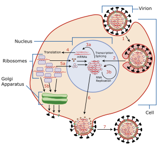
A retrovirus is a type of virus that inserts a copy of its RNA genome into the DNA of a host cell that it invades, thus changing the genome of that cell. Once inside the host cell's cytoplasm, the virus uses its own reverse transcriptase enzyme to produce DNA from its RNA genome, the reverse of the usual pattern, thus retro (backwards). The new DNA is then incorporated into the host cell genome by an integrase enzyme, at which point the retroviral DNA is referred to as a provirus. The host cell then treats the viral DNA as part of its own genome, transcribing and translating the viral genes along with the cell's own genes, producing the proteins required to assemble new copies of the virus.

<i>Rhabdoviridae</i> Family of viruses in the order Mononegavirales

Rhabdoviridae is a family of negative-strand RNA viruses in the order Mononegavirales. Vertebrates, invertebrates, and plants serve as natural hosts. Diseases associated with member viruses include rabies encephalitis caused by the rabies virus, and flu-like symptoms in humans caused by vesiculoviruses. The name is derived from Ancient Greek rhabdos, meaning rod, referring to the shape of the viral particles. The family has 40 genera, most assigned to three subfamilies.

<i>Mimivirus</i> Genus of viruses

Mimivirus is a genus of giant viruses, in the family Mimiviridae. Amoeba serve as their natural hosts. This genus contains a single identified species named Acanthamoeba polyphaga mimivirus (APMV). It also refers to a group of phylogenetically related large viruses.

Cauliflower mosaic virus (CaMV) is a member of the genus Caulimovirus, one of the six genera in the family Caulimoviridae, which are pararetroviruses that infect plants. Pararetroviruses replicate through reverse transcription just like retroviruses, but the viral particles contain DNA instead of RNA.

<i>Papillomaviridae</i> Family of viruses

Papillomaviridae is a family of non-enveloped DNA viruses whose members are known as papillomaviruses. Several hundred species of papillomaviruses, traditionally referred to as "types", have been identified infecting all carefully inspected mammals, but also other vertebrates such as birds, snakes, turtles and fish. Infection by most papillomavirus types, depending on the type, is either asymptomatic or causes small benign tumors, known as papillomas or warts. Papillomas caused by some types, however, such as human papillomaviruses 16 and 18, carry a risk of becoming cancerous.

Viral protein

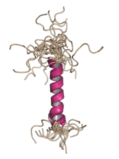
A viral protein is both a component and a product of a virus. Viral proteins are grouped according to their functions, and groups of viral proteins include structural proteins, nonstructural proteins, regulatory proteins, and accessory proteins. Viruses are non-living and do not have the means to reproduce on their own, instead depending on their host cell's resources in order to reproduce. Thus, viruses do not code for many of their own viral proteins, and instead use the host cell's machinery to produce the viral proteins they require for replication.

<i>Herpesviridae</i> Family of DNA viruses

Herpesviridae is a large family of DNA viruses that cause infections and certain diseases in animals, including humans. The members of this family are also known as herpesviruses. The family name is derived from the Greek word herpein 'to creep', referring to spreading cutaneous lesions, usually involving blisters, seen in flares of herpes simplex 1, herpes simplex 2 and herpes zoster (shingles). In 1971, the International Committee on the Taxonomy of Viruses (ICTV) established Herpesvirus as a genus with 23 viruses among four groups. As of 2020, 115 species are recognized, all but one of which are in one of the three subfamilies. Herpesviruses can cause both latent and lytic infections.

The murine leukemia viruses are retroviruses named for their ability to cause cancer in murine (mouse) hosts. Some MLVs may infect other vertebrates. MLVs include both exogenous and endogenous viruses. Replicating MLVs have a positive sense, single-stranded RNA (ssRNA) genome that replicates through a DNA intermediate via the process of reverse transcription.

The Simian foamy virus (SFV) is a species of the genus Spumavirus, which belongs to the family of Retroviridae. It has been identified in a wide variety of primates, including pro-simians, New World and Old World monkeys as well as apes, and each species has been shown to harbor a unique (species-specific) strain of SFV, including African green monkeys, baboons, macaques and chimpanzees. As it is related to the more well-known retrovirus human immunodeficiency virus (HIV), its discovery in primates has led to some speculation that HIV may have been spread to the human species in Africa through contact with blood from apes, monkeys, and other primates, most likely through bushmeat hunting practices.

<i>Molluscum contagiosum virus</i> Species of virus

Molluscum contagiosum virus (MCV) is a DNA poxvirus that causes the human skin infection molluscum contagiosum. Molluscum contagiosum affects about 200,000 people a year, about 1% of all diagnosed skin diseases. Diagnosis is based on the size and shape of the skin lesions and can be confirmed with a biopsy, as the virus cannot be routinely cultured. Molluscum contagiosum virus is the only species in the genus Molluscipoxvirus. MCV is a member of the subfamily Chordopoxvirinae of family Poxviridae. Other commonly known viruses that reside in the subfamily Chordopoxvirinae are variola virus and monkeypox virus.

Viral entry

Viral entry is the earliest stage of infection in the viral life cycle, as the virus comes into contact with the host cell and introduces viral material into the cell. The major steps involved in viral entry are shown below. Despite the variation among viruses, there are several shared generalities concerning viral entry.

Phycodnaviridae is a family of large (100–560 kb) double-stranded DNA viruses that infect marine or freshwater eukaryotic algae. Viruses within this family have a similar morphology, with an icosahedral capsid. As of 2014, there were 33 species in this family, divided among 6 genera. This family belongs to a super-group of large viruses known as nucleocytoplasmic large DNA viruses. Evidence was published in 2014 suggesting that specific strains of Phycodnaviridae might infect humans rather than just algal species, as was previously believed. Most genera under this family enter the host cell by cell receptor endocytosis and replicate in the nucleus. Phycodnaviridae play important ecological roles by regulating the growth and productivity of their algal hosts. Algal species such Heterosigma akashiwo and the genus Chrysochromulina can form dense blooms which can be damaging to fisheries, resulting in losses in the aquaculture industry. Heterosigma akashiwo virus (HaV) has been suggested for use as a microbial agent to prevent the recurrence of toxic red tides produced by this algal species. Phycodnaviridae cause death and lysis of freshwater and marine algal species, liberating organic carbon, nitrogen and phosphorus into the water, providing nutrients for the microbial loop.

Virus Small non-cellular infectious agent that only replicates in cells

A virus is a submicroscopic infectious agent that replicates only inside the living cells of an organism. Viruses infect all life forms, from animals and plants to microorganisms, including bacteria and archaea. Since Dmitri Ivanovsky's 1892 article describing a non-bacterial pathogen infecting tobacco plants and the discovery of the tobacco mosaic virus by Martinus Beijerinck in 1898, more than 9,000 virus species have been described in detail of the millions of types of viruses in the environment. Viruses are found in almost every ecosystem on Earth and are the most numerous type of biological entity. The study of viruses is known as virology, a subspeciality of microbiology.

Sputnik virophage

Mimivirus-dependent virus Sputnik is a subviral agent that reproduces in amoeba cells that are already infected by a certain helper virus; Sputnik uses the helper virus's machinery for reproduction and inhibits replication of the helper virus. It is known as a virophage, in analogy to the term bacteriophage.

<i>Cafeteria roenbergensis virus</i> Species of virus

Cafeteria roenbergensis virus (CroV) is a giant virus that infects the marine bicosoecid flagellate Cafeteria roenbergensis, a member of the microzooplankton community.

Megavirus Genus of viruses

Megavirus is a viral genus containing a single identified species named Megavirus chilensis, phylogenetically related to Acanthamoeba polyphaga Mimivirus (APMV). In colloquial speech, Megavirus chilensis is more commonly referred to as just “Megavirus”. Until the discovery of pandoraviruses in 2013, it had the largest capsid diameter of all known viruses, as well as the largest and most complex genome among all known viruses.

Agnoprotein InterPro Family

Agnoprotein is a protein expressed by some members of the polyomavirus family from a gene called the agnogene. Polyomaviruses in which it occurs include two human polyomaviruses associated with disease, BK virus and JC virus, as well as the simian polyomavirus SV40.

Positive-strand RNA virus Class of viruses in the Baltimore classification

Positive-strand RNA viruses are a group of related viruses that have positive-sense, single-stranded genomes made of ribonucleic acid. The positive-sense genome can act as messenger RNA (mRNA) and can be directly translated into viral proteins by the host cell's ribosomes. Positive-strand RNA viruses encode an RNA-dependent RNA polymerase (RdRp) which is used during replication of the genome to synthesize a negative-sense antigenome that is then used as a template to create a new positive-sense viral genome.

<i>Tupanvirus</i> Proposed genus of viruses


Tupanvirus is a genus of viruses first described in 2018. The genus is composed of two species of virus that are in the giant virus group. Researchers discovered the first isolate in 2012 from deep water sediment samples taken at 3000m depth off the coast of Brazil. The second isolate was collected from a soda lake in Southern Nhecolândia, Brazil in 2014. They are named after Tupã (Tupan), a Guaraní thunder god, and the places they were found. These are the first viruses reported to possess genes for amino-acyl tRNA synthetases for all 20 standard amino acids.

<i>Woolly monkey hepatitis B virus</i> Species of virus

The woolly monkey hepatitis B virus (WMHBV) is a viral species of the Orthohepadnavirus genus of the Hepadnaviridae family. Its natural host is the woolly monkey (Lagothrix), an inhabitant of South America categorized as a New World primate. WMHBV, like other hepatitis viruses, infects the hepatocytes, or liver cells, of its host organism. It can cause hepatitis, liver necrosis, cirrhosis, and hepatocellular carcinoma. Because nearly all species of Lagothrix are threatened or endangered, researching and developing a vaccine and/or treatment for WMHBV is important for the protection of the whole woolly monkey genus.

References

  1. 1 2 3 4 Netherton C, Moffat K, Brooks E, Wileman T (2007). "A guide to viral inclusions, membrane rearrangements, factories, and viroplasm produced during virus replication". Advances in Virus Research. 70: 101–82. doi:10.1016/S0065-3527(07)70004-0. ISBN   9780123737281. PMC   7112299 . PMID   17765705.
  2. 1 2 3 4 5 6 7 8 9 10 11 12 13 14 Novoa RR, Calderita G, Arranz R, Fontana J, Granzow H, Risco C (February 2005). "Virus factories: associations of cell organelles for viral replication and morphogenesis". Biology of the Cell. 97 (2): 147–72. doi:10.1042/bc20040058. PMC   7161905 . PMID   15656780.
  3. Xiong C, Muller S, Lebeurier G, Hirth L (1982). "Identification by immunoprecipitation of cauliflower mosaic virus in vitro major translation product with a specific serum against viroplasm protein". The EMBO Journal. 1 (8): 971–6. doi:10.1002/j.1460-2075.1982.tb01280.x. PMC   553144 . PMID   16453427.
  4. Nilsson M, von Bonsdorff CH, Weclewicz K, Cohen J, Svensson L (March 1998). "Assembly of viroplasm and virus-like particles of rotavirus by a Semliki Forest virus replicon". Virology. 242 (2): 255–65. doi: 10.1006/viro.1997.8987 . PMID   9514960.
  5. Szajner P, Weisberg AS, Wolffe EJ, Moss B (July 2001). "Vaccinia virus A30L protein is required for association of viral membranes with dense viroplasm to form immature virions". Journal of Virology. 75 (13): 5752–61. doi:10.1128/JVI.75.13.5752-5761.2001. PMC   114291 . PMID   11390577.
  6. Wei T, Kikuchi A, Suzuki N, Shimizu T, Hagiwara K, Chen H, Omura T (September 2006). "Pns4 of rice dwarf virus is a phosphoprotein, is localized around the viroplasm matrix, and forms minitubules". Archives of Virology. 151 (9): 1701–12. doi:10.1007/s00705-006-0757-4. PMID   16609816. S2CID   22657012.
  7. Sodeik B, Doms RW, Ericsson M, Hiller G, Machamer CE, van 't Hof W, et al. (May 1993). "Assembly of vaccinia virus: role of the intermediate compartment between the endoplasmic reticulum and the Golgi stacks". The Journal of Cell Biology. 121 (3): 521–41. CiteSeerX   10.1.1.274.2733 . doi:10.1083/jcb.121.3.521. PMC   2119557 . PMID   8486734.
  8. Salas ML, Andrés G (April 2013). "African swine fever virus morphogenesis". Virus Research. 173 (1): 29–41. doi:10.1016/j.virusres.2012.09.016. PMID   23059353.
  9. 1 2 Suzan-Monti M, La Scola B, Barrassi L, Espinosa L, Raoult D (March 2007). "Ultrastructural characterization of the giant volcano-like virus factory of Acanthamoeba polyphaga Mimivirus". PLOS ONE. 2 (3): e328. Bibcode:2007PLoSO...2..328S. doi: 10.1371/journal.pone.0000328 . PMC   1828621 . PMID   17389919. Open Access logo PLoS transparent.svg
  10. Abergel C, Legendre M, Claverie JM (November 2015). "The rapidly expanding universe of giant viruses: Mimivirus, Pandoravirus, Pithovirus and Mollivirus". FEMS Microbiology Reviews. 39 (6): 779–96. doi: 10.1093/femsre/fuv037 . PMID   26391910. Open Access logo PLoS transparent.svg
  11. Tourís-Otero F, Cortez-San Martín M, Martínez-Costas J, Benavente J (August 2004). "Avian reovirus morphogenesis occurs within viral factories and begins with the selective recruitment of sigmaNS and lambdaA to microNS inclusions". Journal of Molecular Biology. 341 (2): 361–74. doi:10.1016/j.jmb.2004.06.026. PMID   15276829.
  12. Fontana J, López-Iglesias C, Tzeng WP, Frey TK, Fernández JJ, Risco C (September 2010). "Three-dimensional structure of Rubella virus factories". Virology. 405 (2): 579–91. doi:10.1016/j.virol.2010.06.043. PMC   7111912 . PMID   20655079.
  13. Lahaye X, Vidy A, Pomier C, Obiang L, Harper F, Gaudin Y, Blondel D (August 2009). "Functional characterization of Negri bodies (NBs) in rabies virus-infected cells: Evidence that NBs are sites of viral transcription and replication". Journal of Virology. 83 (16): 7948–58. doi:10.1128/JVI.00554-09. PMC   2715764 . PMID   19494013.
  14. Evans AB, Peterson KE (August 2019). "Throw out the Map: Neuropathogenesis of the Globally Expanding California Serogroup of Orthobunyaviruses". Viruses. 11 (9): 794. doi: 10.3390/v11090794 . PMC   6784171 . PMID   31470541.
  15. Karczewski MK, Strebel K (January 1996). "Cytoskeleton association and virion incorporation of the human immunodeficiency virus type 1 Vif protein". Journal of Virology. 70 (1): 494–507. doi:10.1128/JVI.70.1.494-507.1996. PMC   189838 . PMID   8523563.
  16. 1 2 3 4 Bak A., Gargani D., Macia J-L., Malouvet E., Vernerey M_S., Blanc S. and Drucker, M. Virus factories of Cauliflower mosaic virus are virion reservoirs that engage actively in vector-transmission. 2013 journal of Virology
  17. 1 2 3 4 5 Moshe A, Gorovits R (October 2012). "Virus-induced aggregates in infected cells". Viruses. 4 (10): 2218–32. doi: 10.3390/v4102218 . PMC   3497049 . PMID   23202461.
  18. Shalla TA, Shepherd RJ, Petersen LJ (April 1980). "Comparative cytology of nine isolates of cauliflower mosaic virus". Virology. 102 (2): 381–8. doi:10.1016/0042-6822(80)90105-1. PMID   18631647.
  19. Wileman T (May 2006). "Aggresomes and autophagy generate sites for virus replication". Science. 312 (5775): 875–8. Bibcode:2006Sci...312..875W. doi:10.1126/science.1126766. PMID   16690857. S2CID   26616991.
  20. Kopito RR (December 2000). "Aggresomes, inclusion bodies and protein aggregation". Trends in Cell Biology. 10 (12): 524–30. doi:10.1016/s0962-8924(00)01852-3. PMID   11121744.