Rubella virus

Last updated

Rubivirus rubellae
Rubella virus TEM B82-0203 lores.jpg
Transmission electron micrograph of Rubella virus virions
Virus classification OOjs UI icon edit-ltr.svg
(unranked): Virus
Realm: Riboviria
Kingdom: Orthornavirae
Phylum: Kitrinoviricota
Class: Alsuviricetes
Order: Hepelivirales
Family: Matonaviridae
Genus: Rubivirus
Species:
Rubivirus rubellae
Synonyms [1]
  • Rubella virus

Rubella virus (RuV) is the pathogenic agent of the disease rubella, transmitted only between humans via the respiratory route, and is the main cause of congenital rubella syndrome when infection occurs during the first weeks of pregnancy.

Contents

Rubella virus, scientific name Rubivirus rubellae, is a member of the genus Rubivirus and belongs to the family of Matonaviridae, whose members commonly have a genome of single-stranded RNA of positive polarity which is enclosed by an icosahedral capsid.

As of 1999 the molecular basis for the causation of congenital rubella syndrome was not yet completely clear, but in vitro studies with cell lines showed that rubella virus has an apoptotic effect on certain cell types. There is evidence for a p53-dependent mechanism. [2]

Taxonomy

Rubella virus (Rubivirus rubellae) is assigned to the Rubivirus genus. [1]

Matonaviridae family

Until 2018, Rubiviruses were classified as part of the family Togaviridae , but have since been changed to be the sole genus of the family Matonaviridae. This family is named after George de Maton, who in 1814 first distinguished rubella from measles and scarlet fever. [3] The change was made by the International Committee on the Taxonomy of Viruses (ICTV), the central governing body for viral classification. Matonaviridae remains part of the realm that it was already in as Togaviridae, Riboviria , because of its RNA genome and RNA dependent RNA polymerase . [3]

Other rubiviruses

In 2020, Ruhugu virus and Rustrela virus joined Rubella virus as second and third of only three members of the genus Rubivirus. [4] Neither of them are known to infect people. [5]

Morphology

While alphavirus virions are spherical and contain an icosahedral nucleocapsid, RuV virions are pleiomorphic and do not contain icosahedral nucleocapsids. [3]

Phylogeny

ICTV analyzed the sequence of RuV and compared its phylogeny to that of togaviruses. They concluded:

Phylogenetic analysis of the RNA-dependent RNA polymerase of alphaviruses, rubella virus and other positive-sense RNA viruses shows the two genera within the Togaviridae are not monophyletic. In particular, rubella virus groups more closely with members of the families Benyviridae , Hepeviridae and Alphatetraviridae , along with several unclassified viruses, than it does with members of the family Togaviridae belonging to the genus Alphavirus. [3]

Structure

The spherical virus particles (virions) of Matonaviridae have a diameter of 50 to 70 nm and are covered by a lipid membrane (viral envelope), derived from the host cell membrane. There are prominent "spikes" (projections) of 6 nm composed of the viral envelope proteins E1 and E2 embedded in the membrane. [6]

The E1 glycoprotein is considered immunodominant in the humoral response induced against the structural proteins and contains both neutralizing and hemagglutinating determinants.

GenusStructureSymmetryCapsidGenomic arrangementGenomic segmentation
RubivirusIcosahedralT=4EnvelopedLinearMonopartite

[7]

Capsid protein

Inside the lipid envelope is a capsid of 40 nm in diameter. The capsid protein (CP) has different functions. [8] Its main tasks are the formation of homooligomeres to form the capsid, and the binding of the genomic RNA. Further is it responsible for the aggregation of RNA in the capsid, it interacts with the membrane proteins E1 and E2 and binds the human host-protein p32 which is important for replication of the virus in the host. [9]

As opposed to alphaviruses the capsid does not undergo autoproteolysis, rather is it cut off from the rest of the polyprotein by the signal-peptidase. Production of the capsid happens at the surface of intracellular membranes simultaneously with the budding of the virus. [10]

Genome

The genome has 9,762 nucleotides and encodes 2 nonstructural polypeptides (p150 and p90) within its 5′-terminal two-thirds and 3 structural polypeptides (C, E2, and E1) within its 3′-terminal one-third. [11] Both envelope proteins E1 and E2 are glycosylated.

There are three sites that are highly conserved in Matonaviruses: a stem-and-loop structure at the 5' end of the genome, a 51-nucleotide conserved sequence near the 5' end of the genome and a 20-nucleotide conserved sequence at the subgenomic RNA start site. Homologous sequences are present in the rubella genome. [11]

The genome encodes several non-coding RNA structures; among them is the rubella virus 3' cis-acting element, which contains multiple stem-loops, one of which has been found to be essential for viral replication. [12]

The only significant region of homology between rubella and the alphaviruses is located at the NH2 terminus of non structural protein 3. This sequence has helicase and replicase activity. In the rubella genome these occur in the opposite orientation to that found in the alphaviruses indicating that a genome rearrangement has occurred.

The genome has the highest G+C content of any currently known single stranded RNA virus (~70%). [13] Despite this high GC content its codon use is similar to that of its human host.

Replication

The viruses attach to the cell surface via specific receptors and are taken up by an endosome being formed. At the neutral pH outside of the cell the E2 envelope protein covers the E1 protein. The dropping pH inside the endosome frees the outer domain of E1 and causes the fusion of the viral envelope with the endosomal membrane. Thus, the capsid reaches the cytosol, decays and releases the genome

The +ssRNA (positive, single-stranded RNA) at first only acts as a template for the translation of the non-structural proteins, which are synthesized as a large polyprotein and are then cut into single proteins. The sequences for the structural proteins are first replicated by the viral RNA polymerase (Replicase) via a complementary ssRNA as a template and translated as a separate short mRNA. This short subgenomic RNA is additionally packed in a virion. [14]

Translation of the structural proteins produces a large polypeptide (110 Dalton). This is then endoproteolytically cut into E1, E2 and the capsid protein. E1 and E2 are type I transmembrane proteins which are transported into the endoplasmatic reticulum (ER) with the help of an N-terminal signal sequence. From the ER the heterodimeric E1·E2-complex reaches the Golgi apparatus, where the budding of new virions occurs (unlike alpha viruses, where budding occurs at the plasma membrane. The capsid proteins on the other hand stay in the cytoplasm and interact with the genomic RNA, together forming the capsid. [15]

GenusHost detailsTissue tropismEntry detailsRelease detailsReplication siteAssembly siteTransmission
RubivirusHumansNoneClathrin-mediated endocytosisSecretionCytoplasmCytoplasmAerosol

[7]

Transmission

RuV is transmitted via respiration between humans. [3]

Epidemiology

On the basis of differences in the sequence of the E1 protein, two genotypes have been described which differ by 8 - 10%. These have been subdivided into 13 recognised genotypes - 1a, 1B, 1C, 1D, 1E, 1F, 1G, 1h, 1i, 1j, 2A, 2B and 2C.

For typing, the WHO recommends a minimum window that includes nucleotides 8731 to 9469. [16]

Genotypes 1a, 1E, 1F, 2A and 2B have been isolated in China.

Genotype 1j has only been isolated from Japan and the Philippines.

Genotype 1E is found in Africa, the Americas, Asia and Europe.

Genotype 1G has been isolated in Belarus, Cote d'Ivoire and Uganda.

Genotype 1C is endemic only in Central and South America.

Genotype 2B has been isolated in South Africa.

Genotype 2C has been isolated in Russia.

Literature

Related Research Articles

<span class="mw-page-title-main">Retrovirus</span> Family of viruses

A retrovirus is a type of virus that inserts a DNA copy of its RNA genome into the DNA of a host cell that it invades, thus changing the genome of that cell. After invading a host cell's cytoplasm, the virus uses its own reverse transcriptase enzyme to produce DNA from its RNA genome, the reverse of the usual pattern, thus retro (backwards). The new DNA is then incorporated into the host cell genome by an integrase enzyme, at which point the retroviral DNA is referred to as a provirus. The host cell then treats the viral DNA as part of its own genome, transcribing and translating the viral genes along with the cell's own genes, producing the proteins required to assemble new copies of the virus. Many retroviruses cause serious diseases in humans, other mammals, and birds.

<i>Hepadnaviridae</i> Family of viruses

Hepadnaviridae is a family of viruses. Humans, apes, and birds serve as natural hosts. There are currently 18 species in this family, divided among 5 genera. Its best-known member is hepatitis B virus. Diseases associated with this family include: liver infections, such as hepatitis, hepatocellular carcinomas, and cirrhosis. It is the sole accepted family in the order Blubervirales.

<i>Papillomaviridae</i> Family of viruses

Papillomaviridae is a family of non-enveloped DNA viruses whose members are known as papillomaviruses. Several hundred species of papillomaviruses, traditionally referred to as "types", have been identified infecting all carefully inspected mammals, but also other vertebrates such as birds, snakes, turtles and fish. Infection by most papillomavirus types, depending on the type, is either asymptomatic or causes small benign tumors, known as papillomas or warts. Papillomas caused by some types, however, such as human papillomaviruses 16 and 18, carry a risk of becoming cancerous.

<span class="mw-page-title-main">Mumps virus</span> Viral agent that causes mumps

The mumps virus (MuV) is the virus that causes mumps. MuV contains a single-stranded, negative-sense genome made of ribonucleic acid (RNA). Its genome is about 15,000 nucleotides in length and contains seven genes that encode nine proteins. The genome is encased by a capsid that is in turn surrounded by a viral envelope. MuV particles, called virions, are pleomorphic in shape and vary in size from 100 to 600 nanometers in diameter. One serotype and twelve genotypes that vary in their geographic distribution are recognized. Humans are the only natural host of the mumps virus.

<span class="mw-page-title-main">Hepatitis C virus</span> Species of virus

The hepatitis C virus (HCV) is a small, enveloped, positive-sense single-stranded RNA virus of the family Flaviviridae. The hepatitis C virus is the cause of hepatitis C and some cancers such as liver cancer and lymphomas in humans.

Pseudoviridae is a family of viruses, which includes three genera.

<i>Alphavirus</i> Genus of viruses

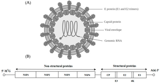
Alphavirus is a genus of RNA viruses, the sole genus in the Togaviridae family. Alphaviruses belong to group IV of the Baltimore classification of viruses, with a positive-sense, single-stranded RNA genome. There are 32 alphaviruses, which infect various vertebrates such as humans, rodents, fish, birds, and larger mammals such as horses, as well as invertebrates. Alphaviruses that could infect both vertebrates and arthropods are referred dual-host alphaviruses, while insect-specific alphaviruses such as Eilat virus and Yada yada virus are restricted to their competent arthropod vector. Transmission between species and individuals occurs mainly via mosquitoes, making the alphaviruses a member of the collection of arboviruses – or arthropod-borne viruses. Alphavirus particles are enveloped, have a 70 nm diameter, tend to be spherical, and have a 40 nm isometric nucleocapsid.

<i>Aphthovirus</i> Genus of viruses

Aphthovirus is a viral genus of the family Picornaviridae. Aphthoviruses infect split-hooved animals, and include the causative agent of foot-and-mouth disease, Foot-and-mouth disease virus (FMDV). There are seven FMDV serotypes: A, O, C, SAT 1, SAT 2, SAT 3 and Asia 1, and four non-FMDV serotypes belonging to three additional species Bovine rhinitis A virus (BRAV), Bovine rhinitis B virus (BRBV) and Equine rhinitis A virus (ERAV).

<i>Pestivirus</i> Genus of viruses

Pestivirus is a genus of viruses, in the family Flaviviridae. Viruses in the genus Pestivirus infect mammals, including members of the family Bovidae and the family Suidae. There are 11 species in this genus. Diseases associated with this genus include: hemorrhagic syndromes, abortion, and fatal mucosal disease.

The murine leukemia viruses are retroviruses named for their ability to cause cancer in murine (mouse) hosts. Some MLVs may infect other vertebrates. MLVs include both exogenous and endogenous viruses. Replicating MLVs have a positive sense, single-stranded RNA (ssRNA) genome that replicates through a DNA intermediate via the process of reverse transcription.

<span class="mw-page-title-main">Viral envelope</span> Outermost layer of many types of the infectious agent

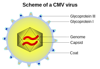
A viral envelope is the outermost layer of many types of viruses. It protects the genetic material in their life cycle when traveling between host cells. Not all viruses have envelopes. A viral envelope protein or E protein is a protein in the envelope, which may be acquired by the capsid from an infected host cell.

<i>Lipothrixviridae</i> Family of viruses

Lipothrixviridae is a family of viruses in the order Ligamenvirales. Thermophilic archaea in the phylum Thermoproteota serve as natural hosts. There are 11 species in this family, assigned to 4 genera.

Vesivirus is a genus of viruses, in the family Caliciviridae. Swine, sea mammals, and felines serve as natural hosts. There are two species in this genus. Diseases associated with this genus include: respiratory disease, Feline calicivirus (FCV); conjunctivitis, and respiratory disease.

<i>Lagovirus</i> Genus of viruses

Lagovirus is a genus of viruses, in the family Caliciviridae. Lagomorphs serve as natural hosts. There are two species in this genus. Diseases associated with this genus include: necrotizing hepatitis leading to fatal hemorrhages.

Mason-Pfizer monkey virus (M-PMV), formerly Simian retrovirus (SRV), is a species of retroviruses that usually infect and cause a fatal immune deficiency in Asian macaques. The ssRNA virus appears sporadically in mammary carcinoma of captive macaques at breeding facilities which expected as the natural host, but the prevalence of this virus in feral macaques remains unknown. M-PMV was transmitted naturally by virus-containing body fluids, via biting, scratching, grooming, and fighting. Cross contaminated instruments or equipment (fomite) can also spread this virus among animals.

Tristromaviridae is a family of viruses. Archaea of the genera Thermoproteus and Pyrobaculum serve as natural hosts. Tristromaviridae is the sole family in the order Primavirales. There are two genera and three species in the family.

<i>Middelburg virus</i> Species of virus

Middelburg virus (MIDV) is an alphavirus of the Old World Group that has likely endemic and zoonotic potential. It is of the viral family Togaviridae. It was isolated from mosquitos in 1957 in South Africa, MDIV antigens have now been found in livestock, horses, and humans.

<i>Avian metaavulavirus 2</i> Species of virus

Avian metaavulavirus 2, formerly Avian paramyxovirus 2, is a species of virus belonging to the family Paramyxoviridae and genus Metaavulavirus. The virus is a negative strand RNA virus containing a monopartite genome. Avian metaavulavirus 2 is one of nine species belonging to the genus Metaavulavirus. The most common serotype of Avulavirinae is serotype 1, the cause of Newcastle disease (ND). Avian metaavulavirus 2 has been known to cause disease, specifically mild respiratory infections in domestic poultry, including turkeys and chickens, and has many economic effects on egg production and poultry industries. The virus was first isolated from a strain in Yucaipa, California in 1956. Since then, other isolates of the virus have been isolated worldwide.

Yokose virus (YOKV) is in the genus Flavivirus of the family Flaviviridae. Flaviviridae are often found in arthropods, such as mosquitoes and ticks, and may also infect humans. The genus Flavivirus includes over 50 known viruses, including Yellow Fever, West Nile Virus, Zika Virus, and Japanese Encephalitis. Yokose virus is a new member of the Flavivirus family that has only been identified in a few bat species. Bats have been associated with several emerging zoonotic diseases such as Ebola and SARS.

Rio Negro virus is an alphavirus that was first isolated in Argentina in 1980. The virus was first called Ag80-663 but was renamed to Rio Negro virus in 2005. It is a former member of the Venezuelan equine encephalitis complex (VEEC), which are a group of alphaviruses in the Americas that have the potential to emerge and cause disease. Río Negro virus was recently reclassified as a distinct species. Closely related viruses include Mucambo virus and Everglades virus.

References

  1. 1 2 Bennett AJ, Paskey AC, Ebinger A, Kuhn JH, Bishop-Lilly KA, Beer M, Goldberg TL (31 July 2020). "Create two new species and rename one species in genus Rubivirus (Hepelivirales: Matonaviridae)" (docx). International Committee on Taxonomy of Viruses (ICTV). Archived from the original on 21 December 2022. Retrieved 15 May 2021.
  2. Megyeri K, Berencsi K, Halazonetis TD, et al. (June 1999). "Involvement of a p53-dependent pathway in rubella virus-induced apoptosis". Virology. 259 (1): 74–84. doi: 10.1006/viro.1999.9757 . PMID   10364491.
  3. 1 2 3 4 5 ICTV. "Create a new family Matonaviridae to include the genus Rubivirus, removed from the family Togaviridae Release" . Retrieved 29 October 2019.[ dead link ]
  4. Bennett, Andrew J.; Paskey, Adrian C.; Ebinger, Arnt; Pfaff, Florian; Priemer, Grit; Höper, Dirk; Breithaupt, Angele; Heuser, Elisa; Ulrich, Rainer G.; Kuhn, Jens H.; Bishop-Lilly, Kimberly A. (7 October 2020). "Relatives of rubella virus in diverse mammals". Nature: 1–5. doi:10.1038/s41586-020-2812-9. ISSN   1476-4687. PMC   7572621 . PMID   33029010.
  5. Gibbons, Ann (7 October 2020). "Newly discovered viruses suggest 'German measles' jumped from animals to humans". science. doi:10.1126/science.abf1520. Archived from the original on 8 December 2022. Retrieved 30 June 2022.
  6. Bardeletti G, Kessler N, Aymard-Henry M (1975). "Morphology, biochemical analysis and neuraminidase activity of rubella virus". Arch. Virol. 49 (2–3): 175–86. doi:10.1007/BF01317536. PMID   1212096.
  7. 1 2 "Viral Zone". ExPASy. Archived from the original on 24 June 2015. Retrieved 15 June 2015.
  8. Chen MH, Icenogle JP (April 2004). "Rubella virus capsid protein modulates viral genome replication and virus infectivity". Journal of Virology. 78 (8): 4314–22. doi:10.1128/jvi.78.8.4314-4322.2004. PMC   374250 . PMID   15047844.
  9. Beatch MD, Everitt JC, Law LJ, Hobman TC (August 2005). "Interactions between rubella virus capsid and host protein p32 are important for virus replication". J. Virol. 79 (16): 10807–20. doi:10.1128/JVI.79.16.10807-10820.2005. PMC   1182682 . PMID   16051872.
  10. Beatch MD, Hobman TC (June 2000). "Rubella virus capsid associates with host cell protein p32 and localizes to mitochondria". J. Virol. 74 (12): 5569–76. doi:10.1128/JVI.74.12.5569-5576.2000. PMC   112044 . PMID   10823864.
  11. 1 2 Dominguez G, Wang CY, Frey TK (July 1990). "Sequence of the genome RNA of rubella virus: evidence for genetic rearrangement during togavirus evolution". Virology. 177 (1): 225–38. doi:10.1016/0042-6822(90)90476-8. PMC   7131718 . PMID   2353453.
  12. Chen, MH; Frey TK (1999). "Mutagenic analysis of the 3' cis-acting elements of the rubella virus genome". J Virol. 73 (4): 3386–3403. PMC   104103 . PMID   10074193.
  13. Zhou Y, Chen X, Ushijima H, Frey TK (2012) Analysis of base and codon usage by rubella virus. Arch Virol
  14. "Togaviridae- Classification and Taxonomy". Archived from the original on 5 August 2012. Retrieved 10 May 2009.
  15. Garbutt M, Law LM, Chan H, Hobman TC (May 1999). "Role of rubella virus glycoprotein domains in assembly of virus-like particles". J. Virol. 73 (5): 3524–33. PMC   104124 . PMID   10196241.
  16. "Standardization of the nomenclature for genetic characteristics of wild-type rubella viruses" (PDF). Wkly Epidemiol Rec. 80 (14): 126–132. 2005. PMID   15850226. Archived from the original (PDF) on 18 May 2009.